www.fm.viamedica.pl
Address for correspondence: Prof. M.J. Golalipour, Department of Embryology and Histology, Gorgan University of Medical Sciences, Gorgan, Iran, P.O. Box: 49175-553, tel./fax: +98 (171) 4425165, 4425660, e-mail: mjgolalipour@yahoo.com
Morphometric alterations to the rat spleen
following formaldehyde exposure
M.J. Golalipour
1, H. Kord
1, S. Ghafari
1, A.M. Gharravi
1, A. Davarian
1,
S.A. Fazeli
1, R. Azarhoush
21Department of Embryology and Histology, Gorgan University of Medical Sciences, Gorgan, Iran
2Department of Pathology, Gorgan University of Medical Sciences, Gorgan, Iran
[Received 20 August 2007; Revised 19 December 2007; Accepted 3 January 2008]
Formaldehyde is commonly used in the production of various industrial and medical products. At room temperature formaldehyde easily evaporates. Expo-sure to formaldehyde can be hazardous to human health. Studies show that the vapour can be the cause of clinical symptoms such as throat, eye, skin and nasal irritation. It can also decrease the production of IgM in the spleen cells. This study was designed to determine the morphometric changes to the spleen in rats when samples were exposed to formaldehyde for 18 weeks.
A total of 28 albino Wistar rats aged 6–7 postnatal weeks were divided into the following three case groups according to their exposure to formaldehyde: E1 (2 h/day, 2 days/week), E2 (2 h/day, 4 days/week), E3 (4 h/day, 4 days/week) and one control group. When the exposure period had expired the animals were anaesthetised with chloroform. After cervical dislocation, the abdomen was dissected and spleen specimens were taken. These were sectioned and stained with the haematoxylin and eosin technique for morphometric study. Data was obtained from an Olympus light microscope and then analysed with SPSS (version 11.5) and one-way ANOVA test.
The white pulp area and diameter and the marginal zone diameter were greater in group E3 than those in the other groups. The germinal centre area and diameter and the diameter of the periarterial lymphoid sheaths (PALS) were greater in group E2 than in other groups, although there was no significant difference between groups in the area of white pulp and the PALS diameter (p < 0.05).
This study showed that formaldehyde vapour can cause morphometric chang-es in the white pulp of the spleen in rats (Folia Morphol 2008; 67: 19–23).
Key words: white pulp, periarterial lymphoid sheaths, follicle, germinal centre area, marginal zone diameter, mantle zone
INTRODUCTION
Formaldehyde (CH2O) is a flammable colourless reactive gas, readily polymerised at normal room temperature and pressure, with a relative molecu-lar mass of 30.03 and a pungent odour. Formalde-hyde is soluble in water, ethanol and diethyl ether.
It is also used in polymerised from as paraformalde-hyde [22].
the daytime, while in the presence of nitrogen diox-ide it drops to 35 minutes [22].
Formaldehyde is used in the manufacture of melamine, polyacetal and phenolic resins. Phenol-formaldehyde resins are used in the production of polywood, particle board, foam insulation and a wide variety of moulded or extruded plastic items [5]. Major anthropogenic sources affecting humans are indoor environments. Others include direct emis-sions, especially from the production and use of formaldehyde [22].
Its potential to act as an electrophile and act with macromolecules such as DNA, RNA and protein to from reversible adducts or irreversible cross-links [10] makes it a conventional tissue fixative, particularly for cadaver fixation. Acute formaldehyde exposure mainly produces mucosal irritation of the eye and upper respiratory tract in humans [23], and long-term exposure leads to the production of nasal tu-mours in rodents [14]. Formaldehyde also causes pulmonary function impairment [3] and asthmatic reactions in sensitised individuals [4, 9]. It can also decrease human body weight and increase lymph node weight during oral exposure, but there is no effect on lymphoid tissue cellularity and formalde-hyde decreases the production of immunoglobin from the spleen cell [20].
In the dissection laboratory and during cadaver dissection instructors of anatomy and medical stu-dents are exposed to formaldehyde vapour derived from fixed cadavers. This study on 28 albino Wistar rats was carried out in order to study the morpho-metric changes in the spleen due to formaldehyde exposure in the dissection laboratory and to deter-mine its relationship with the duration of exposure.
MATERIAL AND METHODS
The study was performed in 2004 in the Faculty of Medicine, Gorgan University of Medical Science on 28 albino Wistar rats aged 6–7 postnatal weeks provided by the Iranian Pasteur Institute. The rats were randomly divided into three equal case groups on the basis of the differences in the periods of ex-posure to formaldehyde:
— E1 — 2 h/day, for 2 days/week for 18 weeks (2 h/day, 2 days/week);
— E2 — 2 h/day, 4 days/week for 18 weeks (2 h/day, 4 days/week);
— E3 — 4 h/day, 4 days/week for 18 weeks (4 h/day, 4 days/week).
There was also a control group that did not un-dergo any exposure. The mean weights for the
groups were found by means of a digital scale to be as follows: 222 g (E1), 209 g (E2), 252 g (E3) and 195 g (control group).
Approval for this study was gained from the An-imal Care and Ethics Committee of the Gorgan Uni-versity of Medical Sciences.
The concentration of formaldehyde vapour in the dissection room (the place of exposure) was mea-sured at the beginning, during and at the end of the study by means of a detector tube and dragger pump (model 31, made in Germany) after the cov-ers of the cadavcov-ers had been removed. The mean vapour concentration of the dissection room was 1.5 ppm. The temperature of the dissection room was 20–26°C and the air pressure was 760–763 atm. At non-exposure times all groups of animals were kept in the laboratory animal quarters, which were far removed from the place of exposure and where no formaldehyde was detected. The animal quar-ters were ventilated and the temperature kept at about 21°C; air-conditioning and adequate light were supplied. All groups were fed with a similar standard diet (provided by the Iranian Pasteur Insti-tute) twice a day (morning and afternoon), and water was available ad libitum.
In the time of exposure the cages of the case groups were transferred to the dissection room, placed at the same height as a cadaver and separat-ed by a distance of 15 cm. This procseparat-edure was fol-lowed during the 18 weeks of exposure according to the time protocols described above. During each period of exposure the control group was kept in the animal quarters. When the exposure period had expired, each rat of the three experimental groups and the control group was anaesthetised with ether, and spleen specimens were taken.
After tissue processing and paraffin embedding, vertical serial sections were cut from each specimen at 5 µm and approximately 20 were sections select-ed from each spleen. Sections were arrangselect-ed in a number of sequences and stained with haematoxy-lin and eosin. All of the sections were studied by an Olympus light microscope and Olysia program with a magnification of 400¥.
In this study the morphometric variables of white pulp were used to measure the light microscopy changes to the following:
— the white pulp subdivided into the periarterial lym-phoid sheaths (PALS), the follicles (germinal centres and mantle zone) and the marginal zone [16, 21]; — area: the total area of the white pulp and
— diameter: the diameter of the white pulp, mar-ginal zone, germinal centre, PALS and mantle layer. The data were analysed with SPSS (version 11.5) and compared by ANOVA (p < 0.05).
RESULTS
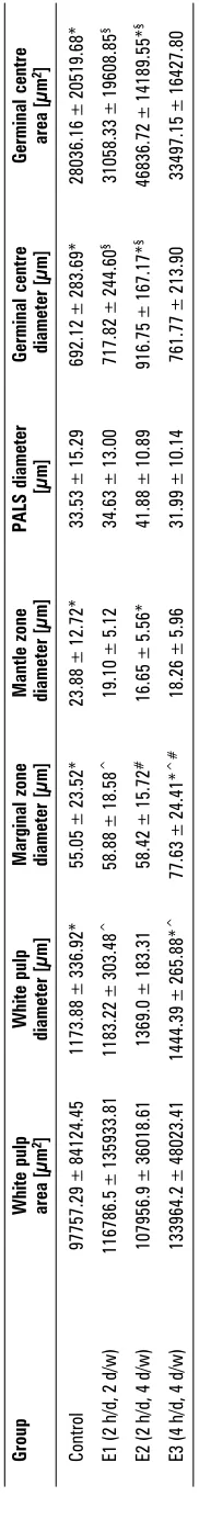
The morphometric variables of white pulp in the experimental and control group are given in Table 1. The white pulp area and diameter in group E3 were greater than in the other groups (133964 ± ± 48023 µm2 and 1444.39 ± 265.88 µm respecti-vely). However, there was no significant difference between groups in the area of white pulp (p > 0.05). The mean ± SD of marginal zone diameters in E1, E2, E3 and the control group were 58.88 ± 18.58 µm, 58.42 ± 15.72 µm, 77.63 ± 24.41 µm and 55.05 ± ± 23.52 µm respectively. This difference was signifi-cant between E3 and other the groups (p < 0.05; Table 1).
The mean ± SD of the germinal centre diameter in group E2 (916.75 ± 167.17 µm) was greater than in the other groups (p < 0.05).
Furthermore, the mantle zone diameter in the control group (23.88 ± 12.72 µm) was significantly greater than in group E2 (16.65 ± 5.56 µm; p < 0.05). There were no significant differences between any of the groups in the diameter of the PALS.
DISCUSSION
The study showed that formaldehyde vapour can cause morphometric changes in the spleen of rats. Formaldehyde vapour exposure of greater duration increased germinal centre diameter, germinal cen-tre area and marginal zone diameter and decreased the mantle layer diameter.
The data obtained in the experiment by Thrasher et al. [18, 19] showed chronic exposure to formal-dehyde to be associated with immunological hy-persensitivity, as measured by elevated circulat-ing IgG and IgE autoantibodies to human serum albumin. In addition, a decrease in the proportion of T-cells was observed, indicating altered immu-nity [16].
Other studies have shown that formaldehyde can decrease human body weight and increase lymph node weight during oral consumption, although there is no effect on the cellularity of lymphoid tis-sue [20].
Formaldehyde induced an increase in bacterial survival in the lungs of mice exposed to 15 ppm (18 mg/m3) [11] as well as a reduction in serum IgM
titers in animals orally administered 40 or 80 mg/kg Table 1.
Morphometric evaluation of the spleen of rats in the three experimental groups and the control group
Group White pulp White pulp Marginal zone Mantle zone PALS diameter Germinal centre Germinal centre area [µm 2] diameter [µm] diameter [µm] diameter [µm] [µm] diameter [µm] area [µm 2] Control 97757.29 ± 84124.45 1173.88 ± 336.92*
55.05 ± 23.52*
23.88 ± 12.72*
33.53 ± 15.29
692.12 ± 283.69* 28036.16 ± 20519.68*
E1 (2 h/d, 2 d/w)
116786.5 ± 135933.81 1183.22 ± 303.48 ^
58.88 ± 18.58
^
19.10 ± 5.12
34.63 ± 13.00
717.82
±
244.60
§
31058.33 ± 19608.85
§
E2 (2 h/d, 4 d/w)
107956.9 ± 36018.61 1369.0 ± 183.31 58.42 ± 15.72 #
16.65 ± 5.56*
41.88 ± 10.89
916.75
±
167.17*
§
46836.72 ± 14189.55*
§
E3 (4 h/d, 4 d/w)
133964.2 ± 48023.41 1444.39 ± 265.88* ^
77.63 ± 24.41*
^#
18.26 ± 5.96
31.99 ± 10.14
761.77 ± 213.90 33497.15 ± 16427.80
Results are expressed as mean ± SD (standard deviation) of the mean; P-value < 0.05; *experimental group
were compared with control group;
§group E2 was compared with group E1; ^group E3 was compared with group E1; #group E3 was
body weight per day for 5 days per week over 4 weeks [20]. Formaldehyde administered at 20, 40, 80 mg//kg/day for 4 weeks by gavage caused a dose--dependent reduction of antibody response (IgG, IgM), reduced phagocytic activity, dose-dependent depres-sion of haemagglutinin titers in the liver (vacuolisa-tion of hepatocytes) and spleen histology (for exam-ple, narrowed thymus-dependent zones of PALS) [15]. A study of the bone marrow and spleen of ani-mals injected with 6.25–25 mg/kg formaldehyde IP showed no induction of chromosomal aberration [15]. The immunotoxicity of formaldehyde in mice was studied by Dean et al. [6]. Female B6C3F1 mice un-derwent inhalational exposure to 18 mg/m3 for 6 h/day, 5 days/week, over 3 weeks. Most of the immune functions involving T and B lymphocytes and macrophages were not impaired and there was enhanced resistance to Listeria monocytogenes [6]. In another study by Adams et al. [1] formaldehyde exposure of mice to 18 mg/m3
(15 ppm) for 6 h/day over 3 weeks caused an approximately twofold in-crease in competence of the peritoneal macrophag-es in the release of hydrogen peroxide (H2O2) [1]. Enhanced function of the macrophages may be re-sponsible for the increased resistance reported by Dean et al. [6].
Epidemiological investigations into the effects of exposure to formaldehyde on the immune system have focused mainly upon allergic reactions [2, 8, 17]. Formaldehyde can interact with molecules on cell membranes and in body tissues and fluids (for in-stance, proteins and DNA) and disrupt cellular func-tions. High concentrations cause precipitation of pro-teins, which results in cell death. Absorption from the respiratory tract is very rapid. Once absorbed, formaldehyde is metabolised to formic acid, which may lead to acid-base imbalance and a number of other systemic effects [13].
According to Elmore study [7] formaldehyde may act as antigens. A severe immune reaction to anti-gens could result in an increased cellularity in the B-cell areas and an increase in secondary follicles with major germinal centres. These effects lead to an in-crease in the white pulp area and follicle diameter. Immature B cells or immunoblasts will multiply in response to antigenic stimuli that result in an in-crease in the diameter of the germinal centre mar-ginal zone and mantle zone.
Lino dos Santos Franco et al. [12] also indicated that a significant increase in the total cell count due to formaldehyde inhalation (1% formalin solution
for 90 min once a day over 4 days) was found in the spleen of formaldehyde-treated rats [12].
In conclusion, the results of this study showed that chronic formaldehyde exposure at the concen-tration and duration described can cause profound morphometric alterations in the follicles and margin-al zone of the rat spleen. In addition, it seems that there is no relationship between the severity of expo-sure-induced changes and the duration of exposure. The mechanism of its action remains, therefore, to be elucidated. Further study is required to determine the relationship between the severity of the expo-sure-induced changes and the duration of exposure.
ACKNOWLEDGEMENTS
The authors would like to express their apprecia-tion of the Department of Research, Gorgan Univer-sity of Medical Sciences for financial support, and are particularly grateful to Mr. Moludi and Ms Irani. In addition, we are indebted to Prof. Boldaji, who guided us through the study.
REFERENCES
1. Adams DO, Hamilton TA, Lauer LD, Dean JH (1987) The effect of formaldehyde exposure upon the mono-nuclear phagocyte system of mice. Toxicol Appl Phar-macol, 88: 165–174.
2. Bardana EJ JR, Montanaro A (1991) Formaldehyde: an analysis of its respiratory, cutaneous, and immunolog-ic effects. Ann Allergy, 66: 441–452.
3. Berbstein RS, Staynedr LT, Elliott LJ, Kimbrough R (1984) Inhalation exposure to formaldehyde: an over-view of its toxicology, epidemiology, monitoring and control. Am Ind Hyg Assoc J, 45: 778–785.
4. Burge PS, Harries MG, Lam WK, O’Brien IM, Patchett PA (1985) Occupational asthma due to formaldehyde. Thorax, 40: 255–260.
5. CARB (1999) Air toxics emissions data collected in the Air Toxics Hot Spots Program CEIDARS; database as of January 29.
6. Dean JH, Lauer LD, House RB, Murray MJ, Stillman WS, Irons RD, Steinhagen WH, Phelps MC, Adams DO (1984) Studies of immune function and host resistance in B6C3F1 mice exposed to formaldehyde. Toxicol Appl Pharmacol, 72: 519–529.
7. Elmore SA (2006) Enhanced histopathology of the spleen. Toxicol Pathol 34: 648–655.
8. Feinman SE (1988) Formaldehyde sensitivity and tox-icity. Boca Raton, FL, CRC Press.
9. Gorski P, Krakowiak A (1991) Formaldehyde induced bronchial asthma: does it really exist? Pol J Occup Med, 4: 317–320.
11. Jakab GJ (1992) Relationship between carbon black particulate-bound formaldehyde, pulmonary antibac-terial defenses, and alveolar macrophage phagocy-tosis. Inhal Toxicol, 4: 325–342.
12. Lino dos Santos Franco A, Damazo AS, Beraldo de Souza HR, Domingos HV, Oliveira-Filho RM, Oliani SM, Costa SK, Tavares de Lima W (2006) Pulmonary neutrophil recruit-ment and bronchial reactivity in formaldehyde-exposed rats are modulated by mast cells and differentially by neuropeptides and nitric oxide. Toxicol Appl Pharma-col, 214: 35–42.
13. Medical Management Guidelines for Formaldehyde Agen-cy for Toxic Substance and Disease Registry (2002) Divi-sion of Toxicology (http://www.atsdr.cdc.gov/substanc-es/formaldehyde/).
14. Monticello TM, Swenberg JA, Gross EA, Leininger JR, Kimbell JS, Seilkop S, Starr TB, Gibson JE, Morgan KT (1996) Correlation of regional and nonlinear formal-dehyde-induced nasal cancer with proliferating popu-lations of cells. Cancer Res, 56: 1012–1022.
15. Organization for Economic Cooperation and Develop-ment (OECD) (2002) Screening Information Data Set (SIDS) Initial Assessment Report For SIAM 14. United Nations Environment Programme (UNEP), Paris. 16. Saito H, Yokoi Y, Watanabe S, Tajima J, Kuroda H,
Namihisa T (1988) Reticular meshwork of the spleen in
rats studied by electron microscopy. Am J Anat, 181: 235–252.
17. Stenton SC, Hendrick DJ (1994) Formaldehyde. Immunol Allergy Clin NA, 14: 635–657.
18. Thrasher JD, Wojdani A, Cheung G, Heuser G (1987) Evidence for formaldehyde antibodies and altered cel-lularity immunity in subjects exposed to formaldehyde in mobile homes. Arch Environ Health, 42: 347–350. 19. Thrasher JD, Broughton A, Madison R (1990) Immune
activation and autoantibodies in humans with long-term inhalation exposure to formaldehyde. Arch Envi-ron Health, 45: 217–223.
20. Vargova M, Wagnerova J, Liskova A, Jakubovsky J, Gajdova M, Stolcova E, Kubova J, Tulinska J, Stenclova R (1993) Subacute immunotoxicity study of formalde-hyde in male rats. Drug Chem Toxicol, 16: 255–275. 21. Ward JM, Mann PC, Morishima H, Frith CH (1999)
Thy-mus, Spleen, and Lymph Nodes. In: Maronpot RR (ed.) Pathology of the mouse. Cache River Press, Vienna, Illinois, pp. 333–360.
22. World Health Organization (1989) Environmental health criteria for formaldehyde. No. 89, Geneva.