www.fm.viamedica.pl
Address for correspondence: M. Topol, Department of Angiology, Chair of Anatomy, Medical University, G. Narutowicza 60, 90–136 Łódź, Poland, tel: +48 42 630 49 49, e-mail: mtopol@toya.net.pl
The femoral artery and its branches
in the baboon Papio anubis
Dyl Ł., Topol M.
Department of Angiology, Chair of Anatomy, Medical University, Łódź, Poland
[Received 13 July 2007; Revised 19 October 2007; Accepted 19 October 2007]
The aim of the research was to examine the anatomy of the arterial system in the inguinal region, hip and thigh of Papio anubis. No description of this was found in the available scientific literature, although, at the same time, the baboon is con-sidered to be a good animal model in biomedical research.
Macroscopic anatomical research was carried out on 20 hind limbs (10 cadav-ers: 9 male and 1 female) of adult Papio anubis and the results were then compared with the anatomy of the arterial hind limb systems of other apes as described in the literature. The circulatory system of the whole body was filled with coloured latex via the common carotid artery and internal jugular vein, and traditional methods were then used to prepare the vessels. The arterial system in the hind extremity of Papio anubis was recorded. The anatomical names of human arteries were used as well as the names of those of apes as applied in the literature.
The femoral artery was the only artery supplying the hind limb of Papio anubis. It started under the inguinal ligament as a continuation of the external iliac artery. It went down and divided into the popliteal artery, which coursed in the popliteal fossa, and the saphenous artery, which passed on the medial side of the thigh and leg. The number of smaller branches and the way in which they issued from the larger arteries were documented. The external diameter and length of the hind limb arteries were measured.
It was observed that the cutaneous branches of the femoral artery supplied the inguinal and genital regions and the abdominal wall, while the deep artery of the thigh was the main vessel of the hip and thigh.
Key words: anatomy, baboon, Papio anubis, femoral artery
INTRODUCTION
The aim of the research was to examine the anat-omy of the arterial system in the inguinal region, hip and thigh of Papio anubis. No description of the baboon’s arterial system was found among docu-ments dealing with the anatomy of apes, although the baboon is considered to be good animal model in biomedical research [4]. However, several similar-ities and differences were found between the
MATERIAL AND METHOD
A macroscopic anatomical study was conduct-ed on 20 hind limbs (10 cadavers: 9 male and 1 female) of adult Papio anubis. The circulatory system of the whole body was filled with coloured latex via the common carotid artery and the inter-nal jugular vein and the hind limbs were then pre-pared by traditional methods. Details of the arte-rial system of the hind limb of Papio anubis were recorded. In this research the anatomical names for the arteries of apes were used according to their use in the literature of the subject. Observa-tion was focused on the beginning of the vessels, their course and division and the regions they sup-plied. The external diameter at the beginning of the arteries was measured. Next, the results were compared with those presented in literature deal-ing with the arterial hind limb systems of other apes and of humans.
RESULTS
All the arteries of the hind limb of Papio anubis were single vessels and the femoral artery was the only artery to supply the limb. This had its origin under the inguinal ligament, in the femoral sheet and was the continuation of the external iliac artery. It ran downwards and finished in the medial part of the femur, dividing into the popliteal artery and the saphenous artery. At the proximal part of its course the femoral artery gave off the following cutaneous (superficial) branches:
— the superficial epigastric artery in 8 specimens (40%) branching separately, in 12 specimens (60%), with other arteries;
— the external pudendal artery in 8 specimens (40%) branching separately, in 12 specimens (60%), with other arteries;
— the superficial circumflex iliac artery in 12 speci-mens (60%), branching separately in 8 specispeci-mens (40%), with other arteries.
It was difficult to estimate the leading branch when at least two arteries started with a trunk from the femoral artery. The following branches of the femoral artery were muscular (deep) arteries: — the deep artery of the thigh in 4 specimens (20%)
started at the same level of the femoral artery as other arteries, while in 16 specimens (80%) it was the main trunk for other arteries;
— the medial circumflex femoral artery in only 4 specimens (20%) started directly from the fem-oral artery;
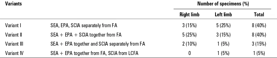
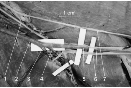
— the lateral circumflex femoral artery was a branch of the femoral artery in 8 specimens (40%). The femoral artery was 4–6 mm wide (Fig. 1). We distinguished 4 variants in the origin of the cutaneous branches of the femoral artery (Table 1): — variant I: The superficial epigastric artery, the
ex-ternal pudendal artery and the superficial circum-flex iliac artery arose from the femoral artery sep-arately (8 cases, 40%);
Figure 1. The right femoral triangle in Papio anubis. 1 — the inguinal ligament, 2 — the external pudendal artery, 3 — the superficial epigastric artery, 4 — the superficial circumflex iliac artery, 5 — the lateral circumflex femoral artery, 6 — the femoral artery, 7 — the femoral vein, 8 — the deep artery of the thigh, 9 — the medial circumflex femoral artery. The medial view of the right femur in Papio anubis. Bare 1 cm.
Table 1. Variants of the origin of the cutaneous branches of the femoral artery in Papio anubis (for details refer to the text)
Variants Number of specimens (%)
Right limb Left limb Total
Variant I SEA, EPA, SCIA separately from FA 3 (15%) 5 (25%) 8 (40%)
Variant II SEA + EPA + SCIA together from FA 5 (25%) 3 (15%) 8 (40%)
Variant III SEA + EPA together and SCIA separately from FA 2 (10%) 1 (5%) 3 (15%)
Variant IV SEA + EPA together from FA, SCIA from LCFA 0 1 (5%) 1 (5%)
— variant II: The superficial epigastric artery, the ex-ternal pudendal artery and the superficial circum-flex iliac artery branched from the femoral artery together (8 cases, 40%);
— variant III: The superficial epigastric artery and the external pudendal artery arose together from the femoral artery and the superficial circumflex iliac artery branched from the femoral artery sep-arately (3 cases, 15%);
— variant IV: The superficial epigastric artery and the external pudendal artery arose together from the femoral artery and the superficial circumflex iliac artery branched from the lateral circumflex femoral artery (1 case, 5%).
The superficial epigastric artery branched off the femoral artery separately in 8 specimens (40%), in the trunk with the external pudendal and the super-ficial circumflex iliac arteries in 8 specimens (40%) and in the trunk with the external pudendal artery in 4 specimens (20%). It rose just below the inguinal ligament and supplied the skin of the inguinal re-gion and the wall of the abdomen. It was less than 1 mm wide.
The external pudendal artery arose from the femo-ral artery separately in 8 specimens (40%), in the trunk with the superficial epigastric and the superficial cir-cumflex iliac arteries in 8 specimens (40%) and in the trunk with the superficial epigastric artery in 4 speci-mens (20%). It went medially towards the genitals, supplying the skin of this region. It was 1 mm wide.
The superficial circumflex iliac artery was a branch of the femoral artery in 19 specimens (95%) and the lateral circumflex femoral artery in 1 specimen (5%). It left these vessels separately in 12 specimens (60%) and in the trunk with the superficial epigastric and external pudendal artery in 8 specimens (40%). After leaving the femoral artery, the lateral circum-flex femoral artery or trunk, it passed laterally and surrounded the hip. It was 1–2 mm wide.
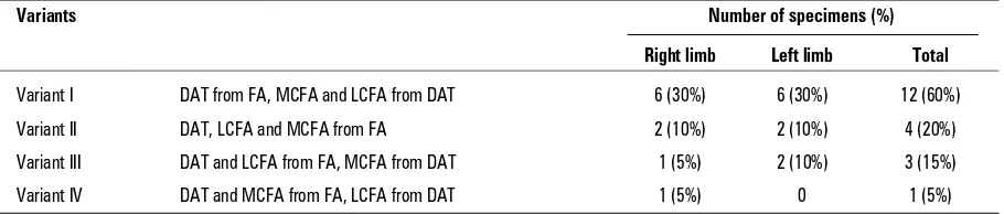
We distinguished four variants of the muscular branches of the division of the femoral artery (Table 2): — variant I: The deep artery of the thigh began from the femoral artery, while the medial and lateral circumflex femoral arteries branched from the deep artery of the thigh (12 cases, 60%); — variant II: The deep artery of the thigh and the
lateral and medial circumflex femoral arteries branched from the femoral artery (4 cases, 20%); — variant III: The deep artery of the thigh and the lateral circumflex femoral artery branched from the femoral artery, while the medial circumflex femoral artery branched from the deep artery of the thigh (3 cases, 15%);
— variant IV: The deep artery of the thigh and the medial circumflex femoral artery branched from the femoral artery, while the lateral circumflex femoral artery branched from the deep artery of the thigh (1 case, 5%).
The deep artery of the thigh mainly supplied the thigh muscles. It started in the proximal third of the femoral artery at 2.0–4.9 cm (mean 3.4 cm) below the inguinal ligament and descended posteriorly to the femoral artery, passing between the anterior and medial muscle compartments of the thigh. Both the medial and lateral circumflex femoral arteries were branches of this in 12 specimens (60%), while in 3 specimens (15%) only the former was a branch of it and in 1 specimen (5%) only the latter was a branch of it. The deep artery of the thigh was 3 mm wide.
The medial circumflex femoral artery arose from the femoral artery in 5 specimens (25%) and from the deep artery of the thigh in 15 specimens (75%). At the beginning it went medially. It then turned pos-teriorly and ended in the muscles of the posterior fas-cial compartment of the thigh. It was 1 mm wide.
The lateral circumflex femoral artery arose from the femoral artery in 7 specimens (35%) and from the deep artery of the thigh in 13 specimens (65%). It followed
Table 2. Variants of the muscular branches of the division of the femoral artery in Papio anubis (for details refer to the text)
Variants Number of specimens (%)
Right limb Left limb Total
Variant I DAT from FA, MCFA and LCFA from DAT 6 (30%) 6 (30%) 12 (60%)
Variant II DAT, LCFA and MCFA from FA 2 (10%) 2 (10%) 4 (20%)
Variant III DAT and LCFA from FA, MCFA from DAT 1 (5%) 2 (10%) 3 (15%)
Variant IV DAT and MCFA from FA, LCFA from DAT 1 (5%) 0 1 (5%)
into the anterior thigh muscles, running posteriorly to the quadriceps femoris. It was 2 mm wide.
The observations showed that the cutaneous branches of the femoral artery supplied the inguinal, abdominal, iliac and genital regions, while the deep artery of the thigh was the main vessel of the hip and the thigh. The terminal branches of the femoral artery, namely the popliteal artery and the saphen-ous artery, reached and supplied the knee, leg and foot of Papio anubis (Fig. 2).
DISCUSSION
The research showed many similarities between the arterial systems of the hind limb of Papio anubis and those of other apes. Although there were differences from the human lower extremi-ty, especially in the distal part, several similarities were found between the human and baboon ar-terial systems. On these grounds the baboon may be considered a good animal model in biomedical research [4].
The femoral artery has been seen to be the con-tinuation of the external iliac artery in Papio anubis, Macaca rhesus, Macaca cynomolgus, Macaca cyclop-sis and humans [3, 6, 9, 13]. In all these species the branches of the femoral artery have been reported as supplying the inferior part of the abdominal wall, the inguinal and genital regions and the hind leg [12, 13]. During the phylogenesis of primates changes in anatomy are observable. The cutaneous (super-ficial) branches of the femoral artery are the su-perficial epigastric artery, the susu-perficial circum-flex iliac artery and the external pudendal arteries. In most cases of baboons and humans these ar-teries had separate beginnings [13]. The cutane-ous branches in Prosimii, Ceboidea and the lower species of Cercopithecoidea (Macaca) began from one trunk [13]. Our study of the baboon’s vessels showed four variants of the cutaneous branches of the femoral artery. The most frequently encoun-tered were two opposite variants: variant I, where three branches left the femoral artery in sequence, and variant II, with all branches leaving the femo-ral artery together.
The superficial epigastric artery branches off the femoral artery in all species. The human artery is separate (in 58%), more often than that of Macaca rhesus (in 14%) or Macaca cynomolgus (in 8%). In humans this artery branches off the femoral artery in a trunk with the superficial circumflex iliac artery (36%) or, only in 2%, with the external pudendal artery [13].
In Macaca the external pudendal artery was a branch of the femoral artery or medial circum-flex femoral artery and in most cases (66%) formed a common trunk with other arteries [6, 13]. In hu-mans this artery is mainly (96%) a separate branch of the femoral artery [13]. The origin of this artery in Papio anubis is often in a trunk with other ar-teries (60%).
The superficial circumflex iliac artery branches off the femoral artery, the external iliac artery or the lateral circumflex femoral artery to make a common trunk with other arteries more frequently in Macaca (66%) than in humans, where it makes a common trunk with the superficial epigastric artery (36%), but was a separate artery from the femoral artery (36%) or, in 4%, from the external iliac artery [13].
The deep artery of the thigh and the medial and lateral circumflex femoral arteries give off the deep muscle branches of the thigh. It was found in Pros-imii, Ceboidea and lower species of Cercopithecoidea that in the majority of cases these arteries were sep-arate branches of the femoral artery. In most cases in Macaca and baboons the medial and lateral cir-cumflex femoral arteries branched off the deep ar-tery of the thigh [1, 11, 13]. As reported by Bloda et al. [1], the most frequently encountered version in humans is one in which the deep artery of the thigh, together with the lateral circumflex femoral artery, arises from the femoral artery (37.5%), while the less common cases (32.5%) showed one trunk formed together by the deep artery of the thigh and the lateral and the medial circumflex femoral arteries. Załuska and Urbanowicz [13] found the common Figure 2. The division of the femoral artery in the middle of the thigh in Papio anubis. 1 — the femoral vein, 2 — the femoral artery, 3 — the popliteal artery, 4 — the descending genicular artery (branch of the saphenous artery), 5 — the saphenous artery, 6 — two trunks of the long saphenous vein, 7 — the saphenous nerve. The medial view of the right femur in Papio
trunk formed by the deep artery of the thigh and the lateral and medial circumflex femoral arteries to be frequent (62%) in humans.
According to Suder and Nizankowski [10], the ori-gin of the muscular branches of the femoral artery was as a common trunk in over 50% of human foetuses.
The deep artery of the thigh and its perforating and nutrient arteries supply the muscles of the thigh and the shaft of the human femur, and the medial and lateral circumflex femoral arteries surrounded and supplied the proximal part of the human fe-mur [3].
In humans the deep artery of the thigh was found in all cases to be a branch of the femoral artery [8, 9]. In the majority of cases the lateral circumflex femoral artery branched off the deep artery of the thigh but sometimes arose from the femoral artery [3]. Tanyeli et al. [11] demonstrated that in 79% of cas-es the medial circumflex femoral artery was a branch of the deep artery of the thigh and in 15% of the femoral artery.
The human femoral artery continues as the popliteal artery when the former passes through the adductor hiatus [2], while in Macaca and Papio anubis it splits into the popliteal artery and the saphe-nous artery [12, 13].
As we demonstrated previously in baboons [5, 7], the region of the division of the femoral ar-tery was that of the venous saphenofemoral junc-tions, where two similar trunks of the great saphen-ous vein surrounding the saphensaphen-ous artery reached one femoral vein (Fig. 2).
CONCLUSIONS
1. The main artery in the hind extremity of Papio anubis was the femoral artery.
2. The superficial epigastric artery, the superficial iliac artery and the external pudendal artery arose from the femoral artery separately with the same frequency (variant I, 40%) as they did together (variant II, 40%).
3. Of three variants of the muscular branching of the division of the femoral artery, the most common was variant I, where the medial and lateral cir-cumflex femoral arteries branched from the deep artery of the thigh. This was found in 60% cases.
REFERENCES
1. Bloda E, Sierocinski W, Kling A (1982) Variation of the arteria profunda femoris in man. Folia Morphol, 41: 123–131.
2. Colborn GL, Lumsden AB, Taylor BS, Skandalakis JE (1994) The surgical anatomy of the popliteal artery. Am Surg, 60: 238–246.
3. Colborn GL, Mattar SG, Taylor B, Skandalakis JE, Lums-den AB (1995) The surgical anatomy of the deep fem-oral artery. Am Surg, 61: 336–346.
4. Dormehl IC, Hugo N, Beverly G (1992) The baboon: an ideal model in biomedical research. Anesth Pain Con-trol Dent, 1: 109–115
5. Dyl L, Topol M (2007) Superficial veins of the foot in the baboon Papio anubis. Folia Morphol, 66: 15–19. 6. Fujita J (1963) Pelvic arteries in macacus cyclopsis.
I. The common iliac artery and the external iliac artery. Okajimas Folia Anat Jpn, 39: 85–116.
7. Janowski K, Topol M (2005) The topography of the superficial veins of the hind leg in the baboon Papio anubis in comparison with the superficial veins of the lower limb in humans. Folia Morphol, 64: 287–291. 8. Martin P, Renwick S, Stephenson C (1968) On the surgery
of the profunda femoris artery. Br J Surg, 55: 539–542. 9. Sahin B, Uzun A, Emirzeoglu M, Kosif R, Bilgic S (2003)
A deep femoral artery passing in front of the femoral vein. Folia Morphol, 62: 143–146.
10. Suder E, Nizankowski C (1985) Variations in the origin of the deep femoral arteries in human fetuses. Folia Morphol, 44: 262–269.
11. Tanyeli E, Uzel M, Yildirim M, Celik HH (2006) An ana-tomical study of the origins of the medial circumflex femoral artery in the Turkish population. Folia Mor-phol, 65: 209–212.
12. Załuska S, Urbanowicz Z (1970) The arteries of the leg in man and in Macacus. Ann Univ Mariae Curie Sklodowska Med, 25: 31–42.