www.fm.viamedica.pl O R I G I N A L A R T I C L E
Address for correspondence: Dr Małgorzata Waszak, Department of Functional Anatomy, University of Physical Education, ul. Królowej Jadwigi 27/39, 61–871 Poznań, Poland, tel: +61 835 52 26, fax: +61 833 00 87, e-mail: [email protected]
Sex dimorphism in development dynamics and
in development progression of morphological
features in human foetuses
Małgorzata Waszak, Krystyna Cieślik
Department of Functional Anatomy, Academy of Physical Education in Poznań, Poland
[Received 24 October 2002; Accepted 10 October 2002]
The study material comprised 3889 foetuses of both sexes, aged 20–42 weeks. t-Student test has been applied to evaluate the existence of potential sex-de-pendent differentiation of developmental trends as assessed by weekly mea-surements of selected somatic features and by the weight of internal organs. The regression coefficients of the analysed variables have been compared against the opposite sex. The rate of development of the analysed features in consecu-tive weeks has been found to be sex-related. Highest sex-related differences have been observed for the total body weight and for the weight of internal organs, with the exception of the adrenals, and the differences have been sig-nificant enough to justify the existence of contrasting, sex-dependent patterns of development of the analysed variables. The development of the analysed morphological features has been depicted by curvilinear regression. When de-scribed by various degree polynomials the development course of the analysed features displays sex-related differences. Only the change in the weight of the adrenals is similar for both sexes. The evaluation of the developmental advance-ment of the analysed features has revealed that they are usually at a more ad-vanced development level in female foetuses.
key words: foetal sex differentiation, rate of feature development, developmental maturity, feature development
INTRODUCTION
This study is part of a larger project focusing on sex dimorphism during foetal development in hu-mans. Sex dimorphism in foetal development has been a subject of numerous studies and controver-sy [2, 4, 6–9]. The formation of dimorphic differenc-es in somatic featurdifferenc-es and in the weight of internal organs reported in our earlier study [10] signalled the problem of sex-related differences in foetal de-velopment. In order to get a more complete picture of sex-related differences in foetal development oth-er manifestations of sex dimorphism need to be
in-vestigated. As a result, the aim of this study has been to analyse sex-dependent differences in developmen-tal dynamics and in the developmendevelopmen-tal advancement of somatic features and of the weight of internal organs in humans.
MATERIAL AND METHODS
includ-ed somatic features (1–7) and the weights of inter-nal organs (8–15). The variables comprised the fol-lowing items: 1. total body length; 2. crown-rump length; 3. body weight; 4. circumference of the head; 5. circumference of shoulders; 6. circumfer-ence of the chest; 7. circumfercircumfer-ence of the abdo-men; 8. brain weight; 9. heart weight; 10. lung weight; 11. liver weight; 12. spleen weight; 13. kid-ney weight; 14. adrenals weight; 15. thymus weight. None of the stillborn foetuses displayed any signs of developmental pathology and the causes of death have remained unexplained.
T-Student test has been applied to evaluate the existence of potential sex-dependent differentiation in developmental trends and in the rate of develop-ment as assessed by weekly measuredevelop-ments of the selected somatic features and the weight of internal organs. Development rate index (DRI) has been used for comparisons of developmental dynamics between the sets of features. The regression analysis (y = bx + + a) has been used in order to search for the possi-ble existence of sex-related differences in the devel-opment of the somatic features. In order to verify the existence of the above differences the “b” direc-tional coefficients of regression for the analysed fea-tures have been compared between male and fe-male foetuses by “z” statistics:
2 f 2 m f m – Eb Eb – b b z =
where: bm — directional coefficient of linear
regres-sion for a given variable in male foetuses; bf —
di-rectional coefficient of linear regression for a given variable in female foetuses; E — standard error for each of the regression coefficients bm and bf.
In testing for statistical significance of the differ-ence between regression coefficients, the encoun-tered difficulties are related to not knowing the ex-act distribution of “z” statistics. It is, however, well known that the value of b/Eb is characterised by cen-tral distribution of a t-Student type with N grades of freedom, where b = 0.
N t Eb b ≈
N = n – 2 = 23 – 2 = 21 grades of freedom; b — — theoretical directional coefficient of regression. It may, therefore, be assumed that “z” statistics head asymptotically towards normal distribution if bm = bf. As a result, the significance level of a “z”
test for a = 0.05 does not exactly equal this value and remains its approximation. The accuracy of the significance level has been corrected by
construct-ing the significance ranges for bm and bf at the
sig-nificance level of 1 – a = 0.95. L = b – taEb < b < b + taEb = R
where: ta — value from tables; ta = tN(a); tN(a) — is the
upper ?–percentage point of t-Student distribution with N grades of freedom N = n – 2 = 23 – 2 = 21. The subsequent part of the study involved de-picting the development of the analysed morpho-logical features by curvilinear regression. The chang-ing developmental dynamics of various features may be documented by expressing the magnitude of a giv-en feature during consecutive weeks of life as a per-centage of its final value. Such a procedure, which allowed for the evaluation of developmental progress of the analysed variables in consecutive weeks of intrauterine life, has been adopted in this study with the mean values at week 42 having been assumed as 100%.
RESULTS
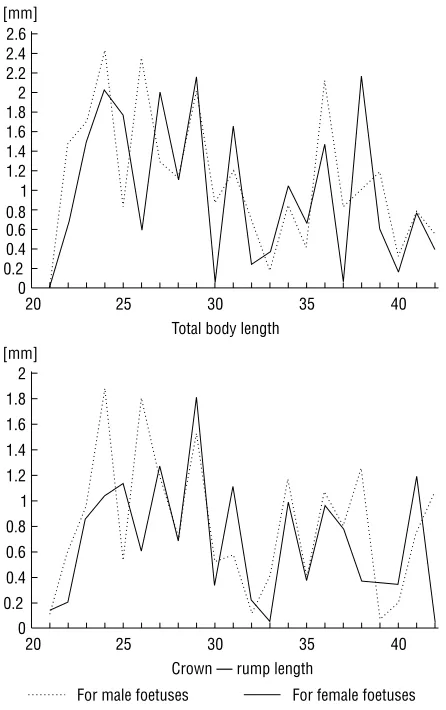
The analysis of weekly changes in the analysed variables has revealed that the periods of most in-tensive accruals of those features, i.e. the periods of most intensive growth of the foetus, are generally the same for both sexes, although it does not mean that the magnitudes of those accruals were the same. The inter-sex analysis of the increases in the analy-sed variables in a given period of foetal ontogenesis (week 20 – week 42) has therefore been carried out (Fig. 1). The analysis of the curves of the accruals, which depicted the rate of developmental changes, has revealed a different, time-dependent magnitude of the increases in the analysed features in male and female foetuses, i.e. a different rate of the develop-ment of the analysed characteristics of both sexes in consecutive weeks of life, yet following a similar change pattern. In some weeks the accruals have been more pronounced in male foetuses, whereas in others in female foetuses. The total increase in the value of each of the analysed features in the period from week 20 to week 42 is significantly more pronounced in male foetuses. The somatic features increase in magnitude almost twice during this peri-od, whereas the increases in the body weight and in the weight of the internal organs are higher and more diversified (Table 1).
21 and 27, between weeks 34 and 36 and during week 42 the values of development rate indices are higher for male foetuses.
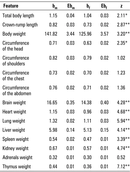
Figure 3 displays the development of each of the analysed features in male and female foetuses be-tween week 20 and week 42. With the exception of shoulder, chest and abdomen circumference and the weight of the adrenals the regression coefficients for the analysed features exhibit a significant sex-related differentiation, which indicates the presence of differing, sex-dependent developmental trends for these features. The sex-related differences between directional regression coefficients and the difference significance have been included in Table 2.
The accuracy of the significance level has been corrected by constructing significance ranges for bm
and bf. Table 3 includes 95% confidence intervals of
linear regression directional coefficients for both sexes. If confidence intervals of directional coeffi-cients for a given feature for both sexes are disjoint-ed, then the theoretical values of those coefficients
differ between one another. This method of deter-mining the sex-related differences in regression co-efficients allowed us to eliminate those features for which the named difference was close to the critical value of the “z” test (i.e. total body length, crown-rump length and head circumference). Disjointed confidence intervals for simple regression direction-al coefficients, which at the level of 0.05 confirm the significance of sex-related differences, charac-terise the body weight and the weights of internal organs with the exception of the adrenals. Signifi-cantly higher directional coefficients in male foetus-es indicate a higher rate of development of thfoetus-ese features in male foetuses.
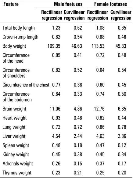
Rectilinear regression depictions of the analysed features significantly divert from the empirical points and as a result they do not reflect exactly the devel-opmental lines of these features (Fig. 3). The poly-nomials provide a more precise matching with the empirical points, which is confirmed by the magni-tude of the standard error of the estimate, which Table 1. Total accruals and multiplicity of the accruals of the analysed features from week 20 to week 42 of foetal life
Feature Total Multiplicity
of accrual of the accrual
Male Female Male Female
foetuses foetuses foetuses foetuses
Total body length 24.47 21.51 1.85 1.74
Crown-rump length 17.71 14.85 1.94 1.77
Body weight 3051.46 2530.68 7.81 6.32
Circumference 15.48 13.10 1.8 1.65
of the head
Circumference 17.49 16.34 1.92 1.86
of shoulders
Circumference 15.68 14.06 2.01 1.82
of the chest
Circumference 16.18 14.36 2.06 1.93
of the abdomen
Brain weight 365.34 296.3 6.83 5.78
Heart weight 23.85 19.56 7.31 6.04
Lung weight 28.59 22.71 5.34 4.87
Liver weight 128.16 109.32 5.95 5.36
Spleen weight 11.24 8.9 12.13 9.9
Kidney weight 14.47 11.99 7.16 6.65
Adrenals weight 6.72 6.44 4.34 4.68
Thymus weight 9.38 8.16 8.63 8.92
reflects the matching of the straight line and the empirical points. The lower the error value the bet-ter the matching. The magnitude of the standard error of the estimate for curvilinear regression is significantly lower compared to rectilinear regres-sion representation (Table 4). Therefore, the devel-opment of the analysed features has been depict-ed by curvilinear regression (Fig. 4). The develop-ment course of the analysed features, as depicted by various polynomials, differentiates between the sexes of the foetuses, with the most significant dif-ferentiation being that of the body weight and the weight of the internal organs, except for the
adrenals. This observation indicates the existence of developmental parallelism in the compared groups of foetuses.
The developmental progression in foetuses of both sexes, as compared to 42-week-old foetuses, has been Table 2. Inter-sex comparison of directional coefficients of linear regression of analysed features
Feature bm Ebm bf Ebf z
Total body length 1.15 0.04 1.04 0.03 2.11*
Crown-rump length 0.82 0.03 0.73 0.02 2.87**
Body weight 141.82 3.44 125.96 3.57 3.20**
Circumference 0.71 0.03 0.63 0.02 2.35*
of the head
Circumference 0.82 0.03 0.79 0.02 1.02
of shoulders
Circumference 0.73 0.02 0.70 0.02 1.23
of the chest
Circumference 0.76 0.02 0.71 0.02 1.36
of the abdomen
Brain weight 16.65 0.35 14.38 0.40 4.28**
Heart weight 1.15 0.03 0.96 0.03 4.68**
Lung weight 1.32 0.02 1.11 0.03 5.94**
Liver weight 5.98 0.14 5.13 0.15 4.14**
Spleen weight 0.54 0.02 0.47 0.01 3.39**
Kidney weight 0.67 0.01 0.57 0.01 4.74**
Adrenals weight 0.32 0.01 0.30 0.01 0.52
Thymus weight 0.44 0.01 0.36 0.01 7.12**
Figure 3. Development of total body length in foetuses of both sexes from week 21 to week 42 as depicted by simple regression model.
Table 3. 95% confidence intervals of directional coeffi-cients of linear regression of the analysed features for both sexes
Feature L-R
Male Female
foetuses foetuses
Total body length 1.07 – 1.23 0.97 – 1.11
Crown-rump length 0.77 – 0.88 0.68 – 0.77
Body weight 134.67 – 148.97 118.54 – 133.38*
Circumference of the head 0.66 – 0.77 0.59 – 0.68
Circumference of shoulders 0.77 – 0.87 0.74 – 0.83
Circumference of the chest 0.68 – 0.79 0.66 – 0.74
Circumference 0.71 – 0.80 0.67 – 0.76
of the abdomen
Brain weight 15.93 – 17.37 13.54 – 15.21*
Heart weight 1.09 – 1.21 0.91 – 1.02*
Lung weight 1.27 – 1.36 1.05 – 1.16*
Liver weight 5.68 – 6.27 4.83 – 5.43*
Spleen weight 0.51 – 0.57 0.44 – 0.50*
Kidney weight 0.64 – 0.70 0.54 – 0.60*
Adrenals weight 0.30 – 0.34 0.27 – 0.32
Thymus weight 0.43 – 0.46 0.35 – 0.38*
depicted in Figure 5. At week 20 the most advanced of the characteristics include, in order of importance, head circumference, longitudinal features and the remaining circumferences, with body weight and the weight of the internal organs being least advanced. Significant differences in developmental advancement of specific features stem from different developmen-tal dynamics (higher for body weight and the weight
Figure 4. Polynomials which describe the accruals of the adrenals weight in both sexes.
Figure 5. Inter-sex comparison of developmental gradients of selected features.
Table 4. Standard estimate errors for rectilinear and cur-vilinear regression
Feature Male foetuses Female foetuses
Rectilinear Curvilinear Rectilinear Curvilinear regression regression regression regression
Total body length 1.23 0.62 1.08 0.65
Crown-rump length 0.82 0.54 0.68 0.46
Body weight 109.35 46.63 113.53 45.33
Circumference 0.85 0.41 0.72 0.48
of the head
Circumference 0.82 0.52 0.64 0.54
of shoulders
Circumference of the chest 0.77 0.38 0.60 0.45
Circumference 0.64 0.33 0.74 0.50
of the abdomen
Brain weight 11.06 4.86 12.76 6.85
Heart weight 0.93 0.48 0.82 0.44
Lung weight 0.72 0.72 0.86 0.78
Liver weight 4.54 2.44 4.63 2.86
Spleen weight 0.48 0.18 0.47 0.12
Kidney weight 0.45 0.38 0.45 0.34
Adrenals weight 0.26 0.15 0.37 0.17
of internal organs, lower for longitudinal features and the circumferences). The evaluation of the develop-mental advancement of selected features has revealed that these features are usually either more develop-mentally advanced in female foetuses or do not ex-hibit any trend towards higher developmental ad-vancement in any of the sexes.
Because of the high number of the analysed mor-phological features, the graphic presentations includ-ed in this article comprise only some of them.
DISCUSSION
Prenatal sex dimorphism manifests not only through absolute and relative differences between the analysed morphological features but also through differing dynamics of the development of somatic features and sex-dependent weight increase of in-ternal organs. Foetal development is determined by multiple factors and, as a result, the development rate changes with time. It may be assumed that as of the 5th month onwards the development rate begins to decrease [11]. Similar observations have been made by Reicher [7] and Scammon and Calkins [8]. The dynamics of the development of morpho-logical features in the time period analysed in our study has been found to vary. Intertwining periods of accelerated and decelerated growth and devel-opment may be distinguished. The periods of most intensive growth are observed generally during the same time in both sexes, yet the rates of growth vary between sexes. The accruals in morphological features may be higher in female foetuses at one time and in male foetuses at another. Such differtiation may be explained by cyclical changes in en-docrine metabolism in the pregnant woman, which may differently affect foetal development [3] in male and female foetuses. Similarly, Bożiłow et al. [1] have found that the increase in head circumference dur-ing foetal development is subject to cyclical, age and sex-dependent changes.
The level of developmental advancement of so-matic features confirms the well-known principle of cephalo-caudal development of morphological char-acteristics in prenatal age. Morphological advance-ment is highest for (in order of importance) head circumference, total body length and crown-rump length, shoulder, chest and abdomen circumferenc-es, with the least developmental advancement be-ing observed for total body weight. Head
circumfer-ence reaches 90% of its final prenatal value as early as in week 34, whereas body weight reaches 50% of its final value in week 31. A similar degree of devel-opmental advancement has been confirmed by Marecki [5]. As regards the analysed morphological features, female foetuses appear developmentally older than male foetuses, except for the weight of the thymus and the adrenals. Similar results have also been obtained by Malinowski et al. [4].
The cross-sectional nature of the analysed mate-rial, which did not allow us to discuss real accruals or decreases in the analysed morphological features, may be considered a limitation of the interpretation capacity of this study (similarly to the majority of other studies on foetal development). Despite great advancement in medical techniques, there is still no possibility of accurate investigation of prenatal de-velopment. The ultrasound examination is routinely applied in situations when some foetal or maternal pathology is suspected. As regards the interpreta-tion of such visualisainterpreta-tions, it cannot be definitively stated whether these are physiological or patholog-ical representations of foetal development. More-over, such data are not sufficiently abundant and do not span regular, weekly time intervals. Our cross-sectional research may be, as a result, slightly con-founded, yet the error is not significant enough to give up the research while waiting for new research techniques.
CONCLUSIONS
The rate of development of the analysed features in consecutive weeks of foetal life exhibits sex-related differences.
Total accruals for each of the analysed features in the period between week 20 and week 42 are significantly higher for male foetuses, yet the ad-vancement of the analysed features makes the fe-male foetuses developmentally older than the fe-male ones.
Body weight and the weight of internal organs (except for the adrenals) are the features character-ised by different, sex-related developmental dynam-ics. The rate of feature formation is significantly high-er in male foetuses.
REFERENCES
1. Bożiłow W, Kurlej W, Gworys B, Poradnik E (1985) Zmienność obwodu głowy podczas rozwoju prena-talnego i okołoporodowego człowieka. Przegl Antr, 51: 103–113.
2. Bożiłow W, Sawicki K, Poradnik E, Kurlej W, Gworys B (1992) Zmienność masy ciała podczas rozwoju prenatal-nego i okołoporodowego. Przegl Antr, 55: 1–2, 45–55. 3. Halberg F (1974) The Necessity for Relating Treatment
to Bodity Rhythms. Chronobiological aspects of endo-crinology, Sympozia Medice Hoechst 9.
4. Malinowski A, Kłosin R, Rucka A (1981) Somatometria płodów żywo i martwo urodzonych. Zróżnicowanie morfologiczne człowieka, 199: 69–99.
5. Marecki B (1989) Development relations between the weight of internal organs and somatic features of fetuses and newborns. Z Morph Anthrop, 78: 107–115.
6. Miller H, Futrakul P (1968) Birth weight, gestational age, and sex as determining factors in the incidence of respiratory distress syndromes of prematurely born infants. J Pediat, 72: 5, 628–635.
7. Reicher M (1923) Rozwój wzrostu i proporcji płodów ludzkich. Arch Nauk Antrop Warszawa – Lwów, 2: 5. 8. Scammon R, Calkins L (1929) The development and
growth of the external dimensions of the human body in the foetal period. Univ. Minnesota Press, Minneapolis.
9. Tanner JM, Thomson AM (1970) Standards for birth weight at gestation periods from 32 to 42 weeks, al-lowing for maternal height and weight. Archiv Dis Child, 45: 566–569.
10. Waszak M, Cieślik K (2001) The extent of sexual differ-ences in somatic traits and mass of internal organs in human foetal life. Papers on Anthropology, 10: 334–349. 11. Wich J (1972) Z badań nad rozwojem płodowym