RESEARCH
Using a marine microalga as a chassis
for polyethylene terephthalate (PET)
degradation
Daniel Moog
1,2*, Johanna Schmitt
1, Jana Senger
3, Jan Zarzycki
3, Karl‑Heinz Rexer
4, Uwe Linne
2,5, Tobias Erb
2,3and Uwe G. Maier
1,2Abstract
Background: The biological degradation of plastics is a promising method to counter the increasing pollution of our planet with artificial polymers and to develop eco‑friendly recycling strategies. Polyethylene terephthalate (PET) is a thermoplast industrially produced from fossil feedstocks since the 1940s, nowadays prevalently used in bottle packag‑ ing and textiles. Although established industrial processes for PET recycling exist, large amounts of PET still end up in the environment—a significant portion thereof in the world’s oceans. In 2016, Ideonella sakaiensis, a bacterium pos‑ sessing the ability to degrade PET and use the degradation products as a sole carbon source for growth, was isolated.
I.sakaiensis expresses a key enzyme responsible for the breakdown of PET into monomers: PETase. This hydrolase might possess huge potential for the development of biological PET degradation and recycling processes as well as bioremediation approaches of environmental plastic waste.
Results: Using the photosynthetic microalga Phaeodactylum tricornutum as a chassis we generated a microbial cell factory capable of producing and secreting an engineered version of PETase into the surrounding culture medium. Initial degradation experiments using culture supernatant at 30 °C showed that PETase possessed activity against PET and the copolymer polyethylene terephthalate glycol (PETG) with an approximately 80‑fold higher turnover of low crystallinity PETG compared to bottle PET. Moreover, we show that diatom produced PETase was active against indus‑ trially shredded PET in a saltwater‑based environment even at mesophilic temperatures (21 °C). The products resulting from the degradation of the PET substrate were mainly terephthalic acid (TPA) and mono(2‑hydroxyethyl) terephthalic acid (MHET) estimated to be formed in the micromolar range under the selected reaction conditions.
Conclusion: We provide a promising and eco‑friendly solution for biological decomposition of PET waste in a saltwater‑based environment by using a eukaryotic microalga instead of a bacterium as a model system. Our results show that via synthetic biology the diatom P.tricornutum indeed could be converted into a valuable chassis for biological PET degradation. Overall, this proof of principle study demonstrates the potential of the diatom system for future biotechnological applications in biological PET degradation especially for bioremediation approaches of PET polluted seawater.
Keywords: Polyethylene terephthalate, PETase, Plastic pollution, Plastic degradation, Diatoms
© The Author(s) 2019. This article is distributed under the terms of the Creative Commons Attribution 4.0 International License (http://creat iveco mmons .org/licen ses/by/4.0/), which permits unrestricted use, distribution, and reproduction in any medium, provided you give appropriate credit to the original author(s) and the source, provide a link to the Creative Commons license, and indicate if changes were made. The Creative Commons Public Domain Dedication waiver (http://creativecommons.org/ publicdomain/zero/1.0/) applies to the data made available in this article, unless otherwise stated.
Open Access
*Correspondence: daniel.moog@biologie.uni‑marburg.de 1 Laboratory for Cell Biology, Philipps University Marburg, Karl‑von‑Frisch‑Str. 8, 35032 Marburg, Germany
Background
Plastic is an extremely useful material with a wide range of applications and seemingly no longer indispensable for our daily life. However, the millions of tons of plas-tic waste produced each year have become a major eco-logical issue on our planet in the last decades, mostly due to inadequate disposal and the high durability of the synthetic material [1]. The consequences of plas-tic pollution for Earth’s ecosystems are so far unfore-seeable, but it becomes more and more evident that the plastic accumulating in nature is harmful for life. One major threat is the formation and distribution of microplastics, particles smaller than 5 mm, especially in waters such as the oceans [2]. Microplastic parti-cles can include harmful additives and, because of their small size and physical properties, can adsorb toxic compounds (e.g., heavy metals and organic pollutants) and enter the food chain at the level of small animals or even microorganisms [3–7].
Polyethylene terephthalate (PET)—a plastic material intensively used for packaging of liquids (bottles)/food and for production of synthetic textile fibres [8]—is pro-duced from crude oil resources (terephthalic acid and ethylene glycol) with highly increasing rates predicted to exceed more than 70 million tons per year by 2020 (see [9] and references therein). As for many other plastics, the steadily growing demand for PET necessitates an effi-cient and comprehensive global manufacturing and waste management system with the goal of adverting damage from nature. Although efficient processes for industrial PET (synthesis and) recycling are established, a signifi-cant fraction of PET waste is still incinerated, landfilled or ends up in the environment as macro-, meso-, micro-, and nano-particles due to improper disposal [10–12].
Recently, a bacterium named Ideonella sakaiensis 201-F6 has been isolated from PET waste sources in Japan that is capable of utilizing this plastic as sole carbon source [13]. I. sakaiensis expresses a whole enzymatic pathway for PET biodegradation and uptake, with two enzymes, PET hydrolase (PETase) and mono(2-hydroxyethyl) tere-phthalic acid hydrolase (MHETase), having the ability to decompose PET into its environmentally non-hazardous monomers—terephthalic acid (TPA) and ethylene glycol (EG). I. sakaiensis exhibits the highest natural PET deg-radation efficiency known so far and PETase as well as MHETase are improved by protein engineering continu-ously (see e.g., [9, 14–16]). The key enzyme PETase is nat-urally secreted by I. sakaiensis, which might adhere to the surface of PET to initiate its biodegradation [13], showing the potential of PETase for biological PET degradation and bioremediation approaches. Besides I. sakaiensis, several bacterial systems, including Escherichia and
Bacillus, have been utilized to generate synthetic PETase
secreting cell factories with potential application in bio-logical PET recycling by now (see e.g., [13, 17, 18]). How-ever, certain disadvantages, such as the dependence on PET substrate presence or the addition of costly carbon sources (at industrial scale) into the culture/bioreactor for growth, exist for these bacterial systems in biotech-nology approaches that have to be overcome. Moreover,
I. sakaiensis and other microorganisms used so far for PETase production are not well adapted to marine habi-tats (see, e.g., [19])—the environments in which most of the plastic waste accumulates. Thus, these organisms, for example, are not suitable for bioremediation of PET pol-luted saltwater.
The diatom Phaeodactylum tricornutum is a marine photosynthetic single-celled eukaryote with a high potential for biotechnological applications. P. tricornu-tum combines the benefits of a photosynthetic organ-ism that is easily cultivable and rapidly grows under CO2
consumption in a saltwater-based environment, with those of an established laboratory model organism for which a comprehensive genetic toolbox exists. That is, genes can be inserted into (or edited within) the genome of the diatom via standard methods and their products can be expressed with highest efficiency under induc-ible conditions [20–22]. The diatom P. tricornutum is an excellent system for expression of foreign recombi-nant proteins such as antibodies, antigens [22–25] and even whole enzymatic pathways [26–28]. Cultivation of P. tricornutum is cost-efficient, cells can be grown to high densities and since photoautotrophic the organism does not require supplementation of expensive sugars or other carbohydrates as carbon source into the growth medium if a light source is present [22]. The diatom can easily be transformed with multiple constructs and as shown before it has the ability to efficiently secrete syn-thetic recombinant proteins into the medium fraction [24]. These features highlight the potential of P. tricornu-tum as a model organism for synthetic biology and bio-technology and underline the benefits of the diatom over bacterial expression systems with respect to developing a photosynthetic PETase production factory for biological PET decomposition under marine conditions.
Results
Expression and secretion of PETase in the algal system
The gene sequence encoding I. sakaiensis PETase (improved/engineered version: PETaseR280A [14]) was
adapted to the codon usage of P. tricornutum and expressed as fusion with gfp in the diatom to test whether the product (PETaseR280A-GFP) is correctly and efficiently
synthesized. Although the PETaseR280A-GFP construct
was expressed with the endogenous bacterial signal peptide (SP), GFP fluorescence was detected via confo-cal laser scanning microscopy (CLSM) in the ER and most likely other compartments of the secretory path-way (Fig. 1). In addition, secretion of PETaseR280A-GFP
into the medium was investigated. For this approach, three different PETaseR280A-GFP expressing clones were
analyzed for the presence of the enzyme in the culture medium via concentration of the proteins in the medium fraction, using SDS-PAGE and Western Blot (see “ Meth-ods”). To this end, a total volume of 50 ml supernatant/ medium fraction of a diatom culture was used. Besides the concentrated proteins in the medium fraction, a total protein extract was obtained from the cell pellet to analyze both fractions for the presence of the expressed PETaseR280A-GFP fusion protein. As shown in Additional
file 1: Figure S1, at least one of the three clones express-ing the PETaseR280A-GFP fusion construct (clone 24) was
able to secrete the recombinant protein detected in both the medium and cell pellet fraction, whereas no signal
could be detected in the control (wild type). Interest-ingly, when compared to the protein standard the sig-nal for PETaseR280A-GFP (clone 24) appeared at more
than 70 kDa while the calculated molecular mass was 57.7 kDa, which indicated post-translational modifica-tion of the recombinant protein in P. tricornutum (see below). For the two remaining clones, the GFP fusion protein could be detected in the pellet fraction only, indi-cating that the PETaseR280A-GFP protein is expressed, but
cannot be secreted by the diatom efficiently (Additional file 1: Figure S1).
Since both, the relatively bulky GFP moiety and the non-eukaryotic bacterial signal peptide of Ideonella
PETase, might represent factors reducing an efficient secretion and/or enzymatic function of the PETase fusion protein by the diatom, we substituted the endogenous SP with the SP of P. tricornutum alkaline phosphatase (AP) [29] and replaced the 27.5 kDa GFP by a 1 kDa FLAG-tag (DYKDDDDK). After the modified gene was trans-formed into the alga, several clones grew on selection medium and three of them were further analyzed via Western Blot as described above. As depicted in Fig. 2, PETaseR280A-FLAG was secreted into the medium by
P. tricornutum most likely by means of the signal pep-tide of the diatom AP protein as a targeting signal. No FLAG-specific signal was detected in the medium frac-tion of the wild type control. As only weak signals for PETaseR280A-FLAG were present for clone 2 and 3 in the
Fig. 1 Expression and localization of PETase‑GFP in the diatom P. tricornutum. PETaseR280A‑GFP (see schematic of fusion protein) was expressed
successfully in the diatom. Confocal laser scanning microscopy showed that the recombinant protein localized in the ER and most likely other compartments of the secretory pathway. Secretion of the fusion protein could not be analyzed via this method. The lower part of the figure shows a wild type control in which no recombinant protein is expressed. Only plastid autofluorescence but no GFP signal was detectable. SP signal peptide,
cell pellet fraction, secretion of the recombinant proteins by the diatom clones occurred with high efficiency. To test if the positive signals in the medium fraction were actually a result of secretion and not due to lysis of algal cells, we performed a control experiment using an anti-body against alpha-tubulin, a component of the cytoskel-eton of the eukaryotic cell, which has been established as a suitable control protein for secretion analyses of recom-binant proteins before [24]. Signals for alpha-tubulin could be observed exclusively in the cell pellet and not the medium fraction, confirming that cells remained intact and no substantial cell lysis took place during the experiment (Fig. 2).
As already observed for PETaseR280A-GFP, we again
detected a putative mass shift for the expressed protein on the Western Blot. The calculated molecular mass of the AP_SP-PETaseR280A-FLAG is 30.4 kDa, those of the
expected processed form (SP removed) 28.5 kDa. The dominant signals observed on the Western Blot were two bands between 40 and 50–55 kDa, which was approxi-mately 10–25 kDa higher than the predicted molecular mass (Fig. 2). To investigate the nature of the observed signals, the proteins of the medium fraction of a 500 ml
culture expressing AP_SP-PETaseR280A-FLAG (clone
2) were concentrated via 10 kDa cutoff filter units,
precipitated with TCA and separated via SDS-PAGE fol-lowed by a Coomassie-staining. The staining revealed that corresponding bands to both dominant signals observed in the Western Blot were present in the SDS-gel (Additional file 1: Figure S2). A subsequent mass spectro-metric (MS) analysis shed light on the nature of the two bands. The identity of both bands could be unambigu-ously assigned to AP_SP-PETaseR280A-FLAG. Whereas
analysis of the upper band (< 55 kDa, Additional file 1: Figure S2) resulted in detection of 7 unique peptides and a coverage of the protein sequence of 28%, for the lower band (> 40 kDa, Additional file 1: Figure S2) 8 unique peptides were identified via mass spectrometry covering 33% of the protein sequence (see Additional file 2: Tables S1 and S2). These results indicate that the FLAG-tag pro-tein detected in the medium was indeed AP_SP-PETa-seR280A-FLAG. In order to investigate if the observed
mass shift was caused by post-translational modifications of the enzyme, we exemplarily tested N-linked glycosyla-tion of the secreted protein. To this end, the supernatant of a 500 ml culture of AP_SP-PETaseR280A-FLAG clone
2 was concentrated to a volume of 250 µl and a fraction was treated with PNGase F before it was separated on an SDS-gel and analyzed via Western Blot (see “Methods”). As shown in Additional file 1: Figure S3, the signals for
Fig. 2 Secretion analysis of PETase‑FLAG. a Schematic of the expressed recombinant protein AP_SP‑PETaseR280A‑FLAG. b Western Blot after
SDS‑gel separation of the cell pellet (10 µg of total protein) and medium fractions (total precipitated protein fraction) of 50 ml cultures (induced at OD600= 0.4) expressing AP_SP‑PETaseR280A‑FLAG. Detection of recombinant proteins was conducted using an antibody against the FLAG‑tag
(α‑FLAG). As control for intracellular proteins, an alpha‑tubulin antibody (α‑Tubulin) was used. Wild type medium and cell pellet fractions as well as a FLAG positive control lysate (Rockland, FLAG+) served as control protein fractions. AP_SP‑PETaseR280A‑FLAG clone 2 showed the highest
expression and secretion efficiency (middle), whereas complete secretion of the recombinant protein was only achieved by clone 1 (left). As shown by the control via alpha‑tubulin detection (right), presence of PETaseR280A‑FLAG in the medium fraction was not due to cell lysis. A signal in the
range of the calculated molecular mass of AP_SP‑PETaseR280A‑FLAG (30.4 kDa) could only be observed for clone 2 (left and middle). The dominant
signals detected by the FLAG‑tag antibody appeared at molecular masses of approximately 40 and 50–55 kDa in AP_SP‑PETaseR280A‑FLAG clone 1,
2 and 3. Calculated molecular masses: AP_SP‑PETaseR280A‑FLAG: 30.4 kDa; FLAG‑tag, 1 kDa; PETaseR280A‑FLAG: 28.5 kDa; FLAG+, 60 kDa. AP alkaline
phosphatase, SP signal peptide, WT wild type, AP_# AP_SP‑PETaseR280A‑GFP clone #. Numbers beside/on the Western Blots indicate molecular
AP_SP-PETaseR280A-FLAG treated with PNGase F
cor-responded to a significantly lesser molecular mass than the untreated sample (negative control). This indicates that the recombinant protein AP_SP-PETaseR280A-FLAG
is (N-linked) glycosylated when expressed in the diatom.
PET degradation experiments using diatom produced and secreted PETase
Having shown that the diatom efficiently secretes AP_SP-PETaseR280A-FLAG into the culture medium (for analysis
of production and secretion efficiency by clone 2 within a time frame of 7 days see Additional file 1: Figure S4), we next investigated whether the secreted enzymes are able to degrade PET. To this end, we chose two different experimental approaches. In the first one, cells from AP_ SP-PETaseR280A-FLAG clone 1 were grown in contact to
PET film fragments sticking upright in an f/2 (saltwater) agar plate, which was overflowed with 2 ml liquid f/2. The cells were cultivated under inducing conditions (medium supplemented with nitrate) for 2 to 6 weeks. A P. tricor-nutum wild type culture grown under similar conditions served as a negative control. PET film was removed from the culture plate, sputtered with gold and analyzed via scanning electron microscopy (SEM), to monitor PET degradation by PETaseR280A-FLAG (see “Methods”).
Whereas untreated PET film and fragments incubated with wild type cells showed, besides a typical smooth surface, stress marks characteristic of commercially used water bottles (occasional scratches and surface disrup-tions), the PET film incubated with AP_SP-PETaseR280A
-FLAG expressing clone 1 showed a completely different structure (Fig. 3). In these samples, certain areas of the PET film, which were in contact with P. tricornutum cells on the agar plate, showed holes, dents, furrows and cavi-ties clearly visible under the SEM. The small holes were reminiscent to structures observed in earlier PET degra-dation experiments using PETase [13, 15] (see also Addi-tional file 1: Figure S5). The furrows sometimes appeared in a canyon-like shape as if the missing plastic was washed out by a running liquid in a branched manner. In one particular case, we could observe a diatom cell mark in the form of the fusiform morphotype of P. tricornu-tum from which several holes and furrows had their ori-gin (Additional file 1: Figure S6), which overall supports functional secretion and enzymatic activity of AP_SP-PETaseR280A-FLAG synthesized by the diatom.
In a second approach, we used liquid cultures of P. tricornutum wild type and clones expressing AP_SP-PETaseR280A-FLAG (clone 1 and 2) grown in volumes of
50, 150 or 500 ml. For a proof of principle experiment, 1 ml of the medium fraction of an induced (4 days) 50 ml
culture expressing AP_SP-PETaseR280A-FLAG (clone
1) and 1 ml of a wild type control culture were further
incubated with small polyethylene terephthalate glycol (PETG; a highly amorphous PET copolymer, see “ Meth-ods”) commercial film fragments for 1 week at 30 °C. This temperature is near the optimum for PETase activ-ity (~ 35 °C) as reported in the literature [13, 30]. The liquid fractions of the samples were analyzed (expected was mainly MHET and to a minor degree TPA and bis(2-hydroxyethyl) terephthalic acid (BHET) as products) via UHPLC (see “Methods” and below), whereas the small PETG film parts were investigated with the SEM for vis-ible alterations of their surface structure. As shown in Fig. 4, incubation of a small PETG film particle with 1 ml of the medium fraction (sterile filtered, cutoff 0.22 µm) for 7 days at 30 °C led to similar, although more com-prehensive, structures in the surface of the PETG film as observed after the first approach (PET bottle film deg-radation by AP_SP-PETaseR280A-FLAG clone 1 on solid
medium, Fig. 3). Again, we observed holes, furrows and branching canyon-like structures in the upper layer of the PETG film, clearly pointing to an efficient and this time area-wide degradation process of the plastic material. No obvious change in surface structure was observed for a similar PETG substrate incubated with 1 ml of the medium fraction of a wild type culture (Fig. 4, see also Additional file 1: Figure S7). UHPLC analyses of the con-centrated liquid fractions in which the PETG film was incubated revealed the presence of mainly TPA, whereas MHET, the product, which was expected with the high-est abundance, was present in comparably low quantity (Fig. 4). No generation of TPA and MHET was observed in the wild type control. Interestingly, when an identical experiment was performed using PET film from a bottle instead of commercial PETG film (above) as substrate, no significant TPA and MHET production was detect-able under the selected reaction conditions, neither in the sample incubated with 1 ml of the medium fraction of AP_SP-PETaseR280A-FLAG clone 1 nor in the wild type
control (not shown).
A similar experiment was conducted with 1 ml of the medium fraction of the strongly expressing AP_ SP-PETaseR280A-FLAG clone 2 (dense 500 ml culture
induced for 3 days; see Additional file 1: Figure S4 for PETaseR280A-FLAG secretion analysis in relation to
expression time) and bottle PET as well as commercial PETG film. Here the results were highly comparable to the observations made in the experiments with AP_SP-PETaseR280A-FLAG clone 1 (see Additional file 1:
Fig-ures S8, S9 and S10). Essentially, two differences were detected: (i) an almost equal amount of TPA and MHET was identified in the sample where PETG film was incubated with 1 ml of the medium fraction of AP_SP-PETaseR280A-FLAG clone 2 for 7 days at 30 °C (see
and (ii) low, but similar amounts of TPA and MHET were detected in the supernatant (medium fraction) of AP_SP-PETaseR280A-FLAG clone 2 incubated with PET
bottle film (see Additional file 1: Figure S9 and Addi-tional file 2: Table S3). The latter result was supported by structural differences (presence of holes) in the PET bottle film when inspected via SEM (Additional file 1: Figure S9). A rough estimation of the formed total prod-ucts (MHET + TPA) of both approaches (26.32 µm from PETG vs. 0.31 µM from bottle PET) indicated that the turnover of PETG by PETaseR280A-FLAG is approximately
80-fold higher when compared to bottle PET (Additional file 2: Table S3) under the selected reaction conditions.
In a further approach 50 and/or 150 ml cultures were incubated with 5 × 1.5 cm PET and PETG pieces from a bottle or commercial film or shredded PET particles for up to 14 days with and without agitation at 21 or 26 °C (see “Methods”). Samples were collected at different time
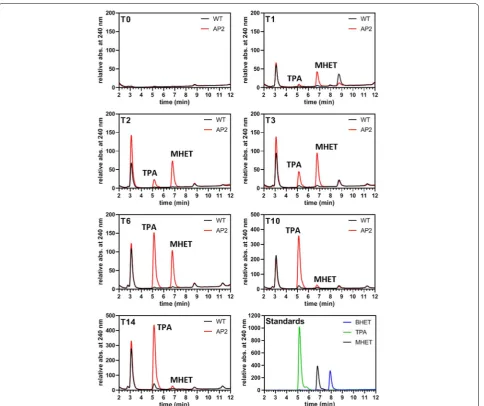
points and the content of the samples was analyzed for the presence of PETase reaction products. The results of these approaches revealed that degradation of PET/ PETG film substrate by PETase-FLAG was very low or even absent under most of the selected conditions (not shown). However, when we used shredded PET—a mix-ture of micro- and macro-plastics up to approximately 1 cm in size (see Additional file 1: Figure S11)—positive results were obtained. In this approach we performed a time series experiment with an around 1 week old 50 ml culture of AP_SP-PETaseR280A-FLAG producing clone
2 and a 50 ml wild type control culture, which were incubated with an industrially shredded PET substrate (approx. 5 g), respectively. 1 ml samples for UHPLC analysis were taken after 0, 1, 2, 3, 6, 10, and 14 days (T0–T14) and filtered (cutoff 0.22 µm). At time points
T3, T6, and T10, the cultures were supplied with new
nitrate to steadily induce expression of the recombinant
Fig. 3 Scanning electron microscopic analysis of PET bottle film degradation by PETase‑FLAG secreted from P.tricornutum. As depicted in the upper left part, untreated PET and PET incubated with wild type cells on an f/2 agar plate overflowed with 2 ml f/2 liquid medium showed, besides a usually smooth surface, occasional stress marks. In contrast, PET incubated for 5 weeks with cells expressing AP_SP‑PETaseR280A‑FLAG (clone 1),
protein AP_SP-PETaseR280A-FLAG by clone 2.
Interest-ingly, we observed a progressive increase of TPA and MHET with a higher amount of MHET generated until T3. After T6 the amount of produced MHET
progres-sively decreased while TPA still significantly increased (Fig. 5, Additional file 2: Table S3). The level of MHET decreased to a minimum at T10, which did not
perceiv-ably change until T14, although the total concentration of
products (TPA + MHET) increased almost linearly until T14 (Additional file 2: Table S3). No production of TPA
or MHET was detected in the wild type control (Fig. 5). The identity of the formed products was confirmed by mass spectrometry; estimated product quantifications in the micromolar range are shown in Additional file 2: Table S3.
A similar approach was taken for AP_SP-PETaseR280A
-FLAG producing clone 1 and shredded PET (approx. 10 g) in 150 ml culture volume. 1 ml samples for UHPLC analy-sis were collected after 0, 3, 6, 10, and 14 days (T0–T14).
At the same time, the cultures were supplied with nitrate (see above). As shown in Additional file 1: Figure S11, we obtained very similar results to those shown in Fig. 5. At T3 MHET was more abundant than TPA, whereas at T6
the situation reversed, the TPA level increased progres-sively until T14 and MHET basically completely vanished
at T10 (Additional file 1: Figure S11). The results of these
UHPLC experiments clearly show that TPA and MHET (identity also confirmed by mass spectrometry) were effi-ciently produced from shredded PET substrates when
incubated with AP_SP-PETaseR280A-FLAG producing
Fig. 4 SEM and UHPLC analysis of PETG film degradation by PETase‑FLAG secreted from P.tricornutum. A small piece of PETG film was incubated with 1 ml of supernatant (medium fraction) of a 50 ml culture expressing AP_SP‑PETaseR280A‑FLAG (clone 1, induced for 4 days) and wild type and
analyzed via SEM. As shown in the upper part, similar but more area‑wide changes in the surface of the PETG film as observed in the solid approach (PET bottle film degradation by AP_SP‑PETaseR280A‑FLAG clone 1 on solid medium, Fig. 3) were detected. The wild type control (lower left) did not
show any significant aberrations in the surface structure of the PETG film. UHPLC analysis (lower right) of the medium fractions after 1 week of incubation with the PETG film at 30 °C revealed production of TPA and MHET in sample AP_1 (AP_SP‑PETaseR280A‑FLAG clone 1), which were absent
clones. In contrast, the wild type culture of P. tricornutum
did not produce any PET degradation products in con-trol experiments. BHET, which besides MHET is another potential product of the PETase reaction [13], was not produced in any detectable amounts in neither of the cul-tures during our experiments.
Discussion
If correctly disposed plastic is not waste but instead a valuable resource that can be part of a closed (circular) recycling process. The recycling of used plastic materials
such as PET is often expensive and can involve harmful chemicals and a high demand of temperature, energy and time [31]. Thus, an eco-friendly biological degradation of synthetic PET polymers into reusable monomers (TPA and EG) via bioremediation is desired. Bioremediation is also one of the most promising solutions to counter-act plastic pollution on Earth. Micro- and nano-plastics, once present in the environment, because of their small sizes are very difficult to remove from nature. Especially waterbodies are highly polluted with plastic waste, and Earth’s oceans are accumulating more plastics year in and
Fig. 5 UHPLC analysis of the medium fraction of shredded PET incubated with PETase‑FLAG producing clone 2. The experiment was performed in 50 ml f/2‑medium containing AP_SP‑PETaseR280A‑FLAG expressing clone 2 (AP2) and approximately 5 g of shredded PET. At T
0 the cultures were
adjusted to an OD600 of 0.4 and expression of the recombinant protein (AP_SP‑PETaseR280A‑FLAG) was induced with nitrate. Samples of 1 ml were
taken at individual time points (T, 1 day = 24 h) and fresh nitrate was supplemented to the cultures at T3, T6 and T10. The standards used for UHPLC
year out. Many plastic materials are highly durable and resistant to biological degradation through e.g., micro-organisms. The recently discovered PETase is a bacterial hydrolase possessing the ability for PET degradation [13]. Recent studies on PETase demonstrate the potential of the enzyme for possible applications in biological PET degradation and recycling (see e.g., [13–18, 30, 32]). In this work, we expand the possible range of applications for PETase via generating a synthetic microbial cell fac-tory in the form of a marine microalga capable of produc-ing and secretproduc-ing functional PETase that can efficiently decompose PET in a saltwater-based environment.
By means of expression and localization studies of GFP fusion proteins as well as Western Blot analyses, our studies show that the marine diatom P. tricornutum
is a suitable chassis for the production of recombinant PETaseR280A-FLAG and its secretion into the medium
fraction (Figs. 1, 2, Additional file 1: Figure S1). The lat-ter is possible either by expressing PETase with the origi-nal bacterial SP of I. sakaiensis PETase, or as a synthetic construct in which the SP of a well-known secreted fac-tor of the diatom, alkaline phosphatase (AP) [29], was used. Western Blot analyses of several PETase-GFP and -FLAG producing P. tricornutum clones revealed that the constructs with a FLAG-tag were secreted much more efficiently than those containing a GFP-tag (Fig. 2 and Additional file 1: Figure S1), which is not surprising and might be due to the massive increase in theoretical molecular mass (GFP: + 27.5 kDa; FLAG: + 1 kDa) and size (bulky barrel-like structure of GFP) of the expressed GFP fusion protein probably hindering secretion by the diatom cell. With AP_SP-PETaseR280A-FLAG clone 2 we
obtained a P. tricornutum mutant cell line capable of the highest production and secretion rates of the recombi-nant enzyme upon induction of expression when com-pared to other generated clones (Fig. 2).
As evident from several Western Blots, a mass shift of the diatom-produced recombinant PETase protein was observed during our experiments (Fig. 2 and Addi-tional file 1: Figure S1). Whereas the original I. sakaien-sis PETase protein has a molecular mass of 30.1 kDa, the theoretical values for the here synthesized recombinant versions are 57.7 for PETaseR280A-GFP and 30.4 kDa for
AP_SP-PETaseR280A-FLAG. However, when
immunode-tection with an antibody specific for the FLAG-tag was performed signals with higher molecular masses in the range of at least 10–25 kDa more could be detected. Via mass spectrometry, these bands could be identified as PETaseR280A-FLAG unequivocally (Additional file 1:
Fig-ure S2, Additional file 2: Tables S1 and S2). As already shown for endogenous as well as secreted recombinantly expressed proteins, typical eukaryotic post-translational modifications in the form of N-glycosylation can occur
in P. tricornutum [33–35]. We exemplarily tested the post-translational modification of AP_SP-PETaseR280A
-FLAG via N-glycosylation by PNGase F (cleaves N-linked oligosaccharides) treatment and observed that the mass increase of the specific protein band was indeed lesser when potential N-linked glycans were removed enzy-matically (Additional file 1: Figure S3). This clearly indi-cates that at least a part of the observed mass increase of the recombinant protein was due to N-glycosylation taking place in the diatom cell. This observation also cor-relates with the presence of predicted N-glycosylation sites in the AP_SP-PETaseR280A-FLAG sequence (see
Additional file 1: Figure S12) and the results of the mass spectrometric analyses in which certain peptides (result-ing from tryptic digestion), includ(result-ing predicted N -gly-cosylation sites, likely due to deviating masses could not be identified (Additional file 2: Tables S1 and S2). How-ever, to explain the huge increase in molecular mass of PETaseR280A-FLAG, further post-translational
modifica-tions, such as, for example, O-glycosylation of the protein by P. tricornutum, have to be considered (and postulated) as well. Whether the post-translational modifications of PETaseR280A-FLAG in P. tricornutum have an influence
on its catalytic activity or substrate specificity is so far unknown and will be (in parallel to their identity) investi-gated in more detail in future studies.
In any case, the results of our studies indicate that PETaseR280A-FLAG—an engineered version of I.
sakaien-sis PETase with increased activity [14]—produced and secreted by P. tricornutum possesses catalytic activity against PET as well as PETG copolymer. This could be shown by several PET/PETG degradation experiments with diatom cells growing on solid f/2-saltwater agar as well as in liquid cell culture (Fig. 3, 4, 5 and Additional files 1, 2). The proof of principle of enzyme functionality was provided in the course of a PET bottle film decom-position approach on solid agar (Fig. 3) in which partial degradation of the substrate was achieved after several weeks of incubation. The observed structural changes (holes and cavities) in the PET surface were comparable to those reported in earlier PET degradation experiments using PETase [13, 15], strongly indicating that the dia-tom produced PETaseR280A-FLAG enzyme was capable of
decomposing bottle PET.
As we had generated several promising diatom mutants efficiently producing and secreting PETaseR280A-FLAG
into the medium (Fig. 2), we aimed at scaling up the approach and reducing reaction time and thus changed to a liquid saltwater environment (cell culture) for fur-ther studies. Initially, PET as well as PETG decomposi-tion was tested at 30 °C—temperature condidecomposi-tions nearly optimal for PETase [13, 30], but not for P. tricornutum
culture synthesizing PETaseR280A-FLAG and incubated
the fraction with commercial PETG or bottle PET film. While the approach with commercial highly amorphous PETG copolymer film proved to be highly successful (Fig. 4, Additional file 1: Figures S7, S8), only little PET degradation could be observed for bottle PET incubated with PETaseR280A-FLAG containing f/2-medium
(Addi-tional file 1: Figure S9, Additional file 2: Table S3). A rough estimation of the formed products of the degrada-tion reacdegrada-tion catalyzed by PETaseR280A-FLAG (Additional
file 2: Table S3) under the selected conditions suggested an approximately 80-fold higher turnover of PETG than bottle PET. These results indicate that the diatom-pro-duced PETaseR280A-FLAG possesses different specificities
for different PET substrates, which, among other things, might be due to the degree of crystallinity of the different polymer materials that has an influence on their biodeg-radability (see e.g., [37]). However, the results show that not only PET but also PETG copolymer can be degraded by the PETaseR280A-FLAG produced and secreted by
P. tricornutum.
When we scaled up our liquid PET/PETG degrada-tion experiments to diatom culture size (50–500 ml), we again recognized differences in PET turnover depend-ing on the form of the substrate. Although the liquid approach at P. tricornutum culturing conditions (see “Methods”) using industrially shredded PET as substrate was highly successful (see Fig. 5 and Additional file 1: Figure S11), PET and PETG film (bottle and commercial) substrates were not or rather poorly degraded by the diatom produced and secreted PETaseR280A-FLAG. This
observation is most likely due to several circumstances causing suboptimal reaction conditions for the enzyme. First, the majority of experiments were performed at 21 °C, the optimal growth temperature of P. tricornu-tum, or 26 °C, which is an almost critical temperature point for diatom growth/survival. Both temperatures are suboptimal for PETase activity, which has its tempera-ture optimum near 35 °C [13, 30]. Second, agitation of the culture, which is necessary for optimal growth of P. tricornutum, might also have impaired PETase effectiv-ity, especially to attach to the rather smooth surface of PET/PETG film substrates. Third, the composition of the f/2-medium in which the diatoms grow might have influenced PETase function, although it was shown ear-lier that salt can increase activity of the enzyme [30]. Another factor might be the general nature of the sub-strate. Whereas shredded PET possesses an enhanced, rough surface providing ample contact sites and starting points for PETase activity, larger pieces of PET film with a smooth surface (and a potentially higher polymer crys-tallinity) are most likely much harder to be efficiently attacked by PETase-FLAG (see above). Our findings
thus suggest that so far only shredded PET can be effi-ciently degraded by diatom produced PETase-FLAG in cultures with living cells, while degradation of PET/ PETG film might require conditions more tailored to the enzyme’s needs than to those of the algal cell cul-ture. However, with respect to the overall potential of the diatom produced PETase for degradation of differ-ent PET substrates in culture, especially factors such as the growth rate of P. tricornutum in connection to the PETase expression level as well as enzyme activity and stability (half-life) will have to be analyzed in more detail and adjusted in future studies. Based on the stability of PETase-FLAG produced by the algae (accumulation in the medium fraction and stable for at least 7 days with-out appearance of recognizable protein degradation products; see Additional file 1: Figure S4), one possibility would be to use only the saltwater supernatant contain-ing the enzyme that can be heated up to the temperature optimum of PETase for a more efficient biodegradation of PET/PETG film (see Fig. 4).
As shown by the PET degradation experiments using industrially shredded material as a substrate (Fig. 5), MHET was produced first as a major component of PET degradation by PETase-FLAG accompanied by a lower increase of TPA until T3 (Fig. 5). At T6 this situation
changed in that the amount of TPA present in the sample abruptly passed the MHET level by an increase more than threefold (see also Additional file 2: Table S3). From T6 to
T10 MHET then decreased to a minimum level that did
not change significantly in the last measurement (T14).
A very similar observation was made for an independent experiment using a different clone (AP_SP-PETaseR280A
-FLAG clone 1) in which MHET basically disappeared at T10, while the TPA level was still slightly increasing
(Additional file 1: Figure S11). This observation might be explained by two potential scenarios: in the first one the here synthesized PETaseR280A-FLAG functions similar
to PETase reported in earlier studies producing MHET and TPA as main products, not being able to further decompose generated MHET [14]. The almost complete absence of MHET from T10 might be due to
PETase-independent turnover or removal of the substance by a so far unknown factor. In putative scenario two, the engi-neered version of I. sakaiensis PETase (PETaseR280A [14]),
produced in this experiment might be able not only to degrade PET into TPA under the chosen conditions, but also further decompose MHET into TPA. However, this is in contrast to previous observations that suggested that the original I. sakaiensis PETase is not able to decompose MHET [13, 14]. In addition, while having maintained constant induction of PETaseR280A-FLAG expression by
file 2: Table S3) or rather stagnant level (Additional file 1: Figure S11) of TPA from T10 together with the absence of
MHET suggest that the PET degradation gradually comes to a halt. The reason for this might be the decay of the culture caused by a lack of nutrients, such as phosphate, vitamins and trace elements that have been used up by the growing diatoms. As a consequence, PETase produc-tion is shut down, which might explain the reduced TPA increase/static TPA level and the continuing absence of MHET. Moreover, as mentioned earlier, it cannot be excluded that potential post-translational modifica-tions of PETaseR280A-FLAG (N-glycosylation, etc.) might
have altered the enzymatic activity or substrate specific-ity of the enzyme enabling the further decomposition of MHET into TPA. A potentially extended substrate range of the diatom-produced PETase will be investigated in detail in future experiments.
Conclusions
Taken together, our studies provide the proof of princi-ple that PETaseR280A-FLAG produced by a marine
dia-tom is functional for PET degradation under (mesophilic marine) growth conditions of the model system. In detail we showed efficient production and secretion of the recombinant proteins, enzyme functionality with respect to different PET substrates (PET and PETG film and shredded PET) under varying conditions highlighting the enormous potential for further experiments and applica-tions with respect to biological PET degradation. These include the design and development of photobioreac-tors for PET bioremediation as well as the development of efficient closed- or open-loop recycling strategies for TPA (and EG) to synthetize new PET from its own deg-radation products up to the point of further metabolic engineering of the microalgal metabolism in order to generate cells capable of completely metabolizing PET and use it as a carbon source. Moreover, the results of our studies might be instrumental and pave the way for self-contained applications helping to decrease the currently enormous plastics pollution on our planet, especially with respect to saltwater environments—i.e., the oce-anic ecosystems—(closed bioremediation systems). An important benefit of the model system used in this study for PETase production is that the diatom is a marine, photosynthetic organism. Its habitat is marine water—the place where the majority of non-recycled plastic waste (macro- and micro-plastic) finally ends up on our planet. Thus, the here generated microbial cell factory might not only be useful for climate friendly PET recycling, but also an application in delimited sewage plant-like bioreactor systems for oceanic microplastic decomposition might be conceivable.
Methods
Plasmid construction, transfection of diatoms and confocal microscopy
The gene encoding an engineered version of I. sakaien-sis PETase (IsPETaseR280A) [14] was synthesized by
Syn-bio Technologies (USA) according to the codon usage of
P. tricornutum with a 5′ EcoRI and a 3′BamHI restric-tion site and cloned together with egfp (BamHI/HindIII) into the nitrate inducible pPha-NR shuttle vector (NCBI accession number: JN180663). To generate a version of PETase efficiently secreted by the diatom, the petase gene was modified at the 5′- and 3′-ends. The gene sequence encoding the original bacterial signal peptide was replaced by a gene region encoding the signal peptide of P. tricornutum alkaline phosphatase (Phatr2: 49678, first 30 aa), a protein that is secreted into the extracel-lular environment by the diatom [29]. At the 3′-end, gfp
was replaced by a FLAG-tag (DYKDDDDK) encoding sequence, followed by a stop codon and a HindIII restric-tion site. Both modificarestric-tions were induced via oligonu-cleotide primer sequences synthesized by Sigma-Aldrich specific to petase that were extended for the particular gene region (5′: GAA TTC ATG AAA TTC TCT ACT GCC GTT GTA TCA CTC ATA ACC GTC GCA CCA CTG GTC GTC GGC GCC CAA ACT AAT CCT TAC GCT CGCGG; 3′: AAG CTT ATT TAT CAT CAT CGT CTT TGT AAT CGG AGC AAT TAG CGG TAC GGA AAT CG) in PCR reac-tions using the Q5® High-Fidelity 2X Master Mix (NEB).
Generated artificial gene sequences were cloned into the pJet1.2/blunt plasmid using the CloneJET PCR Cloning Kit (Thermo Fisher Scientific) and validated via sequenc-ing (Macrogen), before they were cloned into pPha-NR (see above). High purity plasmid of a 50 ml Escherichia coli culture was then isolated via the NucleoBond® Xtra
Midi/Maxi kit (Macherey–Nagel) and transformation of
P. tricornutum wild type cells was performed as described previously [38]. To analyze the expression and locali-zation of GFP fusion proteins, confocal laser scanning microscopy (CLSM) was performed. For this, expression of the GFP fusion proteins was induced by incubation of the transformed cells for 24 h in liquid f/2-medium containing 0.9 mM NaNO3 as a nitrogen source
(activa-tion of the nitrate reductase promoter). The cells were analyzed using a Leica TCS SP2 confocal laser scanning
microscope equipped with a HCX PL APO 40×/1.25–
Cell cultivation and PETase secretion analysis via Western Blot
Phaeodactylum tricornutum (strain Bohlin, UTEX646) cells were grown either in liquid or on solid saltwater f/2-medium in constant light (24 h at 8000–10,000 Lux). The f/2-medium was consisting of the following ingredients: 1.66% (w/v) Tropic Marin (Dr. Biener GmbH) salt, 2 mM Tris/HCl (pH 8.0), 36 µM NaH2PO4, either 1.5 mM NH4Cl
or 0.9 mM NaNO3 was used as a nitrogen source, as well
as vitamins and trace elements (see [40]). The pH of liq-uid f/2-medium was adjusted to 8.0. For solid f/2-medium, 1.3% (w/v) agar agar (Roth) was added. Selection and cul-tivation of transformed clones was conducted by using Zeocin (InvivoGen) of a final concentration of 75 µg/ ml as a supplement to the medium. Liquid cultures were grown under constant agitation (120–150 rpm). To analyse the secretion of recombinant proteins into the medium, 50 ml P. tricornutum culture was grown for approximately 1 week in f/2-medium containing 1.5 mM NH4Cl and
adjusted to an optical density of 0.3 to 0.4 (OD600).
Induc-tion of protein expression was achieved by harvesting the cells (1500×g, 5 min., 21 °C) followed by a medium shift using f/2 containing 0.9 mM NaNO3 and further
culti-vation for 24 h to 7 days. The cells were pelletized again as before, washed with PBS or f/2-medium once and the supernatant (medium fraction) was filtrated (0.22 µm pore size). Subsequently the medium fraction was concentrated with centrifugal filter units (Amicon® Ultra-15) withhold-ing proteins larger than 10 kDa (cutoff). Samples were con-centrated to a volume less than 1000 µl and proteins within the concentrated medium fraction were precipitated with 10% (v/v) trichloracetic acid (TCA). After two to three wash steps using 80% (v/v) ice-cold acetone proteins were dried under vacuum and dissolved in 15 µl SDS loading dye containing 8 M urea and 1% (v/v) β-mercaptoethanol.
From the pellet fraction a total protein extract was generated by resuspending the cells with 200 µl alka-line lysis buffer containing 1.7 M NaOH, 7.5% (v/v) β-mercaptoethanol and protease inhibitor cocktail. After incubation for 30 min on ice, the proteins were precipi-tated as described above and dissolved in 100 µl of SDS loading dye. 10 µg protein of the pellet fraction and the total volume of the medium fraction (15 µl) were sepa-rated via SDS-PAGE and transferred to a nitrocellulose membrane using Western Blot. For immunodetection antibodies against GFP (Rockland, 1:3000), α-tubulin (Sigma-Aldrich, 1:2000) and FLAG-tag (DYKDDDDK, Rockland, 1:2000) were used.
PNGase F assay
N-glycosylation of expressed recombinant proteins
was analyzed using PNGase F (NEB) treatment. To this
end, the supernatant (medium fraction) of a 500 ml cul-ture expressing the recombinant protein was filtered and concentrated (see above) to a volume of 250 µl in 50 mM sodium phosphate buffer (pH 7.5). 18 µl of the concentrated protein fraction was incubated with 1 µl of PNGase F enzyme under denaturating reaction condi-tions according to the manufacturer’s protocol. As a neg-ative control, an identical amount of recombinant protein was subjected to the same treatment replacing PNGase F by reaction buffer. Final analysis of the protein fraction was performed via Western Blot (see above).
Plastic degradation experiments
PET degradation was analyzed in a solid and a liquid approach. For the solid approach, PET from a conven-tional bottle (Fiji water, 500 ml, specific polymer prop-erties unknown) was cut into small pieces less than 1 cm2 in area. P. tricornutum clones expressing
AP_SP-PETaseR280A-FLAG were grown on f/2 agar plates
con-taining nitrate (NO3−, inducing conditions) in direct
contact to PET, which was sticking upright in the solid medium. The plate was overflowed with 2 ml nitrate-containing liquid f/2-medium to generate an aqueous environment for enzymatic catalysis and incubated for 2 to 6 weeks under continuous light at 21 °C (see above). A P. tricornutum wild type culture grown in direct contact to PET under similar conditions served as a negative control. The experimented was finished by removing the PET fragments from the plate and fol-lowed by further processing of the material for SEM analysis (see below).
In the liquid approach, P. tricornutum clones
trans-formed with the gene encoding AP_SP-PETaseR280A
-FLAG were grown in conical flasks containing 50, 150 or 500 ml f/2-medium with NH4+ (non-inducing
con-ditions) under agitation for approximately 1 week. As a control, a wild type culture was used. Both cultures
were adjusted to the same optical density (OD600
between 0.3 and 0.6) and cultivated further in
f/2-medium containing NO3− (inducing conditions)
together with 5 to 10 g of shredded PET (specific poly-mer properties unknown), which was kindly provided by ALPLA-Werke Lehner GmbH & Co KG (Gemünden, Germany) at no cost. The shredded PET substrate was consisting of small pieces mostly less than 1 cm2 in size
with a considerable fraction of microplastic (≤ 5 mm) and sterilized by a wash with 70% Ethanol for ≥ 12 h. The ethanol was removed by drying the PET under vac-uum for at least 5 h. During incubation of the PET with the AP_SP-PETaseR280A-FLAG expressing cells and the
also fresh nitrate was added to the culture. The sam-ples were filtrated (0.22 µm pore size), frozen in liquid nitrogen and stored at − 80 °C before they were further analyzed via ultra high performance liquid chromatog-raphy (UHPLC, see below).
In addition, 1 ml supernatant of P. tricornutum clones expressing AP_SP-PETaseR280A-FLAG or wild type was
incubated for 1 to 2 weeks at 30 °C with small pieces (less than 1 cm2 in size) of PET-based copolyester from
a commercial film (LUMEX®A, PETG SPECTAR ® Resin, Polycasa), a highly amorphous material consisting of polyethylene terephthalate glycol (PETG), with a thick-ness of 0.8 mm (specific physical properties unknown), or bottle PET film (see above). The PET fragment was removed and prepared for SEM (see below), whereas the liquid fraction was frozen in liquid nitrogen and stored at − 80 °C prior UHPLC analysis (see below).
Scanning electron microscopy of PETase treated PET samples
PET substrates (disks and square-cut parts) were treated with 1% SDS for at least 12 h and dried for ≥ 5 h under vacuum. The PET samples were than attached to sam-ple stubs (15 × 6 mm, Plano GmbH) and sputtered with gold under vacuum using a sputter coater (Balzers Union, Lichtenstein). The samples were analyzed using a Hitachi S-530 scanning electron microscope. Image processing was performed with ImageJ/Fiji [39].
Ultra high performance liquid chromatography (UHPLC) of PETase treated PET samples
PET degradation products in the growth medium were analyzed by UHPLC using an Infinity II system (Agi-lent Technologies, CA, USA). Samples were either con-centrated between six and eightfold by speedvac or ca. 15-fold by lyophilization and subsequent redissolvation in water before they were applied to UHPLC. Standard compounds and PET degradation products (i.e., TPA and MHET) were monitored at 240 nm employing a diode array detector. TPA and MHET were separated on a reverse phase C18 column (Sonoma, 3 µm C18(2) 100 Å, 10 cm × 2.1 mm; ES industries, NJ, USA) using a mobile phase system comprised of 50 mM phosphoric acid and methanol. Chromatographic separation was carried out using the following gradient conditions at 50 °C and a flow rate of 250 µl/min: 0 min 20% methanol; 2 min 20% methanol, 12 min 40% methanol. Approximations of TPA and MHET concentrations were calculated using the detected peak sizes/areas. Individual product peaks were collected and applied to UHPLC–MS (mass spectrom-etry) to verify their identities. UHPLC–MS analysis was carried out using a 6550 iFunnel Q-TOF LC–MS system
(Agilent) equipped with an electrospray ionization source set to negative ionization mode. The analytes were sepa-rated on a RP-18 column (50 mm × 2.1 mm, particle size 1.7 µm, Kinetex EVO C18, Phenomenex) using a mobile phase system comprised of 0.1% formic acid in water (A) and acetonitrile (B) with the following gradient con-dition at 40 °C and a flow rate of 250 µl/min: 0 min 5% B; 1 min 5% B, 6 min 95% B; 6.5 min 95% B; 7 min 5% B. For the MS, capillary voltage was set at 3.5 kV and nitrogen gas was used as nebulizing (20 psig), drying (13 l/min, 225 °C) and sheath gas (12 l/min, 40 °C). MS data were acquired with a scan range of 50–1100 m/z. LC–MS data were analyzed using MassHunter Qualita-tive Analysis software (Agilent) using the following exact mass traces: TPA [M−H]−= 165.0193 m/z and MHET
[M−H]−= 209.0455 m/z.
Supplementary information
Supplementary information accompanies this paper at https ://doi. org/10.1186/s1293 4‑019‑1220‑z.
Additional file 1: Figure S1. Secretion analysis of PETase‑GFP in P.
tricornutum cultures. Figure S2. Coomassie‑staining and mass spectrom‑ etry analysis of secreted PETase‑FLAG in P.tricornutum cultures. Figure S3. Western Blot of a PNGase F treated protein sample (18 µl) of the total precipitated medium fraction. Figure S4. Expression and secretion effi‑ ciency analysis of AP_SP‑PETaseR280A‑FLAG using Western Blot. Figure S5. Scanning electron microscopic analysis of PET bottle film degradation by AP_SP‑PETase‑FLAG clone 1 (AP_1) secreted from P.tricornutum on a f/2 agar plate for 5 weeks. Figure S6. Scanning electron microscopic image of a P.tricornutum clone AP_SP‑PETase‑FLAG_1 (AP_1) cell imprint on PET bottle film incubated on a f/2 agar plate for 5 weeks. Figure S7. Scanning electron microscopic analysis of amorphous PETG film degradation by PETase‑FLAG tag secreted from P.tricornutum.Figure S8. Scanning elec‑ tron microscopy and UHPLC analysis of amorphous PETG film treated with 1 ml supernatant of a 500 ml culture of a P.tricornutum clone expressing AP_SP‑PETase‑FLAG (clone 2). Figure S9. Scanning electron microscopy and UHPLC analysis of PET (bottle) film treated with 1 ml supernatant of a 500 ml culture of a P.tricornutum clone expressing AP_SP‑PETase‑FLAG (clone 2). Figure S10. UHPLC with 1 ml supernatant of a 500 ml culture of a P.tricornutum clone expressing AP_SP‑PETase‑FLAG_2 and standard measurements. Figure S11. PET degradation experiment (UHPLC) using shredded PET as a substrate and clone AP_SP‑PETase‑FLAG_1. Figure S12. Predicted N‑glycosylation pattern for AP_SP‑PETase‑FLAG by NetNGlyc 1.0.
Additional file 2: Table S1. Mass spectrometry analysis results for
the < 55 kDa band (see Additional file 1: Figure S2). Table S2. Mass spec‑ trometry analysis results for the > 40 kDa band (see Additional file 1: Figure S2). Table S3. Quantification of TPA and MHET production.
Abbreviations
PET: polyethylene terephthalate; PETG: polyethylene terephthalate glycol; TPA: terephthalic acid; EG: ethylene glycol; MHET: mono(2‑hydroxyethyl) tere‑ phthalic acid; BHET: bis(2‑hydroxyethyl) terephthalic acid; SP: signal peptide; AP: alkaline phosphatase; CLSM: confocal laser scanning microscopy; UHPLC: ultra high performance liquid chromatography; MS: mass spectrometry; SEM: scanning electron microscopy.
Acknowledgements
which was used as a main substrate for plastic degradation experiments in this work. We thank Dr. Niña Cortina for help with the LC‑MS analysis.
Authors’ contributions
DM, UGM and TE designed the study. DM and JoS performed the transforma‑ tion, localization, Western Blot and plastic degradation experiments and DM, JoS and UGM interpreted the data. JaS and JZ conducted the UHPLC analyses and JaS, JZ, TE and DM interpreted the results. KHR, DM and JoS performed the scanning electron microscopy experiments and interpreted the obtained observations. Mass spectrometry analyses were carried out by UL; UL and DM interpreted the data. DM wrote the paper. All authors read and approved the final manuscript.
Funding
This work was supported by the Philipps University Marburg and the SYN‑ MIKRO research center. JZ was supported by FET‑Open Grant 686330 (“Future Agriculture”).
Availability of data and materials
All data generated or analyzed during this study are included in this published article and its additional information files.
Ethics approval and consent to participate Not applicable.
Consent for publication Not applicable.
Competing interests
The authors declare that they have no competing interests.
Author details
1 Laboratory for Cell Biology, Philipps University Marburg, Karl‑von‑Frisch‑Str. 8, 35032 Marburg, Germany. 2 SYNMIKRO Research Center, Hans‑Meerwein‑Str. 6, 35032 Marburg, Germany. 3 Max Planck Institute for Terrestrial Microbiology, Karl‑von‑Frisch‑Str. 10, 35043 Marburg, Germany. 4 Department for Mycology, Philipps University Marburg, Karl‑von‑Frisch‑Str. 8, 35032 Marburg, Germany. 5 Gerätezentrum für Massenspektrometrie und Elementanalytik, Philipps University Marburg, Hans‑Meerwein‑Straße 4, 35032 Marburg, Germany.
Received: 28 June 2019 Accepted: 27 September 2019
References
1. Geyer R, Jambeck JR, Law KL. Production, use, and fate of all plastics ever made. Sci Adv. 2017;3:e1700782.
2. Eriksen M, Lebreton LC, Carson HS, Thiel M, Moore CJ, Borerro JC, Gal‑ gani F, Ryan PG, Reisser J. Plastic pollution in the World’s Oceans: more than 5 trillion plastic pieces weighing over 250,000 tons afloat at Sea. PLoS ONE. 2014;9:e111913.
3. Barnes DK, Galgani F, Thompson RC, Barlaz M. Accumulation and frag‑ mentation of plastic debris in global environments. Philos Trans R Soc Lond B Biol Sci. 2009;364:1985–98.
4. Rios LM, Moore C, Jones PR. Persistent organic pollutants carried by synthetic polymers in the ocean environment. Mar Pollut Bull. 2007;54:1230–7.
5. Mato Y, Isobe T, Takada H, Kanehiro H, Ohtake C, Kaminuma T. Plastic resin pellets as a transport medium for toxic chemicals in the marine environment. Environ Sci Technol. 2001;35:318–24.
6. Chae Y, Kim D, Kim SW, An YJ. Trophic transfer and individual impact of nano‑sized polystyrene in a four‑species freshwater food chain. Sci Rep. 2018;8:284.
7. Law KL. Plastics in the Marine environment. Ann Rev Mar Sci. 2017;9:205–29.
8. Wei R, Zimmermann W. Biocatalysis as a green route for recycling the recalcitrant plastic polyethylene terephthalate. Microb Biotechnol. 2017;10:1302–7.
9. Palm GJ, Reisky L, Bottcher D, Muller H, Michels EAP, Walczak MC, Berndt L, Weiss MS, Bornscheuer UT, Weber G. Structure of the
plastic‑degrading Ideonella sakaiensis MHETase bound to a substrate. Nat Commun. 2019;10:1717.
10. Ioakeimidis C, Fotopoulou KN, Karapanagioti HK, Geraga M, Zeri C, Papathanassiou E, Galgani F, Papatheodorou G. The degradation potential of PET bottles in the marine environment: an ATR‑FTIR based approach. Sci Rep. 2016;6:23501.
11. Webb HK, Arnott J, Crawford RJ, Ivanova EP. Plastic degradation and its environmental implications with special reference to poly(ethylene terephthalate). Polymers. 2013;5:1–18.
12. Ragaert K, Delva L, Van Geem K. Mechanical and chemical recycling of solid plastic waste. Waste Manag. 2017;69:24–58.
13. Yoshida S, Hiraga K, Takehana T, Taniguchi I, Yamaji H, Maeda Y, Toyo‑ hara K, Miyamoto K, Kimura Y, Oda K. A bacterium that degrades and assimilates poly(ethylene terephthalate). Science. 2016;351:1196–9. 14. Joo S, Cho IJ, Seo H, Son HF, Sagong HY, Shin TJ, Choi SY, Lee SY, Kim KJ.
Structural insight into molecular mechanism of poly(ethylene tereph‑ thalate) degradation. Nat Commun. 2018;9:382.
15. Austin HP, Allen MD, Donohoe BS, Rorrer NA, Kearns FL, Silveira RL, Pol‑ lard BC, Dominick G, Duman R, El Omari K, et al. Characterization and engineering of a plastic‑degrading aromatic polyesterase. Proc Natl Acad Sci USA. 2018;115:E4350–7.
16. Han X, Liu W, Huang JW, Ma J, Zheng Y, Ko TP, Xu L, Cheng YS, Chen CC, Guo RT. Structural insight into catalytic mechanism of PET hydrolase. Nat Commun. 2017;8:2106.
17. Huang X, Cao L, Qin Z, Li S, Kong W, Liu Y. Tat‑independent secretion of polyethylene terephthalate hydrolase PETase in Bacillus subti-lis 168 mediated by its native signal peptide. J Agric Food Chem. 2018;66:13217–27.
18. Seo H, Kim S, Son HF, Sagong HY, Joo S, Kim KJ. Production of extracel‑ lular PETase from Ideonella sakaiensis using sec‑dependent signal peptides in E. coli. Biochem Biophys Res Commun. 2019;508:250–5. 19. Tanasupawat S, Takehana T, Yoshida S, Hiraga K, Oda K. Ideonella
sakaiensis sp. nov., isolated from a microbial consortium that degrades poly(ethylene terephthalate). Int J Syst Evol Microbiol. 2016;66:2813–8. 20. Daboussi F, Leduc S, Marechal A, Dubois G, Guyot V, Perez‑Michaut C,
Amato A, Falciatore A, Juillerat A, Beurdeley M, et al. Genome engineer‑ ing empowers the diatom Phaeodactylum tricornutum for biotechnol‑ ogy. Nat Commun. 2014;5:3831.
21. Nymark M, Sharma AK, Sparstad T, Bones AM, Winge P. A CRISPR/ Cas9 system adapted for gene editing in marine algae. Sci Rep. 2016;6:24951.
22. Hempel F, Maier UG. Microalgae as solar‑powered protein factories. Adv Exp Med Biol. 2016;896:241–62.
23. Hempel F, Lau J, Klingl A, Maier UG. Algae as protein factories: expres‑ sion of a human antibody and the respective antigen in the diatom Phaeodactylum tricornutum. PLoS ONE. 2011;6:e28424.
24. Hempel F, Maier UG. An engineered diatom acting like a plasma cell secreting human IgG antibodies with high efficiency. Microb Cell Fact. 2012;11:126.
25. Hempel F, Maurer M, Brockmann B, Mayer C, Biedenkopf N, Kelterbaum A, Becker S, Maier UG. From hybridomas to a robust microalgal‑based production platform: molecular design of a diatom secreting mono‑ clonal antibodies directed against the Marburg virus nucleoprotein. Microb Cell Fact. 2017;16:131.
26. Hempel F, Bozarth AS, Lindenkamp N, Klingl A, Zauner S, Linne U, Stein‑ buchel A, Maier UG. Microalgae as bioreactors for bioplastic produc‑ tion. Microb Cell Fact. 2011;10:81.
27. Slattery SS, Diamond A, Wang H, Therrien JA, Lant JT, Jazey T, Lee K, Klassen Z, Desgagne‑Penix I, Karas BJ, Edgell DR. An expanded plas‑ mid‑based genetic toolbox enables Cas9 genome editing and stable maintenance of synthetic pathways in Phaeodactylum tricornutum. ACS Synth Biol. 2018. https ://doi.org/10.1021/acssy nbio.7b001 91.
28. D’Adamo S, Schiano di Visconte G, Lowe G, Szaub‑Newton J, Beacham T, Landels A, Allen MJ, Spicer A, Matthijs M. Engineering the unicel‑ lular alga Phaeodactylum tricornutum for high‑value plant triterpenoid production. Plant Biotechnol J. 2019;17:75–87.
•fast, convenient online submission •
thorough peer review by experienced researchers in your field • rapid publication on acceptance
• support for research data, including large and complex data types •
gold Open Access which fosters wider collaboration and increased citations maximum visibility for your research: over 100M website views per year •
At BMC, research is always in progress.
Learn more biomedcentral.com/submissions
Ready to submit your research? Choose BMC and benefit from:
30. Liu C, Shi C, Zhu S, Wei R, Yin CC. Structural and functional characteriza‑ tion of polyethylene terephthalate hydrolase from Ideonella sakaiensis. Biochem Biophys Res Commun. 2019;508:289–94.
31. Salvador M, Abdulmutalib U, Gonzalez J, Kim J, Smith AA, Faulon JL, Wei R, Zimmermann W, Jimenez JI. Microbial genes for a circular and sustainable Bio‑PET economy. Genes (Basel). 2019;10:373.
32. Fecker T, Galaz‑Davison P, Engelberger F, Narui Y, Sotomayor M, Parra LP, Ramirez‑Sarmiento CA. Active site flexibility as a hallmark for efficient PET degradation by I. sakaiensis PETase. Biophys J. 2018;114:1302–12. 33. Baiet B, Burel C, Saint‑Jean B, Louvet R, Menu‑Bouaouiche L, Kiefer‑
Meyer MC, Mathieu‑Rivet E, Lefebvre T, Castel H, Carlier A, et al. N‑glycans of Phaeodactylum tricornutum diatom and functional charac‑ terization of its N‑acetylglucosaminyltransferase I enzyme. J Biol Chem. 2011;286:6152–64.
34. Mathieu‑Rivet E, Kiefer‑Meyer MC, Vanier G, Ovide C, Burel C, Lerouge P, Bardor M. Protein N‑glycosylation in eukaryotic microalgae and its impact on the production of nuclear expressed biopharmaceuticals. Front Plant Sci. 2014;5:359.
35. Vanier G, Hempel F, Chan P, Rodamer M, Vaudry D, Maier UG, Lerouge P, Bardor M. Biochemical characterization of human anti‑hepatitis B monoclonal antibody produced in the microalgae Phaeodactylum tricornutum. PLoS ONE. 2015;10:e0139282.
36. Bojko M, Brzostowska K, Kuczynska P, Latowski D, Olchawa‑Pajor M, Krzeszowiec W, Waloszek A, Strzalka K. Temperature effect on growth, and selected parameters of Phaeodactylum tricornutum in batch cul‑ tures. Acta Biochim Pol. 2013;60:861–4.
37. Wei R, Breite D, Song C, Gräsing D, Ploss T, Hille P, Schwerdtfeger R, Mat‑ ysik J, Schulze A, Zimmermann W. Biocatalytic degradation efficiency of postconsumer polyethylene terephthalate packaging determined by their polymer microstructures. Adv Sci. 2019;6:1900491.
38. Mix AK, Cenci U, Heimerl T, Marter P, Wirkner ML, Moog D. Identifica‑ tion and localization of peroxisomal biogenesis proteins indicates the presence of peroxisomes in the cryptophyte Guillardia theta and other ‘chromalveolates’. Genome Biol Evol. 2018. https ://doi.org/10.1093/ gbe/evy21 4.
39. Schneider CA, Rasband WS, Eliceiri KW. NIH Image to ImageJ: 25 years of image analysis. Nat Methods. 2012;9:671–5.
40. Guillard RR, Ryther JH. Studies of marine planktonic diatoms I. Cyclotella nana Hustedt, and Detonula confervacea (cleve) Gran. Can J Microbiol. 1962;8:229–39.
Publisher’s Note