R E S E A R C H
Open Access
The composition and stability of the vaginal
microbiota of normal pregnant women is
different from that of non-pregnant women
Roberto Romero
1,2,3*, Sonia S Hassan
1,4, Pawel Gajer
5,6, Adi L Tarca
1, Douglas W Fadrosh
5, Lorraine Nikita
1,
Marisa Galuppi
1,4, Ronald F Lamont
1,7,8, Piya Chaemsaithong
1, Jezid Miranda
1, Tinnakorn Chaiworapongsa
1,4and Jacques Ravel
5,6*A correction to this article has been published: http://www.microbiomejournal.com/content/2/1/10
Abstract
Background:This study was undertaken to characterize the vaginal microbiota throughout normal human pregnancy using sequence-based techniques. We compared the vaginal microbial composition of non-pregnant patients with a group of pregnant women who delivered at term.
Results:A retrospective case–control longitudinal study was designed and included non-pregnant women (n = 32) and pregnant women who delivered at term (38 to 42 weeks) without complications (n = 22). Serial samples of vaginal fluid were collected from both non-pregnant and pregnant patients. A 16S rRNA gene sequence-based survey was conducted using pyrosequencing to characterize the structure and stability of the vaginal microbiota. Linear mixed effects models and generalized estimating equations were used to identify the phylotypes whose relative abundance was different between the two study groups. The vaginal microbiota of normal pregnant women was different from that of non-pregnant women (higher abundance ofLactobacillus vaginalis,L. crispatus,L. gasseriandL. jenseniiand lower abundance of 22 other phylotypes in pregnant women). Bacterial community state type (CST) IV-B or CST IV-A characterized by high relative abundance of species of genusAtopobiumas well as the presence ofPrevotella,Sneathia,Gardnerella,Ruminococcaceae,Parvimonas,Mobiluncusand other taxa previously shown to be associated with bacterial vaginosis were less frequent in normal pregnancy. The stability of the vaginal microbiota of pregnant women was higher than that of non-pregnant women; however, during normal pregnancy, bacterial communities shift almost exclusively from one CST dominated byLactobacillusspp. to another CST dominated byLactobacillusspp.
Conclusion:We report the first longitudinal study of the vaginal microbiota in normal pregnancy. Differences in the composition and stability of the microbial community between pregnant and non-pregnant women were observed.Lactobacillusspp. were the predominant members of the microbial community in normal pregnancy. These results can serve as the basis to study the relationship between the vaginal microbiome and adverse pregnancy outcomes.
Keywords:Community stability, Longitudinal sampling, Pregnancy, Vaginal microbiome,Lactobacillus, Dynamics
* Correspondence:romeror@mail.nih.gov;jravel@som.umaryland.edu 1
Perinatology Research Branch, Program for Perinatal Research and Obstetrics, Division of Intramural Research,Eunice Kennedy ShriverNational Institute of Child Health and Human Development, NIH, Bethesda, MD and Detroit, MI, USA
5
Institute for Genome Sciences, University of Maryland School of Medicine, Baltimore, MD, USA
Full list of author information is available at the end of the article
Background
The human vagina and the bacterial communities that reside therein represent a finely balanced mutualistic as-sociation [1]. Since the report (and discovery) of Lacto-bacillus (Döderlein Bacillus) as common inhabitants of the human vagina in 1892 by Gustav Döderlein, it is common wisdom that Lactobacillusis a keystone genus in the vagina [2-4]. The presence ofLactobacillusspp. is associated with a healthy state and is thought to protect reproductive age women from non-indigenous patho-gens [5-26], certainly by contributing to the maintenance of a low vaginal pH (<4.5) through the production of lac-tic acid [24,27-34]. The vaginal microbiota is unique as it undergoes major compositional changes throughout a women’s lifespan from birth, to puberty and menopause [35-41]. Very little is known about the composition of the vaginal microbiota throughout these transitional stages, but it appears that sex steroid hormones play major roles in driving the composition and stability of the vaginal microbiota [39,42-49].
The development of culture-independent profiling methods to detect fastidious or non-cultivable organisms through the analysis of the sequence of marker genes, such as the 16S rRNA gene, has precipitated a revolution in biology and medicine, by spurring projects such as the National Institutes of Health (NIH)-funded Human Microbiome Project [50-56], the European MetaHit pro-ject [57] and the creation of the International Human Microbiome Consortium. Culture-based analyses have been used for decades and have contributed critical knowledge about the microbes inhabiting the human body, including the vagina, and the understanding of infectious diseases that affect the genital tract [17,58-71]. However, cultivation techniques are laborious, time-consuming, and quantitative microbiology of polymicrobial infection or complex ecosystems is challenging when trying to ac-curately assess the contribution of each organism to the microbial population structure [72-74]. Moreover, many organisms cannot be cultured because the essential re-quirements for growth are not known [72,75,76]. Ad-vances in cultivation techniques continue to occur and are sometimes informed by the results of sequence-based methods [73,77-79].
Culture-independent characterization of bacterial com-munities can be generated using the amplification and se-quencing of the 16S rRNA gene [80-83] or metagenomics approaches in which the sequences of the bacterial commu-nity genes and genomes are obtained [51,57,76-78,84-88]. However, 16S rRNA gene profiling is widespread and has been used for the discovery of important clinically relevant organisms which had resisted cultivation for decades [76,77,89-91]. This method is also affordable and rapid, and results are tractable from an analytical point of view. The use of molecular culture-independent techniques
has increased the knowledge about the complexity of the microbial ecosystem of multiple body sites, including the human vagina [21,26,40,41,47,76,92-110].
Most of the data published to date on the human vagina microbial ecosystem focused on healthy asymptomatic non-pregnant women of reproductive age [100,109,111,112]. These studies have established that at least six types of va-ginal microbiota exist, referred to as community state types (CSTs) [100,109,112,113]. Four of these CSTs are most often dominated by one of four Lactobacillus spp. commonly found in the vagina (L. crispatus, L. iners, L. jenseniiandL. gasseri), while the remaining two lack sub-stantial numbers ofLactobacillusspp. and are composed of a diverse array of anaerobic bacteria including species asso-ciated with bacterial vaginosis such as Prevotella, Mega-sphaera, Gardnerella vaginalis, Sneathia and Atopobium vaginae[13,96,102,105,114-122]. While these two states are found in otherwise healthy asymptomatic women, they are often associated with high Nugent scores [123], a Gram stain method used in the diagnosis of bacterial vaginosis in research settings [61,71,124-126]. High Nugent scores or changes in the vaginal microbiota have been associated with increased risk of sexually transmitted infections [20,127-138], including HIV [10,14,22,99,139-150], pre-term birth [62,108,151-203], and adverse perinatal out-comes such as post-abortal sepsis [204], early and late miscarriage [165,205,206], recurrent abortion [205], histological chorioamnionitis [160,164] and postpartum endometritis [183,207].
Interestingly, in some women the vaginal microbiota is remarkably dynamic (it can change over a short period of time from Lactobacillus dominated CSTs to CSTs lacking a substantial number of Lactobacillus spp.), while in other women it is relatively stable [100,112]. Menstruation and sexual activity have been shown to have negative effects on the stability of the vaginal microbiota [26,42,43,112,208-210]. The secretory phase of the menstrual cycle, which is characterized by high concentrations of estrogen and progesterone, appears to be more stable in terms of microbial community com-position [112].
during pregnancy; this observation is consistent with the results of a recent 16S rRNA gene sequence-based cross-sectional study reported by Aagaard and colleagues [213]. None of these studies examined the degree of stability in the vaginal microbiota during pregnancy using 16S rRNA gene sequence analysis. Stability and resilience of ecosystems are now recognized to be important in un-derstanding the fitness of the community, as well as the response to perturbations [56,214-220]. Therefore, studies of the microbiota in several body sites are characterizing stability and resilience, as well as how they relate to health and disease [221-233].
The purpose of this study was to characterize the changes in the composition of the vaginal microbiota of pregnant women followed longitudinally (over the dur-ation of pregnancy). The control group consisted of non-pregnant women who were frequently sampled. Here we report the use of 16S rRNA gene sequence-based methods to characterize the vaginal microbiota of normal pregnant women and the differences observed between these and non-pregnant subjects. The two major findings were that the microbial composition of the vaginal microbiota in normal pregnancy is different from that of non-pregnant women; moreover we demonstrate, for the first time, that the vaginal microbiota during pregnancy is more stable than in the non-pregnant state.
Methods Study design
This was a prospective longitudinal cohort study to characterize changes in the vaginal microbiota in normal pregnant and non-pregnant women. A normal pregnancy was defined as a woman with no obstetrical, medical or surgical complications, who agreed to participate in this study, provided written signed informed consent, and de-livered at term (38 to 42 weeks) without complications. Non-pregnant women were of reproductive age and free of clinical disease [112]. These patients were enrolled in a prospective study designed to describe the vaginal micro-biota as a function of time. Details of this study have been previously reported [112].
Study procedures
Pregnant women who agreed to participate in the longi-tudinal study had a speculum examination at each visit and a sample of vaginal fluid was collected under direct visualization from the posterior vaginal fornix by an ob-stetrician or a midwife using a Dacron swab (Medical Packaging Swab-Pak™, Camarillo, CA, USA). Samples were collected every 4 weeks until 24 weeks of gestation, and every 2 weeks until the last prenatal visit. Samples were stored at −70°C until assayed. Non-pregnant pa-tients were self-collected sampled twice weekly for 16 weeks using validated methods previously described
[112,234]. All samples were Gram-stained and analyzed using the Nugent score [61]. The use of samples from the longitudinal study of pregnant women was approved by the Human Investigations Committee of Wayne State University and the Institutional Review Board of the Eunice Kennedy ShriverNational Institute of Child Health and Human Development. The data from non-pregnant women are derived from a previous study [112] and are publicly available in the sequence read archive (accession no. SRA026073). The metadata associated with the se-quence data are available in dbGap (dbGap study no. phs000261).
DNA extraction, amplification and pyrosequencing of barcoded 16S rRNA genes
Genomic DNA was extracted from archived vaginal swab specimens. Procedures for the extraction of gen-omic DNA from frozen vaginal swabs have been devel-oped, validated and previously published [109]. Briefly, frozen vaginal swabs were immersed in 1 ml pre-warmed (55°C) cell lysis buffer, composed of 0.05 M po-tassium phosphate buffer containing 50 μl lyzosyme (10 mg/ml), 6 μl mutanolysin (25,000 U/ml; Sigma-Aldrich, St. Louis, MO, USA) and 3 μl lysostaphin (4,000 U/ml in sodium acetate; Sigma-Aldrich). The mixture was incubated for 1 hour at 37°C followed by the addition of 10 μl proteinase K (20 mg/ml), 100 μl 10% SDS, and 20μl RNase A (20 mg/ml), and the mix-ture was incubated for 1 hour at 55°C. The samples were then transferred to a FastPrep Lysing Matrix B tube (MP Biomedicals, Santa Ana, CA, USA) and mi-crobial cells were lysed by mechanical disruption using a bead beater (FastPrep instrument, MP Biomedicals) set at 6.0 m/s for 30 seconds. The lysate was processed using the ZR Fecal DNA extraction kit (ZYMO Research, Irvine, CA, USA) according to the manufacturer’s recom-mendation and omitting the lysis steps (steps 1 to 3). The kit included a column (Zymo-Sin IV-HRC spin filter) spe-cifically designed to remove PCR inhibitors from DNA samples. The DNA was eluted into 100 μl TE buffer, pH 8.0. This procedure provided between 2.5 and 5μg of high quality whole genomic DNA from vaginal swabs.
Universal primers 27F (Forward) and 338R (Reverse) were used for PCR amplification of the V1-V2 hypervari-able regions of 16S rRNA genes [112]. The 338R primer included a unique sequence tag to barcode each sample. The primers were as follows: 27F-5′-GCCTTGCCAGC
CCGCTCAGTCAGAGTTTGATCCTGGCTCAG-3′ and
338R-5′-GCCTC CCTCGCGCCATCAGNNNNNNNN
CATGCTGCCTCCCGTAGGAGT-3′, where the italicized
denoted by eight Ns. Using 96 barcoded 338R primers [109], the V1-V2 regions of 16S rRNA genes were ampli-fied in 96 well microtiter plates using AmpliTaq Gold DNA polymerase (Applied Biosystems, NY, USA) and 50 ng template DNA in a total reaction volume of 50μl. Reactions were run in a PTC-100 thermal controller (BioRad, Hercules, CA, USA) using the following cycling parameters: 5 minutes denaturation at 95°C, followed by 20 cycles of 30 seconds at 95°C (denaturing), 30 seconds at 56°C (annealing) and 90 seconds at 72°C (elongation), with a final extension at 72°C for 7 minutes. Negative con-trols without a template were included for each barcoded primer pair. The presence of amplicons was confirmed by gel electrophoresis on a 2% agarose gel and staining with SYBRGreen (Life Technologies, Carlsbad, CA, USA). PCR products were quantified using the Quant-iT™ PicoGreen® dsDNA assay (Life Technologies). Equi-molar amounts (100 ng) of the PCR amplicons were mixed in a single tube. Amplification primers and reac-tion buffer were removed from each sample using the Agencourt AMPure Kit (Beckman-Coulter, Pasadena, CA, USA). The purified amplicon mixtures were se-quenced by 454 FLX Titanium pyrosequencing using 454 Life Sciences® primer A by the Genomics Resource Center at the Institute for Genome Sciences, University of Maryland School of Medicine using protocols recom-mended by the manufacturer as arecom-mended by the Center and previously described [109].
Sequence analysis
Sequences were binned by samples using the sample-specific barcode sequences and trimmed by removal of the barcode and primer sequences. Sequence read qual-ity check was performed using a bioinformatics pipeline that is in accordance with NIH Human Microbiome Project (HMP) standard operating procedures [109]. Briefly, raw sequence reads were filtered to meet the fol-lowing criteria: 1) minimum and maximum read length of 200 bp and 400 bp; 2) no ambiguous base calls; 3) no homopolymeric runs longer than 8 bp; 4) a read was dis-carded if the average quality value was less than q25 within a sliding window of 50 bp; 5) a read was dis-carded if it was identified as a putative chimeric se-quence by UCHIME [235]. The sese-quences that passed the above filtering procedure were denoised in order to correct for potential sequencing errors at 99% level using UCLUST [235]. Sequences were then taxonomic-ally classified using pplacer version v1.1.alpha08 [236]. pplacer makes taxonomic assignment using a linear time maximum-likelihood method (or alternatively a Bayesian phylogenetic placement method) using a community specific reference tree. Version 0.2 of the vaginal community 16S rRNA gene reference tree was employed. Overall, 86% of all sequence reads that
passed quality control criteria in this study were classi-fied to the species level, and 57% of the reads were taxonomically assigned to the genusLactobacillus.
Statistical analysis
In microbiology, the abundance of bacteria is measured in a logarithmic scale (base 10), given the wide range of bacterial abundance and the exponential nature of bac-terial growth under certain circumstances (for example, in vitro). Therefore, it is the norm to compare microbial abundance over time using the difference of logs, log10
(p) - log10(q), which is the same as the log fold change
log10 (p/q), where p and q are relative abundances of a
given microorganism in two samples.
Estimating changes in abundance of a complex micro-bial ecosystem within a patient at two time points be-comes more challenging, as several microorganism types (phylotypes) need to be considered. In order to address this challenge, we assessed the dissimilarity between two community states (in other words, how divergent two community states are) using the Jensen-Shannon metric [237]. The term“community state”in microbial ecology refers to the relative abundance of all phylotypes at a particular time point in a subject; in our case, a sample of vaginal fluid.
The Jensen-Shannon divergence between two commu-nity states, p and q, is the average of the Kullback–Leibler divergences DKL(p,a) and DKL(q,a):
DJSðp;qÞ ¼
DKLðp;aÞ þDKLðq;aÞ 2
where a is the mean of p and q and DKL(p,q) is the Kullback–Leibler divergence defined as:
DKLðp;qÞ ¼X n
i¼1
pilog pi
qi ;
and where p = (p1,…. , pn) and q = (q1,…, qn). In essence, the Kullback–Leibler divergence DKL(p,q) calculates the mean log fold changes log (pi/qi). While the Kullback– Leibler divergence measure is widely used, it has one drawback: its value becomes infinite if one of the com-ponents of q is zero. In contrast, the Jensen-Shannon divergence always yields a value between 0 and 1. A Jensen-Shannon divergence score of 0 means that two community states are the same. In contrast, a Jensen-Shannon divergence scores of 1 means that the two community states are completely different. The square root of the Shannon divergence is called Jensen-Shannon distance.
reduce dimensionality. Utilizing Jensen-Shannon diver-gence as a measure of dissimilarity among community states and hierarchical clustering with Ward linkage, five CSTs in the combined dataset of pregnant and non-pregnant women have been identified (Figure 1). Three of the CSTs (CST I, II, III) are dominated byLactobacillus spp. and the remaining two (CST IV-A, IV-B) consist of community states with substantially lower number of Lactobacillusspp. than the other CSTs.
Comparison of community state type frequencies in the non-pregnant state and normal pregnancy
In order to assess significance of differences in frequencies of CSTs between pregnant and non-pregnant women, we considered one CST at a time and created an indicator
variable Y, with Y = 1 for samples that belonged to the CST of interest and 0 otherwise. We regressed the CST indicator variable on the pregnancy status using general-ized estimation equations (GEE) considering that the re-sponse is correlated within patients. The model fitting was performed using thegeepack package [238] in R (version 2.15), specifying a binomial distribution for the dependent variable (CST indicator), and assuming an exchangeable correlation structure (the response correlation within a subject is similar between all pairs of time points). The odds ratio of belonging to a given CST given that the woman is pregnant was reported together with the signifi-cance of the effect (determined by default via a Wald test in geepack). P-values false discovery rate adjustment for multiple comparisons across the five CSTs was performed and a q-value <0.05 was deemed significant.
III I IV−B IV−A II
Lactobacillus iners Lactobacillus crispatus Atopobium vaginae
Lactobacillus Lactobacillus jensenii
Lactobacillus gasseri
Clostridiales Parvimonas micra Leptotrichia amnionii
Prevotella genogroup 2 Actinomycetales Gardnerella vaginalis
Streptococcus anginosus Aerococcus christensenii
Finegoldia magna Peptoniphilus Bifidobacteriaceae
Anaerococcus Peptoniphilus lacrimalis Prevotella bivia
Prevotella Mobiluncus Mycoplasmataceae
Megasphaera sp. type 1 Prevotella melaninogenica
Pregnancy Status
NP TD
Pregnancy Status Community State Types
80 100 Relative abundance
60 40 20 0
Identification of phylotypes accounting for differences in the structure of vaginal microbiota between the
non-pregnant state and normal pregnancy
In order to assess which phylotypes account for the differ-ences in the structure of microbial communities, we mod-eled relative abundance of one phylotype at a time as a function of pregnancy status, then selected those phylo-types for which there was a significant effect of pregnancy status. Only phylotypes present (based on at least one read count) in 25% or more for the samples were considered in this analysis.
Read count data obtained from a longitudinal experi-ment design are typically modeled using GEE or linear mixed-effects models by assuming a Poisson or negative binomial distribution of the response. The choice of a Poisson distribution will be justified when the counts vari-ance equals the counts mean, while the negative binomial distribution will be preferred when the mean variance equality cannot be safely assumed.
Several phylotypes were not detected in a large pro-portion of samples; hence, the frequency of 0 count values in the dataset is larger than expected under a Poisson or negative binomial distribution. For such cir-cumstances, models that can allow for zeroes inflation are more suitable.
In general, the zero-inflated version of a distribution D (for example, negative binomial) of a random variable Y has a probability function of the form:
fZIDð Þ ¼y πI yð ¼0Þ þð1−πÞfDð Þy
where fD(y) is the probability function of the distribution D, fZID(y) is the probability function of the zero inflated version of D with an additional parameterπas the pro-portion of additional zeros and I(x) is the indicator func-tion equal to 1 if x is true and equal to 0 otherwise. From the above equation, the probability of y = 0 is equal to π+ (1−π)fD(0), while the probability of y > 0 is (1−π)fD(y). Zero inflated models for count data have been used in statistics for at least 20 years [239].
To ensure a proper fit of the count data of each phylo-type, we have utilized zero-inflated negative binomial mixed-effects (ZINBLME) models in addition to the simpler negative binomial linear mixed effects (NBLME) and Poisson linear mixed effects (PLME) models. These three types of models were fitted to each phylotype and the model with lowest Akaike Information Criterion (AIC) value was retained. The significance P-value for the association between the microbial relative abundance and the group variable was computed only for the best model (smallest AIC).
The mixed effects modeling of the reads count data (dependent variable) on the pregnancy status (independent variable) was performed using the NLMIXED procedure
in SAS (version 9.3; SAS, Cary, NC, USA) as discussed elsewhere [240-242]. All three types of models (PLME, NBLME and ZINBLME) had included an offset term (the log of the total number of reads in a given sample) to allow for a comparison in the relative abundance (and not abso-lute counts) between groups. The random effect in the ZINBLME models was allowed only on the non-zero infla-tion component (negative binomial mean).
For each of the three types of models, the reported co-efficient represents the difference in mean log relative abundance between in samples from pregnant and non-pregnant women that was further converted into a fold change. TheP-value of the model with the best fit (smal-lest AIC) was retained and false discovery rate adjustment was applied across the phylotypes. A q-value <0.1 and fold change >1.5 was used to claim significance.
Results
Characteristics of the study population
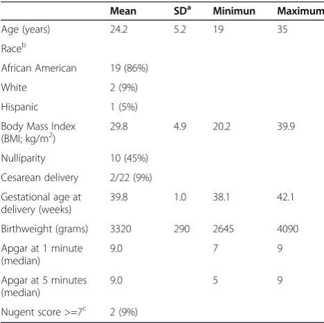
The clinical and demographic characteristics of the preg-nant population are displayed in Table 1. The clinical and demographic characteristics of non-pregnant subjects have been previously reported [112]. The present study in-cluded 32 non-pregnant women and 22 pregnant women who had a term delivery without complications (gesta-tional age at delivery from 38 to 42 weeks). Non-pregnant women self-sampled with a frequency of twice a week for 16 weeks. The median (interquartile range (IQR)) number
Table 1 Descriptive characteristics of the pregnant woman enrolled in the longitudinal study (n=22)
Mean SDa Minimun Maximum
Age (years) 24.2 5.2 19 35
Raceb
African American 19 (86%)
White 2 (9%)
Hispanic 1 (5%)
Body Mass Index
(BMI; kg/m2) 29.8 4.9 20.2 39.9
Nulliparity 10 (45%)
Cesarean delivery 2/22 (9%)
Gestational age at delivery (weeks)
39.8 1.0 38.1 42.1
Birthweight (grams) 3320 290 2645 4090
Apgar at 1 minute (median)
9.0 7 9
Apgar at 5 minutes (median)
9.0 5 9
Nugent score >=7c 2 (9%)
a
Standard Deviation. b
Non-pregnant women: African American (50%), White (40.6%), Hispanic and others (9.4%).
c
of samples with available sequence data was 27.5 samples per participant (IQR: 20.7 to 29). Pregnant women had a median of 6.5 samples per pregnancy (IQR: 6.0 to 7.0).
Characterization of the microbial taxa as a function of depth of coverage
We characterized the vaginal microbiota using pyro-sequencing of barcoded 16S RNA genes. The dataset con-sisted of 2,946,507 high-quality sequences, with an average length of 240 bp. The median number of sequences per sample was 2,878 (IQR: 2,446 to 4,171). Taxonomic assign-ment of the sequences identified a total of 143 taxa in the vaginal microbiota of the women studied; all 143 taxa were observed both in non-pregnant as well as pregnant women who delivered at term. The taxonomic assignments of vaginal bacterial community members are shown in Additional file 1: Table S1.
The vaginal microbiota in the non-pregnant state and normal pregnancy
To study the vaginal bacterial communities of pregnant versus non-pregnant women, we hierarchically clustered the vectors of relative abundances of bacterial phylotypes (one per sample) using the Jensen-Shannon divergence metric and Ward linkage [112]. In this study, we refer to a community state as a vector of relative abundances of bacterial phylotypes for a given sample. Community states were clustered into five groups with similar bac-terial composition and abundance (Figure 1), referred to as CSTs according to the nomenclature established by Gajer and colleagues [112].
Three of these CSTs were most often dominated by L. crispatus(CST I), L. gasseri (CST II) and L. iners (CST III). Communities that clustered in CST IV-A or IV-B lacked a substantial number of Lactobacillus spp. and differed in taxa composition. For example, CST IV-A was characterized by a roughly equal number of Peptoni-philus, Anaerococcus, Corynebacterium, Finegoldia, Pre-votella and a few other taxa. In contrast, those of CST IV-B had higher relative abundance of the genus Atopo-biumand were characterized by the presence ofL. iners (low relative abundance),Prevotella,Sneathia,Gardnerella, Ruminococcaceae,Parvimonas,Mobiluncusand other taxa previously shown to be associated with bacterial vaginosis [96]. These findings are consistent with previous observa-tions indicating that there is no single“core” microbiota of the human vagina [109]. The relationship between
Nugent score and CST was demonstrated. It is note-worthy that CST IV-B was strongly associated with a high Nugent score (defined as 7 to 10) (P= 0.013 using a mixed effect model; odds ratio = 24.3).
Table 2 shows the counts of samples assigned to each CST and corresponding percentages stratified by preg-nancy status. A dramatic difference in the distribution of frequency of CSTs between non-pregnant and pregnant patients who delivered at term was observed (a decrease of 95% in the odds of observing CST IV-B in pregnant women compared to non-pregnant women).
Since Table 2 was generated using correlated samples, standard methods (for example, Fisher tests) cannot be applied to assess significance of differences in frequen-cies of each CST between pregnant and non-pregnant women. Instead, for each CST (T), a logistic regression GEE model was fitted with the binary response variable (T versus non-T) used as a dependent variable and the pregnancy status used as an independent variable. The coefficients, odds ratios, P-values, and q-values for the five GEE models are shown in Table 3. The frequency of CST IV-B (most often dominated by Atopobium) was significantly lower in pregnant compared to non-pregnant women. The relative abundance of CST I (dominated by L. crispatus) was borderline significantly different between pregnant and non-pregnant women (based on unadjusted P= 0.0507 at the 5% significance level).
Constancy of the vaginal microbiota in pregnant and non-pregnant women
Figure 2 shows the profiles of CSTs for pregnant women who delivered at term as a function of gestation time. The CST profiles of pregnant and non-pregnant women are somewhat similar (given smaller number of samples per pregnant woman) except that CST IV-B is rarely present in pregnant women. In particular, none of the pregnant women persist in this CST, which lacks sub-stantial number of Lactobacillus, whereas communities of seven non-pregnant women persist in CST IV-B for 16 weeks [112].
Vaginal bacterial communities of most pregnant and non-pregnant women persist in one CST with some intermittent transitions to other CSTs. Is there a difference in constancy of vaginal bacterial communities between pregnant and non-pregnant women? To address this question, we used an approach in which we computed the mean community state within a subject (mean relative
Table 2 Distribution of samples in each community state-type as a function of pregnancy status (non-pregnant vs normal)
CST/Pregnancy status I II III IV-A IV-B Total
Non-pregnant women 129 (17%) 68 (8.9%) 268 (35.2%) 79 (10.4%) 217 (28.5%) 761
abundance of each bacterial phylotype across all samples of a subject), and then the Jensen-Shannon distance was computed between each community state and the mean community state for each subject. These distances are shown in Figure 3A. This is a measure of instability: the larger the distance, the higher the instability of the mi-crobial community within a subject (in other words, community composition changes often over time). To test if the instability was different between pregnant and non-pregnant women, we modeled the log of these Jensen-Shannon distances using a GEE model. The mean within-subject log Jensen-Shannon distance of pregnant women was significantly lower than that for non-pregnant women (difference in means −0.473 log units; that is, 1.6-fold lower Jensen-Shannon distance, P< 0.001). This means that vaginal bacterial communities are significantly more stable in pregnant than in non-pregnant women. However,
the results indicate that, during pregnancy, the structure of the bacterial community undergoes some change. To characterize the nature of the changes during pregnancy, we evaluated the ability of a community to shift to CST IV (A or B) by computing the Jensen-Shannon distance between each community state and the mean community state of all samples assigned to CST IV-A and CST IV-B (mean relative abundance of each bacterial phylotype across all samples in CST IV-A and CST IV-B). We modeled the log of these Jensen-Shannon distances using a GEE model and found that the mean log Jensen-Shannon distance of pregnant women was significantly higher (further away from CST IV-A or CST IV-B) than that for non-pregnant women (difference in means 0.13 log units; that is, 1.14-fold,P< 0.001) (Figure 3B). Altogether, these results indi-cate that bacterial communities in pregnancy do shift from one CST dominated byLactobacillusspp. to another CST dominated byLactobacillus spp., but rarely to CST IV-A or CST IV-B.
Identification of phylotypes accounting for differences in the structure of vaginal microbiota between the non-pregnant state and normal pregnancy
Table 3 provides evidence that the vaginal microbiota in women who deliver at term is different from the vaginal microbiota of non-pregnant women. Nonetheless, this analysis does not identify explicitly the phylotypes responsible for differences in the structure of the vagi-nal microbiota between pregnant and non-pregnant women.
In order to identify phylotypes whose relative abundances were significantly different between pregnant and non-pregnant women, we used statistical models that: 1) were designed for count data modeling (assuming Poisson and negative binomial distributions); and 2) allowed correlated observations from the same individuals (for example, linear mixed effect models); while 3) allowing for extra zeroes in Table 3 Coefficient estimates, odds ratios, p-values and
q-values for the association between each community state type with the pregnancy status
Community
state typea Estimate b
Odds ratio p-value q-valuec
IV-B −3.06 0.047 0.00000 0.00001
I 1.09 2.986 0.05076 0.12689
III 0.76 2.136 0.11344 0.18907
IV-A −1.23 0.292 0.16958 0.21198
II −0.73 0.482 0.48193 0.48193
a
Community state type:a group of community states with similar microbial
phylotype composition and abundance identified via unsupervised clustering (Figure1).
b
Estimate:the value of the coefficient in the logistic regression model for a
binary variable indicating whether (1) or not (0) a given sample was assigned in the community state named in column 1. The value of the coefficient represents the log of the odds ratio that the sample belongs to the community state indicated in column 1 given that the sample belongs to a pregnant woman (as opposed to a non-pregnant woman).
c
q-value:the False Discovery Rate adjusted p-value across all 5 community
types that were tested.
Community State Type
Gestational Age (weeks)
10 15 20 25 30 35 40
N018 N007 N013 N004 N011 N016 N008 N010 N002 N019 N001 N009 N021 N017 N012 N015 N014 N020 N003 N005 N022 N006 III IV−B
I II V Delivery Subject ID
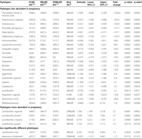
the data since some phylotypes were frequently un-detected. Three types of models were fitted for each phy-lotype, including PLME, NBLME and ZINBLME models. The model type with the smallest AIC value was retained for each phylotype and the P-value for group variable (pregnant versus non-pregnant) was computed only for this model. Only phylotypes that were present in at least 25% of all samples were included in the analysis, restrict-ing the number of phylotypes to 28. Table 4 shows the AIC statistics for all three types of models for each phy-lotype, as well as the estimate, confidence interval and
P-value for the best (smallest AIC) model. Of interest, out of the 28 phylotypes tested, the relative abundance of 26 was significantly different between the two groups (q-value <0.1 and fold change >1.5). Four of the signifi-cant phylotypes (L. vaginalis,L. crispatus,L. gasseriandL. jensenii) were more abundant in pregnant than non-pregnant women (Additional file 2: Figures S1 show box plots of the relative abundances of all significant phylo-types listed in Table 4). The NBLME model provided the optimal fit for a majority of phylotypes, indicating that there is over-dispersion in the sequence count data and, Subject ID
400 401 402 403 404 405 406 407 408 410 411 412 414 415 416 420 423 424 429 430 431 432 434 435 436 437 439 442 443 444 445 446
Non−pregnant Pregnant
Log Jensen−Shannon distance
−4
−3
−2
−1
N001 N002 N003 N004 N005 N006 N007 N008 N009 N010 N011 N012 N013 N014 N015 N016 N017 N018 N019 N020 N021 N022
Log Jensen−Shannon distance
Subject ID
−1.4
−1.2
−1.0
−0.8
−0.6
400 401 402 403 404 405 406 407 408 410 411 412 414 415 416 420 423 424 429 430 431 432 434 435 436 437 439 442 443 444 445 446
Non−pregnant Pregnant
N001 N002 N003 N004 N005 N006 N007 N008 N009 N010 N011 N012 N013 N014 N015 N016 N017 N018 N019 N020 N021 N022
A
B
hence, the Poisson distribution may be too restrictive for the observed count data. This finding is in agreement with previous observations [243]. About a quarter of the significant phylotypes showed zero inflation; therefore, the zero-inflation version of the negative binomial model (ZINBLME) provided the optimum fit based on AIC values. The SAS code and input dataset used to generate
the results presented in Table 4 are provided in Additional files 3 and 4, respectively.
Some of the selected phylotypes are defined at the genus and some at the species level (for example, Anaerococcus and Anaerococcus vaginalis), respectively. A genus level phylotype corresponds to a set of sequences that could not be reliably identified at the species level for any known Table 4 Differential relative abundance of microbial phylotypes between pregnant and non-pregnant women and statistics for the phylotype level analysis
Phylotypes PLME
AICa,d
NBLME AICb,d
ZINBLME AICc,d
Best AICd
Estimate Lower 95% CI
Upper 95% CI
Fold change
p-value q-valuee
Phylotypes less abundant in pregnancy
Clostridiales Family XI Incertae Sedis
3488.3 2996.9 2998.9 NBLME −7.095 −9.020 −5.169 −1205.6 0.0001 0.0002
Anaerococcus vaginalis 3043.8 2748.2 2759.9 NBLME −5.873 −7.260 −4.486 −355.4 0.0001 0.0002
Anaerococcus 5252.6 3804.3 3806.3 NBLME −5.557 −6.605 −4.509 −259.0 0.0001 0.0002
Prevotella genogroup 2 5510.3 4498.2 4504.2 NBLME −5.019 −5.655 −4.384 −151.3 0.0001 0.0002
Peptoniphilus 4707.8 4431.3 4433.3 NBLME −4.921 −5.670 −4.171 −137.1 0.0001 0.0002
Streptococcus anginosus 3285.4 2565.8 2583.8 NBLME −4.629 −5.748 −3.511 −102.4 0.0001 0.0002
Actinomycetales 5672.8 5110.1 5112.1 NBLME −4.546 −5.281 −3.811 −94.2 0.0001 0.0002
Leptotrichia amnionii 7299.3 3889.1 3893.3 NBLME −4.490 −5.359 −3.621 −89.1 0.0001 0.0002
Finegoldia magna 4687.1 4300.6 4302.6 NBLME −4.174 −4.958 −3.391 −65.0 0.0001 0.0002
Prevotella 4540 4094.7 4096.7 NBLME −3.870 −4.502 −3.238 −48.0 0.0001 0.0002
Clostridiales 5864.5 4852.6 NA NBLME −3.373 −4.274 −2.472 −29.2 0.0001 0.0002
Atopobium 3853.1 3275 3261.6 ZINBLME −3.268 −3.943 −2.593 −26.3 0.0001 0.0002
Bacteria 3167.5 3033 3040.1 NBLME −3.083 −3.921 −2.245 −21.8 0.0001 0.0002
Prevotella.bivia 4178.9 3043 3045 NBLME −3.038 −4.089 −1.986 −20.9 0.0001 0.0002
Eggerthella 3149.7 3083.7 3065.2 ZINBLME −1.936 −2.813 −1.060 −6.9 0.0001 0.0002
Gardnerella vaginalis 5472 5105.1 5076.7 ZINBLME −1.760 −2.253 −1.266 −5.8 0.0001 0.0002
Dialister 4048.5 3939.6 3940.9 NBLME −1.399 −2.147 −0.651 −4.1 0.0003 0.0004
Ureaplasma 2819 2700.6 2707.8 NBLME −1.153 −1.817 −0.490 −3.2 0.0007 0.0010
Lactobacillus 10572 9170.2 9172.2 NBLME −0.726 −1.169 −0.283 −2.1 0.0013 0.0017
Atopobium vaginae 12734 6971.9 7024.5 PLME −2.381 −3.946 −0.816 −10.8 0.0029 0.0037
Parvimonas micra 4512.2 3835.1 3821.7 ZINBLME −4.202 −7.609 −0.795 −66.8 0.0157 0.0183
Bifidobacteriaceae 4056.8 3989.9 3991.9 NBLME −0.660 −1.429 0.110 −1.9 0.0927 0.0998
Phylotypes more abundant in pregnancy
Lactobacillus vaginalis 2489.2 2467.9 2458.2 ZINBLME 1.704 1.190 2.218 5.5 0.0001 0.0002
Lactobacillus jensenii 6544.1 5564.1 5549.7 ZINBLME 1.549 1.453 1.645 4.7 0.0001 0.0002
Lactobacillus crispatus 11702 8094 8263.2 NBLME 0.754 0.212 1.295 2.1 0.0064 0.0078
Lactobacillus gasseri 6917.3 4412.4 NA NBLME 1.193 0.214 2.172 3.3 0.0170 0.0190
Non-significantly different phylotypes
Lactobacillus iners 18755 12576 12604 NBLME 0.165 −0.136 0.466 1.2 0.2824 0.2929
Aerococcus christensenii 4321.2 3986.9 3957.1 ZINBLME −0.425 −1.251 0.401 −1.5 0.3132 0.3132
a
PLME: Poisson Linear Mixed Effects Model. b
NBLME: Negative Binomial Linear Mixed Effects. c
ZINBLME: Zero-Inflated Negative Binomial Mixed-Effects Model. d
AIC: Akaike Information Criterion. e
species of the given genus. Thus, in the case of Anaerococ-cusandAnaerococcus vaginalis, the first phylotype corre-sponds to reads that cannot be taxonomically assigned to any known species of Anaerococcus and might represent uncharacterized species ofAnaerococcus, whereas the phy-lotype Anaerococcus vaginalis consists of reads that are classified as corresponding to that species.
Discussion
Principal findings of the study
Using sequence-based methods (rather than cultivation techniques) to characterize the vaginal microbiota in a longitudinal study of normal pregnant women and non-pregnant women, we established that: 1) at the bacterial community level, CST IV-B (characterized by high relative abundance of species of Atopobium as well as the pres-ence of Prevotella, Sneathia, Gardnerella, Ruminococca-ceae, Parvimonas, Mobiluncus and other taxa previously shown to be associated with bacterial vaginosis) was rarely observed in pregnant women who delivered at term; 2) the vaginal microbiota of normal pregnant women who deliver at term was different from that of non-pregnant women (higher abundance ofL. vaginalis,L. crispatus,L. gasseri and L. jensenii and lower abundance of 22 other phylotypes in normal pregnancy); 3) the stability of the va-ginal microbiota of pregnant women was higher than that of non-pregnant women; and 4) during normal pregnancy, bacterial communities do shift from one CST dominated byLactobacillusspp. to another CST dominated by Lacto-bacillusspp. but rarely to CST IV-A or CST IV-B.
The vaginal microbiota of normal pregnant women
This is the first longitudinal study of the vaginal micro-biota in normal pregnancy where samples have been fre-quently collected and microbial composition has been characterized using high-throughput pyrosequencing of the 16S rRNA gene. Previous studies have used a cross-sectional approach [213] and sparse sampling [212]. Some have used low resolution microbiological and mo-lecular techniques [211,212] to characterize the micro-bial communities. The methodology used in the present study provides a less biased, in-depth characterization of the bacterial composition and abundance of the vaginal microbiota. The major finding of this study is that nor-mal pregnant women maintain (throughout the entire pregnancy) vaginal CSTs dominated by Lactobacillus spp. This is in contrast with the observations made in the non-pregnant state, in which there were fluctuations between CSTs lacking a substantial number of Lactoba-cillusspp. and those that are dominated by members of this genus [112].
In a previous study, we focused on non-pregnant women and characterized five different CSTs (CST I to V); CST I, II, III and V were characterized by a predominance of
Lactobacillus spp. CST IV was characterized by a low abundance of Lactobacillus spp. and a predominance of other phylotypes, mainly of anaerobic bacteria. This CST was further subdivided into IV-A and IV-B based on hier-archical clustering [112]. The major difference between the two is that CST IV-B has a higher abundance of Ato-pobium, while CST IV-A has a more even microbial com-position including the following phylotypes:Peptoniphilus, Anaerococcus, Corynebacterium, Finegoldia and Prevo-tella. We have also reported that CST IV-A and CST IV-B were more common in certain ethnic groups (African-American and Hispanic) and were associated with a higher vaginal pH and high Nugent score [109]. In the current study focusing on pregnant women, we identified five of the six CSTs previously described: I, II, III, IV-A and IV-B. We did not find CST V. The most likely ex-planation for this is that the majority of women enrolled in the present study were African-American, and CST V was previously observed in only 1% of such women [109]. Given the sample size of the current study (n = 22 pregnant women) and the ethnic composition (90% African-American), the lack of representation of CST V is not unexpected. Therefore, these findings do not mean that other studies of the microbiota of pregnant women using a different population would not identify CST V.
Stability of the vaginal microbiota during pregnancy
During normal pregnancy, bacterial communities are more stable than in the non-pregnant state; however, some changes do occur. For example, bacterial commu-nities commonly transitioned from one Lactobacillus-dominated CST to another, but rarely to CST IV-A or CST IV-B. This is a reflection of the importance of Lactobacillus spp. in the vaginal ecosystem during preg-nancy. Such an interesting feature can be interpreted to represent an adaptation of the microbial community and the host to maximize reproductive fitness. We propose that the enhanced stability confers greater resilience and has a protective role against ascending infection of the genital tract, which is risk factor for preterm delivery [244-246] and other conditions such as a sonographic short cervix [247-249], cervical insufficiency [250-254], preterm labor in twin gestations [255-257], vaginal bleed-ing in the third trimester [258], placenta previa [259,260], or some cases of fetal death [261-265]. The mechanisms by which bacterial community stability promotes health in the vaginal niche remain to be determined.
Is the vaginal microbiota unique during pregnancy?
identified another 22 phylotypes that had lower relative abundance in pregnant than non-pregnant women (Table 4); many of these phylotypes are associated with CST IV-A and CST IV-B. Interestingly, the relative abundance ofL. inerswas not significantly different be-tween the two groups. This finding might reflect a lack of optimal protection by this commonLactobacillussp. [109] and deserves further investigation. Aagaard and colleagues [213] have proposed that there is a microbiota signature of pregnancy based upon a cross-sectional study of pregnant (n = 24) and non-pregnant women (n = 60). Using a random forest algorithm, pregnancy was well pre-dicted by relative abundances of different phylotypes in vaginal fluid. At this point, even though there are differ-ences in microbial compositions between the pregnant and non-pregnant state, there is no evidence that these differences are specific to pregnant women. Further, it is unclear if a microbial signature of pregnancy could have utility for diagnostic purposes.
It is possible that the composition of the vaginal microbiome associated with pregnancy may have func-tional (that is, metabolic, immune) implications for the host [266]. An alternative interpretation is that changes in the microbiota are a consequence of the physiological state of pregnancy. During the course of the menstrual cycle, stability of microbial communities is higher at the time when estrogen concentrations are high (14 and 21 days) [112]. This has been attributed to the effect of es-trogens on the maturation of the vaginal epithelium, resulting in the accumulation of glycogen on the upper layer of the epithelium [267-270]. Glycogen is a carbon source metabolized to lactic acid by Lactobacillus spp., causing a low vaginal pH [24,26,29]. Further research is required to determine if the relationship between high estrogens and increased stability is causal.
Strengths and limitations
The major strengths of this work are: 1) the longitudinal nature of the study, which allows characterization of the vaginal microbiota over time; 2) the frequent sampling protocol - this allowed characterization of the dynamics of the bacterial communities in pregnancy to an extent not done before; 3) the quality of the sequence-based techniques (16S rRNA) which reduced bias over other methods, including cultivation techniques; 4) the analyt-ical methods that took into consideration changes over time on the same subject, therefore increasing the power of detection of differences between clinical groups; and 5) inclusion of relevant clinical groups: non-pregnant and normal pregnant women. These strengths allowed meaningful differences to be found among these clinical groups. The use of primer 27 F could be a limitation of this study; this primer may have underestimated the true rela-tive abundance of 16S rRNA genes of Bifidobacteriaceae in
general, and those of the genusG. vaginalis, a bacterium commonly found in the vagina of women who experience bacterial vaginosis. The selection of optimal PCR primers is a subject of considerable ongoing discussion in the field of microbiome studies. Unfortunately, there is no consen-sus, nor a perfect set of primers. In this study, we followed the recommendations of the NIH-funded Human Microbiome Project (http://www.hmpdacc.org/). An-other potential limitation of the study is the sample size, which included 22 pregnant women who delivered at term. Yet, despite the apparently limited sample size, the identification of significant differences provides evi-dence that the study of the vaginal microbiota during pregnancy can yield important insights into the relation-ship between the structure and dynamics of microbial communities and pregnancy outcome. Further studies are required to confirm these findings, extend the observa-tions and elucidate the role of microorganisms in adverse pregnancy outcome.
Conclusion
This is the first longitudinal study of the human vaginal microbiota in pregnancy. We demonstrate differences in the vaginal bacterial community structure between normal pregnant and non-pregnant women and show that preg-nancy is characterized by a greater degree of stability than observed in non-pregnant women. We established the baseline stability patterns of the vaginal microbiota in pregnancy. This could serve as the basis to study the relationship between the vaginal microbiota and adverse pregnancy outcomes. The characterization of the vagi-nal microbiota in pregnancy has the potential to yield information of prognostic, diagnostic and therapeutic value.
Additional files
Additional file 1: Table S1.Taxonomic assignments, total 16S rRNA gene sequences assigned to each taxa, and metadata.
Additional file 2: Figure S1.Box plots of relative abundances of all phylotypes that have statistically significantly different relative abundance between pregnant and non-pregnant women.
Additional file 3:SAS code used to generate the results presented in Table 4.
Additional file 4:Input dataset for SAS code used to generate the results presented in Table 4.
Abbreviations
AIC:Akaike Information Criterion; bp: base pair; CST: community state type; GEE: generalized estimation equation; IQR: interquartile range;
NBLME: negative binomial linear mixed effect; NIH: National Institutes of Health; PCR: polymerase chain reaction; PLME: Poisson linear mixed effect; ZINBLME: zero-inflated negative binomial mixed-effect.
Competing interests
Authors’contributions
RR, SSH, PG, AT and JR conceived the study. RR, SSH, LN, MG, RFL, PC, JM and TC performed the clinical sampling and samples management. DWF performed DNA extraction, 16S rRNA gene amplification and sequencing. DWF, PG and JR processed the sequence data. PG and AT performed the statistical analyses. RR, SSH, PG, AT and JR wrote the manuscript. All authors read and approved the final manuscript.
Acknowledgements
This work was funded, in part, by theEunice Kennedy ShriverNational Institute of Child Health and Human Development, NIH, DHHS. The authors wish to acknowledge the contributions of the patients who volunteered for these studies, the medical and healthcare personnel involved in the research effort, and colleagues who contributed to the discussions which eventually led to the conduct of the study. We are particularly grateful to Dr Sharon Hillier of the University of Pittsburgh, Dr Jack Sobel of Wayne State University, Dr David Relman of Stanford University and Dr Sorin Draghici of Wayne State University.
Author details
1
Perinatology Research Branch, Program for Perinatal Research and Obstetrics, Division of Intramural Research,Eunice Kennedy ShriverNational Institute of Child Health and Human Development, NIH, Bethesda, MD and Detroit, MI, USA.2Department of Obstetrics and Gynecology, University of Michigan, Ann Arbor, MI, USA.3Department of Epidemiology and
Biostatistics, Michigan State University, East Lansing, MI, USA.4Department of Obstetrics and Gynecology, Wayne State University School of Medicine, Detroit, MI, USA.5Institute for Genome Sciences, University of Maryland School of Medicine, Baltimore, MD, USA.6Department of Microbiology and Immunology, University of Maryland School of Medicine, Baltimore, MD, USA. 7
Department of Obstetrics and Gynaecology, University of Southern Denmark, Odense, Denmark.8Division of Surgery, University College, Northwick Park Institute for Medical Research Campus, London, UK.
Received: 12 September 2013 Accepted: 18 December 2013 Published: 3 February 2014
References
1. Ma B, Forney LJ, Ravel J:Vaginal microbiome: rethinking health and disease.Annu Rev Microbiol2012,66:371–389.
2. Döderlein A:Das Scheidensekret und seine Bedeutung fuerdas Puerperalfieber.Die Arten des Scheidensekretes1892,11:699.
3. Thomas S:Döderlein Bacillus: lactobacillus acidophilus.J Infect Dis1928, 43:218–227.
4. Rogosa M, Sharpe ME:Species differentiation of human vaginal lactobacilli.J Gen Microbiol1960,23:197–201.
5. Redondo-Lopez V, Cook RL, Sobel JD:Emerging role of lactobacilli in the control and maintenance of the vaginal bacterial microflora.Rev Infect Dis1990,12:856–872.
6. Klebanoff SJ, Coombs RW:Viricidal effect of Lactobacillus acidophilus on human immunodeficiency virus type 1: possible role in heterosexual transmission.J Exp Med1991,174:289–292.
7. Hillier SL, Krohn MA, Klebanoff SJ, Eschenbach DA:The relationship of hydrogen peroxide-producing lactobacilli to bacterial vaginosis and genital microflora in pregnant women.Obstet Gynecol1992,79:369–373. 8. Hillier SL, Krohn MA, Rabe LK, Klebanoff SJ, Eschenbach DA:The normal
vaginal flora, H2O2-producing lactobacilli, and bacterial vaginosis in pregnant women.Clin Infect Dis1993,16(Suppl 4):S273–S281. 9. Curtis H:What is normal vaginal flora?Genitourin Med1997,73:230. 10. Sewankambo N, Gray RH, Wawer MJ, Paxton L, McNaim D,
Wabwire-Mangen F,et al:HIV-1 infection associated with abnormal vaginal flora morphology and bacterial vaginosis.Lancet1997,350:546–550. 11. Gupta K, Stapleton AE, Hooton TM, Roberts PL, Fennell CL, Stamm WE:
Inverse association of H2O2-producing lactobacilli and vaginal Escherichia coli colonization in women with recurrent urinary tract infections.J Infect Dis1998,178:446–450.
12. Sobel JD:Is there a protective role for vaginal flora?Curr Infect Dis Rep
1999,1:379–383.
13. Pybus V, Onderdonk AB:Microbial interactions in the vaginal ecosystem, with emphasis on the pathogenesis of bacterial vaginosis.Microbes Infect
1999,1:285–292.
14. Martin HL, Richardson BA, Nyange PM, Lavreys L, Hillier SL, Chohan B,et al: Vaginal lactobacilli, microbial flora, and risk of human immunodeficiency virus type 1 and sexually transmitted disease acquisition.J Infect Dis
1999,180:1863–1868.
15. Donders GG, Bosmans E, Dekeersmaecker A, Vereecken A, Van Bulck B, Spitz B: Pathogenesis of abnormal vaginal bacterial flora.Am J Obstet Gynecol2000, 182:872–878.
16. van De Wijgert JH, Mason PR, Gwanzura L, Mbizvo MT, Chirenje ZM, Iliff V,
et al:Intravaginal practices, vaginal flora disturbances, and acquisition of sexually transmitted diseases in Zimbabwean women.J Infect Dis2000, 181:587–594.
17. Larsen B, Monif GR:Understanding the bacterial flora of the female genital tract.Clin Infect Dis2001,32:e69–e77.
18. Vasquez A, Jakobsson T, Ahrne S, Forsum U, Molin G:Vaginal lactobacillus flora of healthy Swedish women.J Clin Microbiol2002,40:2746–2749. 19. Ocana VS, Nader-Macias ME:Vaginal lactobacilli: self- and co-aggregating
ability.Br J Biomed Sci2002,59:183–190.
20. Wiesenfeld HC, Hillier SL, Krohn MA, Landers DV, Sweet RL:Bacterial vaginosis is a strong predictor of Neisseria gonorrhoeae and Chlamydia trachomatis infection.Clin Infect Dis2003,36:663–668.
21. De Backer E, Verhelst R, Verstraelen H, Alqumber MA, Burton JP, Tagg JR,
et al:Quantitative determination by real-time PCR of four vaginal Lactobacillus species. Gardnerella vaginalis and Atopobium vaginae indicates an inverse relationship between L. gasseri and L. iners.
BMC Microbiol2007,7:115.
22. Lai SK, Hida K, Shukair S, Wang YY, Figueiredo A, Cone R,et al: Human immunodeficiency virus type 1 is trapped by acidic but not by neutralized human cervicovaginal mucus.J Virol2009,
83:11196–11200.
23. Leppaluoto PA:Bacterial vaginosis: what is physiological in vaginal bacteriology? An update and opinion.Acta Obstetricia Et Gynecologica Scandinavica2011,90:1302–1306.
24. Linhares IM, Summers PR, Larsen B, Giraldo PC, Witkin SS:Contemporary perspectives on vaginal pH and lactobacilli.Am J Obstet Gynecol2011, 204(120):e1–e5.
25. Mirmonsef P, Gilbert D, Zariffard MR, Hamaker BR, Kaur A, Landay AL,et al: The effects of commensal bacteria on innate immune responses in the female genital tract.Am J Reprod Immunol2011,65:190–195.
26. Witkin SS, Ledger WJ:Complexities of the uniquely human vagina.
Science Translational Medicine2012,4:132fs11.
27. Kashket ER:Bioenergitcs of lactic acid bacteria: cytoplasmic pH and osmotolerance.FEMS Microbiol1987,46:233–244.
28. Russell JB, Diez-Gonzalez F:The effects of fermentation acids on bacterial growth.Adv Microb Physiol1998,39:205–234.
29. Boskey ER, Telsch KM, Whaley KJ, Moench TR, Cone RA:Acid production by vaginal flora in vitro is consistent with the rate and extent of vaginal acidification.Infect Immun1999,67:5170–5175.
30. Alakomi HL, Skytta E, Saarela M, Mattila-Sandholm T, Latva-Kala K, Helander IM: Lactic acid permeabilizes gram-negative bacteria by disrupting the outer membrane.Appl Environ Microbiol2000,66:2001–2005.
31. Saling E, Schreiber M, al-Taie T:A simple, efficient and inexpensive program for preventing prematurity.J Perinat Med2001,29:199–211. 32. Boskey ER, Cone RA, Whaley KJ, Moench TR:Origins of vaginal acidity:
high D/L lactate ratio is consistent with bacteria being the primary source.Hum Reprod (Oxford, England)2001,16:1809–1813. 33. Hoyme UB, Saling E:Efficient prematurity prevention is possible by
pH-self measurement and immediate therapy of threatening ascending infection.Eur J Obstet Gyn R B2004,115:148–153.
34. O’Hanlon DE, Moench TR, Cone RA:In vaginal fluid, bacteria associated with bacterial vaginosis can be suppressed with lactic acid but not hydrogen peroxide.BMC Infect Dis2011,11:200.
35. Hammerschlag MR, Alpert S, Onderdonk AB, Thurston P, Drude E, McCormack WM,et al:Anaerobic microflora of the vagina in children.
Am J Obstet Gynecol1978,131:853–856.
36. Hillier SL, Lau RJ:Vaginal microflora in postmenopausal women who have not received estrogen replacement therapy.Clin Infect Dis1997, 25(Suppl 2):S123–S126.
38. Farage M, Maibach H:Lifetime changes in the vulva and vagina.
Arch Gynecol Obstet2006,273:195–202.
39. Gupta S, Kumar N, Singhal N, Kaur R, Manektala U:Vaginal microflora in postmenopausal women on hormone replacement therapy.Indian J Pathol Microbiol2006,49:457–461.
40. Yamamoto T, Zhou X, Williams CJ, Hochwalt A, Forney LJ:Bacterial populations in the vaginas of healthy adolescent women.J Pediatr Adolesc Gynecol2009,22:11–18.
41. Hickey RJ, Zhou X, Pierson JD, Ravel J, Forney LJ:Understanding vaginal microbiome complexity from an ecological perspective.Transl Res2012, 160:267–282.
42. Johnson SR, Petzold CR, Galask RP:Qualitative and quantitative changes of the vaginal microbial flora during the menstrual cycle.Am J Reprod Im Mic1985,9:1–5.
43. Eschenbach DA, Thwin SS, Patton DL, Hooton TM, Stapleton AE, Agnew K,
et al:Influence of the normal menstrual cycle on vaginal tissue, discharge, and microflora.Clin Infect Dis2000,30:901–907.
44. Devillard E, Burton JP, Hammond JA, Lam D, Reid G:Novel insight into the vaginal microflora in postmenopausal women under hormone replacement therapy as analyzed by PCR-denaturing gradient gel electrophoresis.Eur J Obstet Gyn R B2004,117:76–81.
45. Heinemann C, Reid G:Vaginal microbial diversity among postmenopausal women with and without hormone replacement therapy.Can J Microbiol
2005,51:777–781.
46. Bezirtzoglou E, Voidarou C, Papadaki A, Tsiotsias A, Kotsovolou O, Konstandi M: Hormone therapy alters the composition of the vaginal microflora in ovariectomized rats.Microb Ecol2008,55:751–759.
47. Hyman RW, Herndon CN, Jiang H, Palm C, Fukushima M, Bernstein D,et al: The dynamics of the vaginal microbiome during infertility therapy with in vitro fertilization-embryo transfer.J Assist Reprod Genet2012, 29:105–115.
48. Kazi YF, Saleem S, Kazi N:Investigation of vaginal microbiota in sexually active women using hormonal contraceptives in Pakistan.BMC Urol2012, 12:22.
49. Brotman RM, Ravel J, Bavoil PM, Gravitt PE, Ghanem KG:Microbiome, sex hormones, and immune responses in the reproductive tract: challenges for vaccine development against sexually transmitted infections.Vaccine
2013. Epub 2013/10/19.
50. Backhed F, Ley RE, Sonnenburg JL, Peterson DA, Gordon JI:Host-bacterial mutualism in the human intestine.Science2005,307:1915–1920. 51. Turnbaugh PJ, Ley RE, Hamady M, Fraser-Liggett CM, Knight R, Gordon JI:
The human microbiome project.Nature2007,449:804–810.
52. Peterson J, Garges S, Giovanni M, McInnes P, Wang L, Schloss JA,et al:The NIH human microbiome project.Genome Res2009,19:2317–2323. 53. Nelson KE, Weinstock GM, Highlander SK, Worley KC, Creasy HH, Wortman
JR,et al:A catalog of reference genomes from the human microbiome.
Science2010,328:994–999.
54. Conlan S, Kong HH, Segre JA:Species-level analysis of DNA sequence data from the NIH Human Microbiome Project.PloS one2012,7:e47075. 55. Cho I, Blaser MJ:The human microbiome: at the interface of health and
disease.Nat Rev Genet2012,13:260–270.
56. Relman DA:Microbiology: learning about who we are.Nature2012, 486:194–195.
57. Qin J, Li R, Raes J, Arumugam M, Burgdorf KS, Manichanh C,et al:A human gut microbial gene catalogue established by metagenomic sequencing.
Nature2010,464:59–65.
58. Spiegel CA, Amsel R, Holmes KK:Diagnosis of bacterial vaginosis by direct gram stain of vaginal fluid.J Clin Microbiol1983,18:170–177.
59. Eschenbach DA, Hillier S, Critchlow C, Stevens C, DeRouen T, Holmes KK: Diagnosis and clinical manifestations of bacterial vaginosis.Am J Obstet Gynecol1988,158:819–828.
60. Krohn MA, Hillier SL, Eschenbach DA:Comparison of methods for diagnosing bacterial vaginosis among pregnant women.J Clin Microbiol
1989,27:1266–1271.
61. Nugent RP, Krohn MA, Hillier SL:Reliability of diagnosing bacterial vaginosis is improved by a standardized method of gram stain interpretation.J Clin Microbiol1991,29:297–301.
62. Hillier SL, Krohn MA, Nugent RP, Gibbs RS:Characteristics of three vaginal flora patterns assessed by gram stain among pregnant women.Vaginal Infections and Prematurity Study Group. Am J Obstet Gynecol1992, 166:938–944.
63. Hay PE, Morgan DJ, Ison CA, Bhide SA, Romney M, McKenzie P,et al: A longitudinal study of bacterial vaginosis during pregnancy.Br J Obstet Gynaecol1994,101:1048–1053.
64. Rosenstein IJ, Morgan DJ, Sheehan M, Lamont RF, Taylor-Robinson D: Bacterial vaginosis in pregnancy: distribution of bacterial species in different gram-stain categories of the vaginal flora.J Med Microbiol1996, 45:120–126.
65. Schwebke JR, Hillier SL, Sobel JD, McGregor JA, Sweet RL:Validity of the vaginal gram stain for the diagnosis of bacterial vaginosis.Obstet Gynecol
1996,88:573–576.
66. Donders GG:Gram stain method shows better sensitivity than clinical criteria for detection of bacterial vaginosis in surveillance of pregnant, low-income women in a clinical setting.Infect Dis Obstet Gynecol1999, 7:273–275.
67. Curzik D, Drazancic A, Hrgovic Z:Nonspecific aerobic vaginitis and pregnancy.Fetal Diagn Ther2001,16:187–192.
68. Marrazzo JM, Koutsky LA, Eschenbach DA, Agnew K, Stine K, Hillier SL: Characterization of vaginal flora and bacterial vaginosis in women who have sex with women.J Infect Dis2002,185:1307–1313.
69. Wilson JD, Ralph SG, Rutherford AJ:Rates of bacterial vaginosis in women undergoing in vitro fertilisation for different types of infertility.BJOG
2002,109:714–717.
70. Donders GG, Vereecken A, Bosmans E, Dekeersmaecker A, Salembier G, Spitz B: Definition of a type of abnormal vaginal flora that is distinct from bacterial vaginosis: aerobic vaginitis.BJOG2002,109:34–43.
71. Brotman RM, Ravel J, Cone RA, Zenilman JM:Rapid fluctuation of the vaginal microbiota measured by Gram stain analysis.Sex Transm Infect
2010,86:297–302.
72. Bakken LR:Separation and purification of bacteria from soil.Appl Environ Microbiol1985,49:1482–1487.
73. Hugenholtz P, Goebel BM, Pace NR:Impact of culture-independent studies on the emerging phylogenetic view of bacterial diversity.J Bacteriol1998, 180:4765–4774.
74. Kalra A, Palcu CT, Sobel JD, Akins RA:Bacterial vaginosis: culture- and PCR-based characterizations of a complex polymicrobial disease’s pathobiology.Curr Infect Dis Rep2007,9:485–500.
75. Amann RI, Ludwig W, Schleifer KH:Phylogenetic identification and in situ detection of individual microbial cells without cultivation.Microbiol Rev
1995,59:143–169.
76. Eckburg PB, Bik EM, Bernstein CN, Purdom E, Dethlefsen L, Sargent M,et al: Diversity of the human intestinal microbial flora.Science2005, 308:1635–1638.
77. Pace NR:A molecular view of microbial diversity and the biosphere.
Science1997,276:734–740.
78. Torsvik V, Ovreas L:Microbial diversity and function in soil: from genes to ecosystems.Curr Opin Microbiol2002,5:240–245.
79. Martin DH, Zozaya M, Lillis R, Miller J, Ferris MJ:The microbiota of the human genitourinary tract: trying to see the forest through the trees.
Trans Am Clin Climatol Assoc2012,123:242–256.
80. Ward DM, Weller R, Bateson MM:16S rRNA sequences reveal numerous uncultured microorganisms in a natural community.Nature1990, 345:63–65.
81. Zoetendal EG, Akkermans AD, De Vos WM:Temperature gradient gel electrophoresis analysis of 16S rRNA from human fecal samples reveals stable and host-specific communities of active bacteria.Appl Environ Microbiol1998,64:3854–3859.
82. Suau A, Bonnet R, Sutren M, Godon JJ, Gibson GR, Collins MD,et al:Direct analysis of genes encoding 16S rRNA from complex communities reveals many novel molecular species within the human gut.Appl Environ Microbiol1999,65:4799–4807.
83. Ye Y:Identification and Quantification of Abundant Species from Pyrosequences of 16S rRNA by Consensus Alignment.Proceedings (IEEE Int Conf Bioin Biomed)2011,2010:153–157.
84. Handelsman J:Metagenomics: application of genomics to uncultured microorganisms.Microbiol Mol Biol R2004,68:669–685.
85. Petrosino JF, Highlander S, Luna RA, Gibbs RA, Versalovic J:
Metagenomic pyrosequencing and microbial identification.Clin Chem
2009,55:856–866.
86. Pallen MJ, Loman NJ, Penn CW:High-throughput sequencing and clinical microbiology: progress, opportunities and challenges.Curr Opin Microbiol