R E S E A R C H
Open Access
Transcription factor NF
κ
B regulates the
expression of the histone deacetylase SIRT1
Judith Katto
1, Nicole Engel
2, Wasim Abbas
3, Georges Herbein
3and Ulrich Mahlknecht
1,2,4*Abstract
Background:The NAD-dependent protein deacetylase SIRT1 has a wide range of different targets, which may be
regulated either directly through deacetylation and thus potentially altering their activity or localization or indirectly by deacetylation of histones, which in turn alters their transcription rate and availability. SIRT1 is therefore involved in the regulation of many different and fundamental cellular processes such as apoptosis, metabolism, differentiation and cell cycle arrest. It is also involved in the regulation of resistance of cells against oxidative stress and longevity under conditions of caloric restriction. Even though the targets and role of SIRT1 have been studied quite intensively, only little is known about the mechanisms affectingSIRT1transcriptional regulation. The nuclear factor NFκB is a well-studied and widely known transcription factor, which is involved in the regulation of many important cellular activities. The regulation of NFκB by SIRT1 has been reported recently, but it is, however, still unknown whether a feedback mechanism affects the regulation ofSIRT1too, particularly in view of the fact that putative NFκB binding sites within theSIRT1promoter suggest just that.
Results:In the study presented herein we show that there is activation of theSIRT1promoter by overexpression of different NFκB subunits. Direct binding of NFκB to theSIRT1promoter can be demonstrated by an electrophoretic mobility shift assay. Further investigations indicated enhanced expression ofSIRT1on the mRNA levels in cells
overexpressing NFκB. A functional assay showed that acetylation of one of the main target proteins of SIRT1 is reduced in these cells.
Conclusions:These finding together indicate SIRT1 expression to be regulated in a positive feedback loop by NFκB. The putative binding sites for NFκB found within theSIRT1promoter appears to be functional and several NFκB subunits are able to enhance the expression of SIRT1 if they are overexpressed.
Keywords:Protein deacetylase SIRT1, Nuclear factor NF-kappa-B, Apoptosis
Background
The regulation of activity, cellular localization and stabil-ity of nearly all proteins depends on post-transcriptional and/or post-translational modifications. Such modifica-tions are being carried out by a diversity of chemical re-actions including (de)phosphorylation, (de)acetylation or ubiquitination and many others. These highly dynamic regulatory processes are controlled and driven by antag-onizing enzymes and allow cells to react to environmental
changes and to communicate with each other. Histone deacetylases (HDACs) are one such group of enzymes. HDACs are ubiquitous and catalyze the removal of acetyl groups from both histones and non-histone proteins. The most obvious and best examined role of HDACs is their effect on the constitution and maintenance of chromo-somal structure as well as the regulation of global gene ex-pression through modification of the so called histone code and alteration of the accessibility of genes for tran-scription factors [1]. However, apart from their role as superordinative gene regulators and their role in the con-trol of chromatin structure, some proteins of this family are associated with the distinct regulation of single target proteins, which may specifically be deacetylated [2].
Histone deacetylases are divided into four different classes: three groups of classical HDACs known as class I,
* Correspondence:mahlknecht@gmx.de
1José Carreras Center for Immunotherapy and Gene Therapy, Saarland University Medical Center, Kirrbergerstraße Bldg. 45.3, 66421, Homburg/Saar, Germany 2Department of Internal Medicine, University of Heidelberg Medical Center, Im Neuenheimer Feld 410, 69120, Heidelberg, Germany
Full list of author information is available at the end of the article
class II and class IV HDACs and a fourth class, also being referred to as the ‘sirtuins’ [3]. The sirtuins differ from the other classes of HDACs based on their dependence on the presence of the coenzyme NAD+ [4]. Seven human sirtuins, which are homologs of the yeast silent informa-tion regulator 2 (SIR2) protein, have been reported so far. SIR2 is responsible for the transcriptional silencing of silent mating loci, telomeres and ribosomal DNA in yeast. It also suppresses the recombination of rDNA and thereby lowers the formation of toxic extrachromosomal rDNA rings, which extends lifespan in yeast cells dramatically [5]. Yeast SIR2 or mammalian sirtuin overexpression goes along with the global deacetylation of histones [6]. Most of the gene silencing effects that are being associated with sirtuin activity depend on the deacetylation of histone H4 lysine 16 (H4K16) [7] and/or histone H3 lysines 9, 14 or 18 (H3K9, H3K14, H3K18) [8]. Depending on the presence of NAD+, the sirtuins are biological sensors for the cellular energetic and oxidative status. Thus, high levels of NAD+ go along with high sirtuins activity [9].
The human deacetylase sirtuin1 (SIRT1) shares the highest degree of homology of all mammalian sirtuins with the yeast protein SIR2. SIRT1 localizes predominantly to the cell nucleus [10] and deacetylates both histones (preferably H4K16) and non-histone proteins. SIRT1 antag-onizes the histone acetyltransferase p300 [11], which acet-ylates all histones and some non-histone proteins such as p53 and FOXO3a. SIRT1 is involved in a number of cellu-lar regulation mechanisms affecting cellucellu-lar lifespan, stress resistance, apoptosis and cell metabolism [12]. The pro-longation of cellular lifespan under conditions of caloric restriction has been known for quite some time. It has been described for a number of very different organisms ranging from yeast [13] to nematodes [14] to mammals [15,16] and has been reported to be associated with SIRT1. Caloric restriction causes an increase in the production of NAD+ and goes along with increased levels of SIRT1 mRNA [13,16] as well as with increased SIRT1 activity [17]. The positive influence of SIRT1 on longevity in mam-mals has been reported to depend on increased stress resistance and reduced apoptosis. In addition to the modi-fication of histone proteins, SIRT1 also carries the ability to inactivate a number of non-histone proteins by de-acetylation. One of the most prominent examples for this mechanism is the deacetylation of tumor suppressor pro-tein p53. The transcription factor p53 triggers apoptosis in the case of DNA damage or oxidative stress and plays a key role in cellular defense against the development of malignant transformation [18]. The inactivation of p53 by SIRT1 may retard apoptosis and render cells more re-sistant to oxidative stress [19,20]. On the other hand, overexpression of SIRT1 may also promote carcinogenesis because of its inhibitory effect on apoptosis and cell cycle arrest. The forkhead proteins constitute another group of
proteins that is also involved in the regulation of apoptosis, stress resistance and metabolism; these proteins are re-ported to be regulated by SIRT1 [21].
Another protein that may be deacetylated by SIRT1 is the nuclear factor NF-kappa-B (NFκB) [22-24]. NFκB is a ubiquitously distributed transcription factor that con-trols gene expression of many target genes, thereby af-fecting important cellular functions such as cell cycle, angiogenesis, adhesion and apoptosis [25]. NFκB is being constituted by homo- and heterodimers of different NFκB/ Rel-proteins, which all share a highly conserved DNA binding/dimerization domain called ‘Rel homology (RH) domain’ [26]. NFκB/Rel proteins can be divided into two classes. Members of the first class are coded by the genes
NFKB1 and NFKB2 both of them normally being tran-scribed as long isoforms called NFκB-p105 and NFκ B-p100, which are inactive as long as they have inhibiting C-terminal domains. The shorter active forms of this class, the NFκB-p50 (derived from p105) and NFκB-p52 (derived from p100) are produced by arrested transla-tion or partial proteolysis of the longer forms. The sub-unit NFκB-p49 is another isoform that can be generated from an alternatively spliced transcript of p100. It is reported to be involved in the activation of the HIV en-hancer [27]. The second class contains the proteins c-Rel, RelA (also called NFκB-p65) and RelB, which may activate gene expression in homodimers as well as in heterodimers [28]. The most prevalent and best studied dimer is the heterodimer consisting of the p65 and p50 protein. NFκB is constitutively transcribed in many different cell types, but it remains inactive because the dimers are bound to inhibitory proteins of the IκB family, which mask their nu-clear translocation signal and consequently go along with their localization in the cell cytoplasm. In response to diverse stimuli IκB proteins are phosphorylated by IκB kinase (IKK) which triggers their ubiquitination and thus their degradation by the proteasome machinery. NFκB is subsequently translocated to the nucleus were it interacts with promoters, thus activating the expression of specific target genes [29].
Results and discussion
HumanSIRT1gene promoter analysis
Alterations in the SIRT1 expression rate are associated with changes in cell metabolism, differentiation, prolifer-ation and many other fundamental cell features and affect many diseases such as diabetes, cancer or arterio-sclerosis. But at present little information is available on the molecular mechanisms regulating SIRT1 at the pro-moter level. In order to further elucidate the regulation of SIRT1 at the promoter level, a search for putative transcription factor binding sites was carried out using the online tool MOTIF Search (http://motif.genome.jp). The search was based on the alignment with the tran-scription factor database TRANSFAC. Among others, we found the SIRT1 promoter to contain several putative NFκB binding sites. NFκB is a transcription factor that is involved in apoptosis, the regulation of cellular stress, and numerous immunological functions. The NFκB con-sensus sequence GGGAMTTYCC with M and Y being exchangeable bases was found with minor variations in three positions of the SIRT1 promoter. The three men-tioned positions are located at the promoter base-pair positions 1,168 to 1,177 bp, 1,441 to 1,450 bp and 1,838 to 1,847 bp. In accordance with ourin silicoanalyses the latter two NFκB potential binding sites appeared to be specific for the NFκB subunit p65. This suggests that Sirt1 expression could potentially be regulated by NFκB.
NFκB regulates theSIRT1promoter
In order to further investigate whether NFκB was involved in the regulation ofSIRT1at the promoter level, a lucifer-ase reporter plasmid was generated, which contained a 2,228-bp-long fragment of the SIRT1 promoter sequence. This construct was cotransfected in HEK293T cells to-gether with different combinations of expression vectors of the NFκB subunits p49, p50 or p65. Luciferase activity of cell extracts was measured 24 hours after transfection and normalized to the protein content of the cell extracts. Figure 1 plots the results of three independent measure-ments of luciferase activity relative to the constitutive activity. The constitutive activity was determined by trans-fection of the reporter plasmid without cotranstrans-fection of any expression plasmids for NFκB. These results show clearly that the Sirt1 promoter is activated in general by overexpression of NFκB. Statistical P values indicated in the diagram were calculated using a Mann-Whitney U Test and show significance for the difference of each NFκB subunit combination versus control (P ≤0.05). The differ-ent combinations of NFκB subunits we transfected in order to determine the effects of the different homo- and heterodimers of NFκB did not show significant differences among each other (P>0.1).
Our experiments showed overexpression of NFκB p50 subunit or NFκB p65 subunit alone to increase SIRT1 promoter activity by an average of 8.6-foldand 7.3-fold.
Figure 1SIRT1-promoter activation after transfection of NFκB subunits.Reporter plasmids containing theSIRT1promoter were
Overexpression of the two NFκB subunits p50 and p65 in combination was identified to have an activating effect on the SIRT1 promoter with an average of about 7.9-fold, which is very similar to the effects of overexpression of the single subunits. The heterodimer p50/p65 represents the most common and best examined variant of NFκB, which makes these results the most interesting. The over-expression of p49 together with p65 also showed SIRT1 promoter activation. The activating effect of this combi-nation seemed to be lower with an average of only 4.2-fold, but the difference to the other subunit combinations is not significant. Overexpression of all three subunits to-gether goes along with a seven-fold activation of theSIRT1 promoter, which is similar to the other results. Transfec-tion of the empty reporter plasmid (without Sirt1 pro-moter) was used as negative control and did not show any luciferase activity.
NFκB regulatesSIRT1expression
In order to examine whether the effects of promoter ac-tivation through overexpression of NFκB in fact had influ-ence on theSIRT1expression levels, RNA from HEK293T cells was extracted after transient transfection with the NFκB expression constructs. In quantitative real-time PCR we observed a 1.8 to 3-fold upregulation forSIRT1at the mRNA level (Figure 2) in response to NFκB overex-pression for nearly all combinations of NFκB subunits ex-cept in the combination of p50 with p65, which had only minor effects onSIRT1mRNA expression levels in this ex-periment. The little effect of the overexpression of p50 in
combination with p65 on the mRNA level, does not fit with the remarkable promoter activating effects seen for this combination in the luciferase assay. A possible explan-ation may be based on the fact that the p50/p65 dimer is the most common dimer of NFκB, which has numerous physiological functions in the cell. Therefore, other regu-lating mechanisms may also be activated that could affect
SIRT1expression.
Functional assay of SIRT1 activity
A functional assay was carried out not only to investi-gate the levels of SIRT1 expression but also the effects of NFκB on cell metabolism and other proteins in asso-ciation with SIRT1. In order to prove relevant changes in SIRT1 activity after NFκB overexpression we investi-gated the acetylation status of histone H4 lysine residue 16 (H4K16), which is targeted and deacetylated by SIRT1. We isolated the histone fraction of cells after transfec-tion with defined NFκB subunits and analyzed the his-tone H4 acetylation status by western blot technique. Figure 3A shows a remarkable decrease of acetylated H4K16in cells overexpressing NFκB. For quantification of the results, blots were scanned and acetylated histone H4 was normalized to the amount of total histone H4 on the same blot. To be sure, that the effects are not due to the circumstances of cell culture or transfection in general, which may change acetylation status of histones because it induces lots of metabolical reactions and stress, we com-pared the results of cells transfected with NFκB vector with those transfected with an empty vector. Figure 3B
shows that the overexpression of NFκB subunits in all combinations resulted in a significant reduction of the relative amount of acetylated histone H4. The amount of reduction in cells overexpressing two or more NFκB subunits was similar to the reduction caused by direct overexpression of SIRT1. In cells overexpressing only one subunit of NFκB protein the effects are less but still de-tectable. These results suggest that overexpression of NFκB has activating effects on the deacetylation of histone H4, which could be mediated through induction of SIRT1 expression, particularly when two or three of the NFκB subunits are overexpressed, thus allowing for a formation of heterodimers.
Binding of NFκB toSIRT1promoter
To investigate the role of different putative NFκB binding sites, truncations of the SIRT1 promoter were generated (Figure 4). The 2,228 bp and 1,148 bp SIRT1 promoter fragments contained all three putative NFκB binding sites; fragments with length ranging from 1,026 bp, to 921 bp and 887 bp in length contained only two NFκB binding sites and the 668 bp fragment contained only one NFκB binding site.
An electrophoretic mobility shift assay was done to measure the binding of NFκB to the promoter and to in-vestigate the relation of the three potential binding sites. In order to induce NFκB expression, cells were treated
H4K16
total H4
A
B
Figure 3Histone acetylation status in response to SIRT1 activity.Cells were transfected with expression vectors for different NFκB subunits. Expression vector forSIRT1was used as a positive control. Histones were isolated after 24 hours and acetylation status was assessed by western blot. Transfection with empty vector was used as a reference control.(A)Detection was carried out with an antibody specific for acetylated histone H4K16and with another antibody binding to histone H4 independently of the acetylation status (total H4).(B)Detection films of blots were scanned and quantified
with TNFα. Nuclear extracts from both treated and un-treated cells were isolated and incubated with biotin la-beled DNA fragments. The reaction mixture was resolved on a 6% non-denaturing polyacrylamide gel. For all of the tested promoter fragments signals were detected (Figure 5), which proves binding of NFκB. The putative binding site contained in the short fragment is likely to be involved in the NFκB binding of the promoter, but because all of the fragments showed a signal, it is not possible to decide whether the binding is mediated solely by this single site or whether all three sites are involved. Nuclear extracts from untreated cells were used as negative controls. NFκB oligonucleotides were used as a positive control to prove induction of NFκB expression by TNFα and to compare the position of the bands on the blot. The positive control showed a signal that was much stronger than the labeled promoter fragments. This may be caused by the high sen-sitivity of the control, which also could explain the signal seen for the NFκB oligonucleotides when incubated with untreated cells.
Conclusions
NFκB is a transcription factor that can be found in nearly all cell types. NFκB regulates immune reactions and is
involved in the regulation of cellular stress resistance and apoptosis. Also, it has recently been shown that the NFκB binding sequence is a gene regulatory motif that is most essentially involved in the regulation of cellular aging. SIRT1 is a highly conserved protein involved in many cel-lular processes in both health and disease. It has been reported to be involved in the extension of cellular life span under conditions of caloric restriction in many spe-cies. Its positive influence on stress resistance of cells is widely known and its activation prevents apoptosis and cell cycle arrest. Since both proteins, SIRT1 as well as NFκB, are involved in the regulation of immune response, cellular stress, and aging, a regulatory connection between them would not really be very surprising. In some studies SIRT1 has been shown to interact with NFκB and to re-press its activity by deacetylation [22,24]. But at present little information is available on the molecular mecha-nisms regulating the expression of SIRT1at the promoter level. In the study presented herein, we show that in addition to the already known regulation of NFκB by SIRT1, NFκB overexpression enhances SIRT1 promoter ac-tivity. Increased SIRT1 enzymatic activity caused by NFκB overexpression was shown in a functional assay. The direct binding of the transcription factor NFκB to the Sirt1
Figure 4Sirt1 promoter truncations.Schematic illustration of Sirt1 promoter fragments used for electrophoretic mobility shift assay. The positions of NFkB binding sequences are marked by arrows.
promoter was demonstrated by electrophoretic mobility shift assays. With these results a reciprocal feedback regula-tion of SIRT1 at the promoter level through transcripregula-tion factor NFκB is shown to take place.
The conserved NFκB binding motif can be found three times within the humanSIRT1 promoter sequence. Dis-tinguishing which of the three potential NFκB binding sites was responsible for the activation of the promoter could not be made since all of the created SIRT1 pro-moter truncations showed binding of NFκB. With re-porter plasmids containing the human SIRT1 promoter sequence and luciferase assays, we were able to show that high expression levels of NFκB subunits were able to specifically activate SIRT1at the promoter level. Our experiments show that the NFκB binding motifs within the SIRT1 promoter are functional, and that NFκB can act as a positive regulator of SIRT1 at the promoter level. In order to further examine whether NFκB over-expression on SIRT1 promoter activation caused a real increase in SIRT1 expression, we performed RT-PCR ex-periments using cells overexpressing NFκB that showed elevated levels of SIRT1 mRNA for most of the used NFκB subunits. As a functional assay to measure SIRT1 activity the acetylation status of histone H4K16, which is one of the most common targets of the deacetylase SIRT1, was examined. The decrease in acetylation of H4K16 under conditions of NFκB overexpression indi-cates not only that SIRT1 protein levels are increased, but also that this has measurable effects on downstream targets. This means that at least some of the signaling cascades and pathways that activate NFκB may influence the expression of SIRT1 and the acetylation status of its target proteins. The involvement of both of these pro-teins in the development and progression of numerous diseases such as cancer, arteriosclerosis or diabetes ren-ders this regulation mechanism an interesting target for further investigations with regard to the development of novel molecular therapeutics affecting SIRT1 regulation. SIRT1 is one of the most interesting targets for potential epigenetic therapeutics and the ongoing attempts to cre-ate specific activators or deactivators for SIRT1 may benefit from better knowledge about its regulation by intercellular transcription factors. Taken together, these findings provide new insights into the regulation of SIRT1 and interactions between NFκB and SIRT1. They may promote a better understanding of the mechanism involved in regulation of proteins that control proce-dures as important as metabolism and aging.
Methods
Antibodies and plasmids
Anti-histone H4 monoclonal antibody (anti-H4) was pur-chased from Abcam (Cambridge, UK) and antibody specific for Histone H4 acetylated at lysine 16 (anti-H4K16) was
purchased from Upstate via Biomol, (Hamburg, Germany). Secondary antibodies (anti-rabbit and anti-mouse horse-radish peroxidase conjugates) were purchased from Santa Cruz Biotechnology (Santa Cruz, California, USA).
NFκB expression plasmids for subunits p49, p50 and p65 were based on pRSV expression vector systems and were provided by the National Institutes of Health (NIH) AIDS Research and Reference Reagent Program. For con-struction of luciferase reporter plasmids, human SIRT1 promoter from genomic DNA was amplified by PCR. The amplified region spanned the entire SIRT1 promoter gion. The promoter was inserted into the luciferase re-porter plasmid pGL2-basic, which was purchased from Promega (Mannheim, Germany). All plasmids were propa-gated using competent DH5α E. coli and were isolated with a Nucleobond PC100 Kit from Macherey & Nagel (Düren, Germany). All transfection experiments were car-ried out in HEK293T cells.
Luciferase reporter gene assays
HEK293T-cells were cultured in 12-well plates with DMEM supplemented with 10% FCS and were cotransfected with the luciferaseSIRT1promoter or empty pGL2basic vector constructs and different combinations of expression vectors encoding NFκB subunits. In order to normalize the total amount of DNA that was used per transfection, empty vec-tor was added in order to adjust the total amount of DNA. Transfection was done using the nanoparticle-based trans-fection reagent Nanofectin (PAA, Pasching, Austria) and medium was changed 2 hours after transfection. Transfec-tion efficiency was assessed by UV-microscopy after trans-fection of green lantern constructs (Promega, Mannheim, Germany). Cells were harvested after 24 hours and lucif-erase activity was measured using the luciflucif-erase reporter assay system (Promega, Mannheim, Germany) in accord-ance with the manufacturer’s instructions. The activity of each sample was normalized to its protein concentration measured on a UV-Vis spectrophotometer Nanodrop™ ND1000 (PEQLAB, Erlangen, Germany) using DC Protein assay (BioRad, Hercules, California, USA) based on the Lowry method. Relative activity was calculated as fold dif-ference in relation to the control vector pGL2basic. Lu-ciferase assays were performed in duplicates and every experiment was repeated at least twice.
Quantitative real-time PCR
2.0 system and DNA Master SYBR Green Kit both from Roche (Penzberg, Germany). The primers that were used for amplification of humanSIRT1were as follows:
forward primer 5′- ACGCTGGAACAGGTTGCGGG
A - 3′,
reverse primer 5′- AAGCGGTTCATCAGCTGGGCA
C - 3′.
Thermal reaction cycles were as follows: initial cycle at 95°C for 10 min, which was then followed by 45 cycles at 95°C for 10 sec, 60°C for 4 sec and 72°C for 16 sec. GAPDH was used as an internal control to normalize measured Ct values of each sample, and data were ana-lyzed using the delta-delta Ct method.
Extraction of histones and western blot
HEK293T cells were cultured in 12-well plates and were harvested with 100μl lysis buffer after incubation for 24 hours. While cells were being mixed vigorously, 25μl of 2 M sulphuric acid were added drop by drop, and cells were then placed on ice for 1 h. After centrifugation at 13,000 rpm, supernatants were collected, mixed with 400 μl of 20% trichloroacetic acid and incubated on ice for histone precipitation. After 1 h the mixture was spun down and the pellets containing histones were washed twice with acidified acetone (0.1% HCl) and once with pure acetone prior to resuspension in water. Extracted histones were separated on a 15% polyacrylamide gel and blotted onto a nitrocellulose membrane according to the full-wet western blot technique. Acetylated his-tones were detected with anti H4K16 antibody. For detection, the ECL plus western blotting detection re-agent from GE Healthcare (Little Chalfont, UK) was used. After that, an antibody binding to H4 independ-ent of the acetylation status was used in order to detect total histone H4. Blots were scanned and quan-tifications were calculated with the ImageQuant5.1 soft-ware (GE Healthcare, Little Chalfont, UK). Signal of acetylated H4 was normalized to the signal of total H4 in order to eliminate effects depending on the western blot technique itself.
Promoter truncation and electrophoretic mobility shift assay (EMSA)
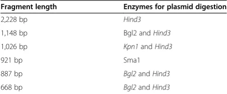
The generation of the different promoter fragments was done with different methods. The fragment with 921 bp was generated by exonuclease digestion. Fragments with 1,148 bp and 1,026 bp were generated through restric-tion enzyme digesrestric-tion and subsequent re-ligarestric-tion. Frag-ments with 887 bp and 668 bp were amplified by PCR from genomic DNA and inserted into an empty vector. Plasmids were transformed intoE. coli DH5afor propa-gation and extracted accordingly. A total of 2μg plasmid DNA were digested with restriction enzymes (Table 1) and then run on a 0.8% agarose gel. Digested fragments from each construct were cut out while the gel was monitored under UV control, and DNA elution was done with a Silica Beads DNA gel extraction kit (Fermentas, Waltham, USA). DNA from each construct was labeled with biotin using the Biotin 3′ End DNA Labeling Kit (Pierce Biotechnology, Waltham, USA). In order to stimu-late NFκB expression, U937 cells were treated with re-combinant TNFα at a concentration of 10 ng/ml for 30 minutes. Control cells were left untreated. Nuclear ex-tracts (10μg) were then incubated with a specific biotin-labeled DNA sample before electrophoretic mobility shift assay (EMSA) was done using a Light Shift Chemilumin-escent EMSA kit (Pierce Biotechnology, Waltham, USA) in accordance with the manufacturer’s instructions.
Abbreviations
NFκB:Nuclear factor of kappa light polypeptide gene enhancer in B-cells; SIRT1: Sirtuin 1.
Competing interests
The authors declare that they have no competing interests.
Authors’contributions
UM, JK, NE, and WA conceived and designed the experiments. JK, NE, and WA analyzed the data. JK wrote the first draft of the manuscript. UM contributed to the writing of the manuscript. UM GH, WA, and NE made critical revisions. All authors reviewed and approved the final manuscript.
Acknowledgements
This work was supported by institutional funds from the Saarland University, grants from the University of Franche-Comté (UFC) and the Region Franche-Comté (RECH-FON12-000013) to G.H. W.A. is a recipient of a doctoral scholarship from the Higher Education Commission, Pakistan. We thank Diasorin SA and Roche for their financial support.
Author details
1José Carreras Center for Immunotherapy and Gene Therapy, Saarland University Medical Center, Kirrbergerstraße Bldg. 45.3, 66421, Homburg/Saar, Germany. 2Department of Internal Medicine, University of Heidelberg Medical Center, Im Neuenheimer Feld 410, 69120, Heidelberg, Germany.3Department of Virology, Pathogens & Inflammation Research Unit UPRES EA4266, SFR FED 4234, University of Franche-Comté, CHU Besançon, Besançon, France.4Department of Haematology/Oncology, St. Lukas Klinik Solingen, Schwanenstraße 132, 42697, Solingen, Germany.
Received: 19 April 2013 Accepted: 25 June 2013 Published: 19 July 2013
Table 1 Enzymes for digestion ofSIRT1 promoter truncations
Fragment length Enzymes for plasmid digestion
2,228 bp Hind3
1,148 bp Bgl2 andHind3
1,026 bp Kpn1andHind3
921 bp Sma1
887 bp Bgl2andHind3
References
1. Yang XJ, Seto E:The Rpd3/Hda1 family of lysine deacetylases: from bacteria and yeast to mice and men.Nat Rev Mol Cell Biol2008,9:206–218. 2. Haberland M, Montgomery RL, Olson EN:The many roles of histone
deacetylases in development and physiology: implications for disease and therapy.Nat Rev Genet2009,10:32–42.
3. Marks PA, Miller T, Richon VM:Histone deacetylases.Curr Opin Pharmacol
2003,3:344–351.
4. Landry J, Slama JT, Sternglanz R:Role of NAD(+) in the deacetylase activity of the SIR2-like proteins.Biochem Biophys Res Commun2000,
278:685–690.
5. Kaeberlein M, McVey M, Guarente L:The SIR2/3/4 complex and SIR2 alone promote longevity in Saccharomyces cerevisiae by two different mechanisms.Genes Dev1999,13:2570–2580.
6. Imai S, Armstrong CM, Kaeberlein M, Guarente L:Transcriptional silencing and longevity protein Sir2 is an NAD-dependent histone deacetylase.
Nature2000,403:795–800.
7. Vaquero A, Scher M, Lee D, Erdjument-Bromage H, Tempst P, Reinberg D:
Human SirT1 interacts with histone H1 and promotes formation of facultative heterochromatin.Mol Cell2004,16:93–105.
8. Feige JN, Auwerx J:Transcriptional targets of sirtuins in the coordination of mammalian physiology.Curr Opin Cell Biol2008,20:303–309. 9. Rajendran R, Garva R, Krstic-Demonacos M, Demonacos C:Sirtuins:
molecular traffic lights in the crossroad of oxidative stress, chromatin remodeling, and transcription.J Biomed Biotechnol2011,2011:368276. 10. Michishita E, Park JY, Burneskis JM, Barrett JC, Horikawa I:Evolutionarily conserved and nonconserved cellular localizations and functions of human SIRT proteins.Mol Biol Cell2005,16:4623–4635.
11. Bouras T, Fu M, Sauve AA, Wang F, Quong AA, Perkins ND, Hay RT, Gu W, Pestell RG:SIRT1 deacetylation and repression of p300 involves lysine residues 1020/1024 within the cell cycle regulatory domain 1.
J Biol Chem2005,280:10264–10276.
12. Michan S, Sinclair D:Sirtuins in mammals: insights into their biological function.Biochem J2007,404:1–13.
13. Lin SJ, Defossez PA, Guarente L:Requirement of NAD and SIR2 for life-span extension by calorie restriction in Saccharomyces cerevisiae.
Science2000,289:2126–2128.
14. Tissenbaum HA, Guarente L:Increased dosage of a sir-2 gene extends lifespan in Caenorhabditis elegans.Nature2001,410:227–230. 15. Boily G, Seifert EL, Bevilacqua L, He XH, Sabourin G, Estey C, Moffat C,
Crawford S, Saliba S, Jardine K, Xuan J, Evans M, Harper ME, McBurney MW:
SirT1 regulates energy metabolism and response to caloric restriction in mice.PLoS One2008,3:e1759.
16. Cohen HY, Miller C, Bitterman KJ, Wall NR, Hekking B, Kessler B, Howitz KT, Gorospe M, de Cabo R, Sinclair DA:Calorie restriction promotes mammalian cell survival by inducing the SIRT1 deacetylase.Science2004,305:390–392. 17. Bordone L, Guarente L:Calorie restriction, SIRT1 and metabolism:
understanding longevity.Nat Rev Mol Cell Biol2005,6:298–305. 18. Levine AJ:p53, the cellular gatekeeper for growth and division.Cell1997,
88:323–331.
19. Vaziri H, Dessain SK:Ng Eaton E, Imai SI, Frye RA, Pandita TK, Guarente L, Weinberg RA: hSIR2(SIRT1) functions as an NAD-dependent p53 deacetylase.Cell2001,107:149–159.
20. Luo J, Nikolaev AY, Imai S, Chen D, Su F, Shiloh A, Guarente L, Gu W:
Negative control of p53 by Sir2alpha promotes cell survival under stress.
Cell2001,107:137–148.
21. Van Der Heide LP, Hoekman MF, Smidt MP:The ins and outs of FoxO shuttling: mechanisms of FoxO translocation and transcriptional regulation.Biochem J2004,380:297–309.
22. Salminen A, Kauppinen A, Suuronen T, Kaarniranta K:SIRT1 longevity factor suppresses NF-kappaB -driven immune responses: regulation of aging via NF-kappaB acetylation?BioEssays2008,30:939–942.
23. Salminen A, Ojala J, Huuskonen J, Kauppinen A, Suuronen T, Kaarniranta K:
Interaction of aging-associated signaling cascades: inhibition of NF-kappaB signaling by longevity factors FoxOs and SIRT1.Cell Mol Life Sci
2008,65:1049–1058.
24. Yeung F, Hoberg JE, Ramsey CS, Keller MD, Jones DR, Frye RA, Mayo MW:
Modulation of NF-kappaB-dependent transcription and cell survival by the SIRT1 deacetylase.EMBO J2004,23:2369–2380.
25. Baldwin AS:Control of oncogenesis and cancer therapy resistance by the transcription factor NF-kappaB.J Clin Invest2001,107:241–246.
26. Hayden MS, Ghosh S:Signaling to NF-kappaB.Genes Dev2004,
18:2195–2224.
27. Schmid RM, Perkins ND, Duckett CS, Andrews PC, Nabel GJ:Cloning of an NF-kappa B subunit which stimulates HIV transcription in synergy with p65.Nature1991,352:733–736.
28. Siggers T, Chang AB, Teixeira A, Wong D, Williams KJ, Ahmed B, Ragoussis J, Udalova IA, Smale ST, Bulyk ML:Principles of dimer-specific gene regulation revealed by a comprehensive characterization of NF-kappaB family DNA binding.Nat Immunol2012,13:95–102.
29. Karin M, Ben-Neriah Y:Phosphorylation meets ubiquitination: the control of NF-[kappa]B activity.Annu Rev Immunol2000,18:621–663.
doi:10.1186/1868-7083-5-11
Cite this article as:Kattoet al.:Transcription factor NFκB regulates the expression of the histone deacetylase SIRT1.Clinical Epigenetics20135:11.
Submit your next manuscript to BioMed Central and take full advantage of:
• Convenient online submission
• Thorough peer review
• No space constraints or color figure charges
• Immediate publication on acceptance
• Inclusion in PubMed, CAS, Scopus and Google Scholar
• Research which is freely available for redistribution