Observations of Lens Epithelium in Cell Cultures*
WALTER J. GEERAETS, M.D.
Professor of Ophthalmology and Associate Professor of Biophysics,
Medical College of Virginia, Health Sciences Division of Virginia
Commonwealth University, Richmond, Virginia
The following investigation had been conducted to evaluate lens epithelial cells in vitro as a poten-tially suitable model for future studies of chronic low dose ionizing radiation effects on this cell type in vitro. Since there is a great range of dose require-ments to produce in vivo radiation cataract in man, monkeys, and rabbits, and an equally wide range in time from radiation exposure until the first minimal lens changes can be biomicroscopically detected ( 3), it may be assumed that in vitro comparison of cells derived from these three species may provide some explanation for the in vivo differences. While con-trolled low dose acute or chronic ionizing radiation exposures and subsequent examinations can easily be achieved in animal experimentation, such data are more difficult to obtain from the human lens for ob-vious reasons. Thus, very minute lens changes can be quite accurately detected through biomicroscopic observation in the rabbit and monkey eye, but they may easily escape attention during routine clinical examination of the human lens after accidental or therapeutic exposure to ionizing radiation. Such mi-nor lens changes are not expected to interfere with visual acuity, thus, the exposed person would not be aware of them. In addition, there is no established pattern for these very early changes in human lenses on which even a trained observer could base his cri-teria. Moreover, exact dosimetry is more difficult on human lenses, and scheduled follow-up examinations are frequently not kept by patients having undergone therapeutic radiation involving the eye.
*This investigation was supported by the National Aeronautics and Space Administration, (NASA), Grant NGR 47-002-005 and in part by the Old Dominion Eye Bank and Research, Inc., Richmond, Virginia, Prevention of Blindness, Inc., New York, New York, and NIH Grant No. 2B5176 and 5 TOI EY00022.
264
The required radiation dose level for production of early in vivo lens changes in rabbits and humans deviates at least by one order of magnitude. Also, the latent periods after .irradiation until lens changes become manifest vary greatly between the two spe-cies. It was hoped that in vitro observations of epi-thelial explants from human, monkey, and rabbit lenses would provide information whether this wide range of clinically observed dose requirements holds true in in vitro systems as well.
H is for those reasons that normal behavior of human, monkey, and rabbit lens epithelial cells in vitro, that is, without irradiation, had to be com-pared to form a base line for further investigation. These observations were focused primarily on mor-phological changes of the three cell strains which possibly might have occurred over the period of ob-servation due to the environment in which the cells of the different species were grown. Thus, if after a prolonged time in in vitro milieu, cells of different species would become indistinguishable from one an-other, as compared with initial findings after ~xplan tation, this model of experimentation would not be applicable to the purpose of studying radiation ef-fects. On the other hand, if species specific charac-teristics remained unchanged from the original ex-plants, the model could be accepted as valid and would possibly allow some guarded extrapolation to in vivo systems.
Literature Review. Attempts to grow lens epi-thelium from different species in vitro had been made by numerous investigators, however, little has been reported on long-term cultivation of lens epithelium. It should be noted that during the early days of tis-sue culture, terminology often varied from one au-thor to the next so that it is frequently difficult to know exactly what is meant by some of the terms.
GEERAETS: OBSERVATIONS OF LENS EPITHELIUM IN CELL CULTURES 265
In our experiments we adhered closely to the nomen-clature recommended by the Tissue Culture Asso-ciation at their 17th annual meeting, May 31-June 3, 1966, San Francisco, California.
In 1922, Fischer, by repeating explantation of different tissues from chick embryo eyes, recorded that his attempts to grow lens epithelium in vitro were unsuccessful. He did succeed in growing epithelium but felt it was from the iris fragments adherent to the lens capsule and not lens epithelium.
In 1926, Kirby reported on various experiments in cultivating lens epithelium of chick embryo. When culturing the anterior one-third of 52-hour-old chick embryo eyes for 72 hours at 37.5°C in homogeneous medium consisting of equal parts of adult chicken plasma and chick embryo extract using the concave slide method of Carrel, he observed differentiation of the posterior portion of the lens vesicle. Kirby subsequently (experiment No. III) attempted to avoid Fischer's uncertainty regarding the source of the outgrowth by first incubating the intact chick embryo lens 24 hours and observing no growth from adherent tissue; he then opened the capsule ("cutic-ula") and obtained epithelial outgrowth in two of four attempts. In his experiment No. IV, lens epi-thelium was explanted on the point of a needle within an embryonic tissue juice plasma clot medium at 37.5°C. After 12-24 hours, lens epithelial cells began to migrate and to divide. Kirby also demonstrated that lens epithelium could be cultivated in media containing extract from the embryonic eye alone and
also from other embryonic tissue after the eyes had been removed. He stated at one point that one strain was carried through "17 generations in six weeks." However, his exact meaning is not quite clear and most likely referred to replications since he reported that the cultures almost doubled in size every 48 hours.
In 1929, Kirby, Estey, and Tabor in another
study using lens epithelium of the chick embryo in vitro demonstrated that during a period of eight
months, 174 of 294 original explants (59%) showed
active growth after 48 hours, that is, survived after
the "first transfer." One hundred twelve (38%)
sur-vived the "second transfer," and 56 (19%) of the
original explants survived the "third transfer." They
reported that it was possible to carry a single strain
through by 112 passages over a period of seven
months. The medium was changed every 48 hours.
They also concluded that cultures of chick embryo
lens epithelium were more successful in the regular
culture media than when the various constituent inorganic salts were increased more than 25 % over the normal. Tyrode's medium with a pH of 8.2 was markedly toxic, but it was possible to revive certain debilitated cultures of lens epithelium by placing them in normal media for several passages.
In 1932, Kirby, Estey, and Wiener studied the effect of changes in the nutrient medium on chick embryo lens epithelium cultured in vitro. They ob-served that glucose proved toxic to the cells when the concentration exceeded 578 mg per 100 ml. Levulose 1660 mg per 100 ml and galactose 333 mg per 100 ml caused growth retardation or cellular death
as did acetone ( 40 mg% ) and betahydroxybutyric acid (120 mg%).
In 1948, Ida Mann reported her experiments on about 70 cultures of mouse lens epithelium. Lenses of young mice up to 10 days old were used first but proved difficult to explant without infection, while those from embryo mice of the same strain were grown successfully. In her study, the culture medium used was composed of rat serum, Tyrode's solution, and mouse embryo extract. Cellular differentiation was observed around 10 days; after that very few cultures survived. No culture remained alive after
12 days, and no further stage of differentiation could be observed. The first stage of differentiation was an increase in the cytoplasm and movement of the nucleus to one side. This resembled the process of normal differentiation of lens epithelial cells into lens fibers in the intact lens where the nucleus moves to the side of the cell next to the capsule. The cell then elongated forming a lens fiber with the nucleus eventually equidistant from the two ends. In a later stage, the voluminous clear cytoplasm apparently flattened in a few cells so that the cell became bluntly pointed at both ends with the nucleus remaining applied to one side of the central part of the elon-gated portion of the cells.
266 GEERAETS: OBSERVATIONS OF LENS EPITHELIUM IN CELL CULTURES
on the plasma covered wall of a roller tube. Rotation
of the tube (9 rev per hour) provided a slow washing action thus promoting adequate exchange of food materials and waste products between fluid and cells. The cultures were kept at 37°C. Evidence of growth observed in cultures of chick lens epithelium occurred
in about 12 hours while in human lens epithelium it took about 48 hours. If the culture was maintained without changing the supernatant fluid, human lens
epithelium survived for approximately three weeks.
No evidence of differentiation of lens epithelium
into lens fibers was observed in their study. They
found that human lens epithelium grew earlier and
more quickly in tissue culture if the explant was obtained from a normal lens. In cultures of chick
embryo lenses, the explants became discolored after
a few days by gradual accumulation of fat within the
cells, followed by progressive loss of a distinguishable cell pattern. The authors also observed that outgrowth had been established. Adult human lens epithelium seemed to be more resistant and less easily destroyed than that derived from the chick embryo.
In 1959, Van der Veen and Heyen reported the continuous culture of lens epithelium from a
3-month-old male calf. The explants, anterior lens
capsule with attached epithelium in small fragments, were supplied with 2 ml of growth medium consist-ing of Hank's solution, 30% calf serum inactivated
at 56°C for 30 minutes, 0.5% lactalbumin
hydroly-sate, 5,000 U penicillin and 5 mg DHS per 100 ml. These cells were cultured in two screw-cap bottles
of 200 ml size without plasma at 36°C. Four milli-liters of fresh growth medium per flask were added
after seven days without removing the old media. After that, 75 % of the culture medium was
"re-newed" every seven days for two weeks and then
every third and fourth day. Initial outgrowth from
some explants was noted four weeks after primary
explantation. After another month, isolated, trans-parent islands of cells were observed with further
growth progressing at a very slow rate. After eleven
weeks the cells were transferred by mechanical de-tachment. Hereafter, the growth rate increased, and three subcultures were made at intervals of two weeks. Later on, cells were detached by using 0.2 %
Versene and 0.5% Trypsin to initiate two other
sublines. Proliferation continued for eight months
at a constant rate. Each culture was then divided into two to four subcultures after 10-14 days incubation. One strain of cells was maintained through 44 pas-sages for two years. Microscopic observation did not
reveal any characteristic cell type. Some cells were polygonal, others pyramidal or irregular in shape.
The nuclei were oval and contained several nucleoli. Under phase-contrast microscopy, intercellular bridges were regularly observed. Dividing lens cells were rarely seen. The authors attributed this to a
probable short period for the completion of mitosis. Their attempts to cultivate the cells in a growth
medium with less than 20% calf serum and to adapt
the cells to a medium containing heterologous serum failed consistently. With their method, they succeeded
in subculturing cells from the two calves lenses, maintaining them through 18 subcultures for one
year. No differences in morphology or growth were
observed in the three strains of calf lens epithelium.
Three other attempts by these investigators to grow
lens cells in continuous culture were unsuccessful. In 1960, Bryan, Leinfelder, and Meltzer studied
the effects of gases upon cell growth of human lens
epithelium. The cultures were maintained as mono-layers in silicon-stoppered T-60 flasks. The nutrient
employed was composed of 4 __:_ 4 - 2 Tyrode's BSS, pooled horse serum and embryonic extract respec-tively and 200 U penicillin and 200 µ, gm
strepto-mycin per milliliter added. The medium was changed
at least once a week, and the flasks were shaken to provide space for new growth. With different gas
mixtures, (95% 02
+
5% C02.; 95% N2+
5% C02; 95% air+
5% C02 and 100% 02 at 1 It perminute) the pH varied. The temperature was kept at
37.5°C. The cell strains (human lens epithelium, embryonic chick heart fibroblasts, and Earle's L-strain) demonstrated the ability to pro\iferate in an
environment low in 02• Growth was determined by initial cell count compared with that after four days. Of the three cell strains tested, only the lens epithe -lium in which the pH of the medium was increased did not show a significant decrease in growth. How-ever, the cells showed more proliferation at a pH of 7.2 (95% 02 ± 5% C02) than the control at a pH
of 7.6.
In 1965, Tamura reported his experimental
re-sults of long-term cultures of rabbit lens epithelium.
He used medium 199 from Chiba Serum Co. Ltd. at
pH 7.2 containing LAH (0.25%) (Difeo Co.) and
calf serum ( 10 % ) . A suspension of the minced tissue of the lens capsule was centrifuged, and the
sediment was cultured at 37°C on coverslips in T-form culture bottles. The medium was changed
GEERAETS: OBSERVATIONS OF LENS EPITHELIUM IN CELL CUL TURES 267
occurred about every five to seven days during culti-vation. Their morphology was variable, but most resembled a stage between fibroblasts or pavement epithelium. A few cells showed nuclear movement to one side and elongation of cytoplasm which sug-gested differentiation of lens epithelial cells into lens fibers. The in vitro growth was steady for about five months. Giant cells were seen after the ninth day in primary cultures. Polynuclear cells appeared in sub-sequent subcultures; whether these were present in primary cultures at any time was not stated.
Zaret ( 13) suggested Q-switched laser irra-diation of lens epithelium in tissue culture to study protein synthesis and polysomal analysis. He had
succeeded in culturing rabbit lens epithelium in tissue culture over seven weeks at the time of his report. The cells were incubated at 37°C in
supple-mented Eagle's medium and 5 % C02 in air. After 10 days, large numbers of mitoses were observed, and the cells began to grow on the glass surface as a monolayer culture. The culture retained its epithelial
identity over the entire observation time. Its karyo-type remained diploid. According to this investigator, these cells remained differentiated and continued to produce lens protein as demonstrated by immuno-fluorescence techniques.
Materials and Methods.
Explant: Lens epithelial explants were obtained
from three different species: human, rabbit, and monkey.
Nutrient: Minimum Essential Medium
=
Eagle (MEM)1 with 5% calf serum and 5% fetal calf serum was used as the medium. Two milliliters ofglutamine and 1 ml of a mixture of 100 U per ml P.G.S., and 0.05 mg per ml of streptomycin S04 were added to each 100 ml MEM. The cultures were kept in a 5 % C02 atmosphere maintaining a pH value of about 7 at a temperature of 37°C ± 1°C.
Method of Explantation. All lenses utilized in this study were removed from the eyes using alpha-chymotrypsin ( 1 : 10,000). Human lenses received
from the operating room were kept in sterile media until explantation which was done on the same day.
Under a dissecting microscope the lens capsule was
separated by an incision through either the anterior or
posterior capsule and removed aseptically from the
cortex. A small portion of the lens capsule with ad-herent lens epithelium was used for initial
electro-1 Grand Island Biological Company
phoretic analysis. The remainder was flattened out, divided into two parts, and each placed on one of two 2.5 x 2.5 cm glass slides within a 5 cm diameter Petri dish. MEM was then added to cover the
ex-plants to about 1 mm above the surface. For the first three days the medium was changed every day
and then twice a week until a satisfactory growth was evident. After a period of about two to three weeks, the original explants were removed for fur-ther electrophoretic studies. When cell growth filled
almost the entire surface of the slides, one half of the culture was again electrophoretically evaluated.
The other half was kept for subculturing or for con-tinuous observation without subculturing. In case of accumulation of metabolic waste products and cellu-lar debris, the cultures were washed as required with MEM without subsequently altering the usual medium change schedule.
A complementary series of cultures was grown in Rose chambers for greater ease of handling,
es-pecially during time-lapse photography. Only human
and rabbit lenses, cataractous and non-cataractous, were used. Explants of lens capsules were made essentially as described; they were divided and placed into separate chambers for parallel observation. One variation consisted in withholding the first medium change until at least a week had elapsed since
ex-plantation. Thereafter, the medium was changed every five to seven days. As a rule, explants remained in their original chambers rather than being trans-ferred to another Rose chamber, although this was done in some cases.
Subculturing.
1. Trypsinization: Standard procedure for
tryp-sinization, as described by Puck ( 9), was modi-fied for preparing subcultures in this study. Trypsin
1-300 2 made up in GKN to a 0.25% solution was
added to the Petri dish after removal of the medium. The cultures were then incubated at 37°C for 15 minutes with occasional gentle agitation. An equal
volume of growth medium was then added and cooled to room temperature to stop the trypsin action. The cells were gently pipetted three to four times to break up larger clumps. The cell suspension was then diluted with growth medium and transferred into the Petri dishes. Growth medium was added in an amount
sufficient to cover the cells to about 1 mm, and the cultures were incubated at 37°C in 5% C02 in air. The medium was changed after three days when
268 GEERAETS: OBSERVATIONS OF LENS EPITHELIUM IN CELL CUL TURES
most of the cells had attached to the glass surface. Thereafter, the medium was changed twice weekly. Trypsinization of Rose chamber cultures was accomplished using a similar technique by injecting the trypsin into the chamber through the silicone gasket and drawing off the cell suspension into a syringe in the same manner after adequate incubation. In fact, suspensions with cell concentrations sufficient for growth could be obtained from moderately or heavily populated Rose chamber cultures utilizing only a vigorous injection, agitation, and withdrawal of medium without trypsin or versene, thereby re-ducing the cellular trauma of transfer.
2. Mechanical trans! er of an original ex plant: When adequate cell growth was. obtained, usually about one to two weeks following explantation, the original explants were transferred to new Petri dishes to produce a new population of cells. This procedure was repeated up to 10 times with continuous good results in obtaining new starting cell growth. Cul-tures were observed regularly by phase contrast microscopy. Time-lapse photographic records were also made under phase contrast on cultures grown in Rose chambers. Cover slips from both Petri dish and Rose chamber cultures at various ages were stained with H & E for more careful study of mitoses, intracellular detail, and comparative morphology.
3. Photography: Phase contrast photomicro-graphs were taken with oblique illumination to obtain a more "three-dimensional" effect of the cells (figs. 1 and 2).
Results. Lens epithelial cultures of three species
were observed in this study: 1) human, 2) monkey, and 3) rabbit. The human lens epithelium was ob-tained from a) cataractous lenses, b) from clear dislocated lenses, and c) from donor eyes received from the Eye Bank. Rabbit lenses were taken from embryonic, adult, and x-ray irradiated eyes. There was a total of 92 original human, 19 monkey, and 38 rabbit lens epithelium explants 'cultured in Petri dishes, and 27 human and 18 rabbit explants in Rose chambers.
There were no observable differences in the mor-phological characteristics of the cells and of the growth pattern among the various specimens, regard-less of whether the epithelium was obtained from cataractous or clear human lenses or frorn the three different sources of rabbit lens epithelium. Conse-quently, with the exception of varying time intervals in the appearance of some morphological changes for the different species, no effort is made to describe
Fig. 1-Human lens epithelium two days after explantation for cell culture in Petri dish. Phase contrast
photomicro-graph.
the results of these cultures separately. This aspect will be discussed under "conclusion."
Some of the original cultures and subcultures were lost for reasons such as pH and temperature changes in the incubator and fungal contamination of the culture medium. Because these "accidental" losses were due to external influences, they are not included in the overall evaluation.
The principal differences between this study and those described in the literature review were in the culture medium used, the frequency of exchanging the culture medium, the difference of techniques including the type of culture chambers, and the methods of examination. /
Evidence of growing lens epithelial cells was
GEERAETS: OBSERVATIONS OF LENS EPITHELIUM IN CELL CULTURES 269
observed within the first 10 days after explantation for human, one to seven days for rabbit, and one to two days for monkey lenses. At this time most of the cells appeared to be flat and of an epithelial type with some showing mitotic figures seen in stained preparations. Others were more of a spindle or fibroblastic type. Further outgrowths from the pri-mary explants were observed in about 3-14 days for human, 2-14 days for rabbit, and 3-6 days for monkey lens epithelium.
Once started, the outgrowing cells steadily mul-tiplied, ultimately forming a relatively solid moho-layer which often covered the entire cover slip. At the same time, cells of the explant and those in the monolayer closer to the explant often exhibited a rounding up of their central areas and engulfment of birefringent droplets which became smaller with time
and appeared finally as small black intracytoplasmic
granules. These cells then became spherical in shape and detached from the glass surface. Thus, a con-currence of multiplication and migration with cell degeneration was common. Degenerative changes tended to occur in focal areas, characterized by progressive intracellular granulation, vacuolization, and inclusion of oil droplets. The initial vacuoles were small and isolated but later became confluent. These areas appeared less transparent and at times showed discoloration. Nuclear fragmentation was frequently seen, after which the cells shrank with
apparent contraction of the cytoplasm; they often detached from the glass. Cells at the periphery of ~he culture were usually fibroblastic and frequently isolated, or they formed a loose network with inter-cellular bridges. In the more closely packed central areas, cells formed a flattened polygonal epithelioid layer, much more quiescent and indistinct. In rriost Rose chamber preparations the cell borders became indefinite, the nuclei ill-defined, and intracellular detail less clear. Time-lapse photographic studies showed continued, though less vigorous, intracellular
activity, however.
Thus, morphology within one cell culture ranged from round epithelial to fusiform and fibro-blastic shapes. It appeared to be a matter of cell
location (environment) as much as age which dis-posed cells to present one form or another. In
addi-tion, all cultures showed numerous multinucleated cells, sometimes with up to 18 nuclei, surrounded by a disproportionately large amount of cytoplasm
(fig. 18a).
Cells from human lenses had a slower rate and
Fig. 3-Spindle cell type observed three weeks after explan-tation.
were less resistant to such trauma as
pH
fluctuation,trypsinization, or mechanical transfer and often ap -peared less active in general than those from rabbits and monkeys. No differences could be detected be-tween cultures from cataractous and normal lenses with regard to the onset of outgrowth, growth rate, vulnerability, and morphology of the cells. All cul-tures grown in Petri dishes showed after several months a tendency to elongation of the cells into a
form which closely resembled the formation of lens fibers. This was enhanced in areas where cells were tightly crowded. No such transition was noted
how-ever, in the Rose chamber cultures of either
s~ecies.
Figures 3 through 1 7 show several forms of observed lens shapes at various times regardless from which species the cultures derived.Conclusion. Growth of lens epithelial cells in
vitro has been described previously by several
Fig. 4-Epithelial cell type with prominent nucleus one year
270 GEERAETS: OBSERVATIONS OF LENS EPITHELIUM IN CELL CULTURES
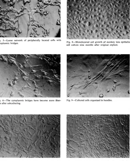
Fig. 5-Loose network of peripherally located cells with
cytoplasmic bridges.
Fig. 6-The cytoplasmic bridges have become more
fiber-like after subculturing.
Fig. 7-More densely packed fibroblastic appearance of ra
b-bit lens epithelial culture four months after first subculture.
Fig. 8-Monolayered cell growth of monkey lens epithelial
cell culture nine months after original explant.
Fig. 9-Cultured cells organized in bundles.
GEERAETS: OBSERVATIONS OF LENS EPITHELIUM IN CELL CULTURES 271
Fig. 11-Two years after original explantation the cells have organized in a directional fashion.
Fig. 12-Three-year-old culture of rabbit lens epithelial cells resembling lens fibers.
Fig. 13-Cell degeneration with several vacuoles six months after explant was placed in Petri dish (human lens epi-thelium).
Fig. 14-Birefringent droplets and small black intracyto-plasmic granules.
Fig. 15-Small "oil droplets" in degeneratnig cells.
272 GEERAETS: OBSERVATIONS OF LENS EPITHELIUM IN CELL CULTURES
Fig. 17-Many swollen cells with cell death observed after third subculture of human lens epithelium, 18 months after original explantation.
investigators. Their techniques and results varied considerably. In 1959, Van der Veen and Heyen reported that in their study no cells grew in medium containing heterologous serum or less than 20%
homologous (calf) serum. In the present study, all epithelial cells from human, rabbit, and monkey lenses did grow in a medium containing heterologous serum (calf) without adding homologous serum. Also, their observation that division of cells was rarely seen could not be confirmed in our study since mitoses could be demonstrated with time-lapse cine-matography, and different states of mitosis were seen in all stained preparations (figs. l 8b and 18c). Furthermore, long-term continuous cultures without subculturing of cells from primary explants have been kept alive for over four-and-a-half years.
The present study thus confirmed that lens epithelial cells can be maintained in vitro with mitotic activity and differentiation over several years. No distinct differences in cellular morphology of the cells derived from human, rabbit, and monkey were noted over the entire observation period. All cultures contained multinucleated cells, though in varying numbers. Human lens epithelium seemed to be less resistant to experimental trauma than that of rabbit, a fact which was particularly obvious after trypsini-zation, but less pronounced after mechanical transfer of cells for subculture.
Whether this cell type represents a suitable in
vitro model to carry out comparative studies on radiation effects is somewhat questionable because of the relatively slow multiplication rate of these cells, for this fact would require prolonged observation
GEERAETS: OBSERVATIONS OF LENS EPITHELIUM IN CELL CULTURES 273
times over which experimental artefacts could easily
be introduced.
In summary, a method for in vitro culturing of
lens epithelial cells from human, rabbit, and monkey has been described. The lens epithelium grown in vitro was carried on for successive generations. The cultures of rabbit and human lens epithelium were maintained for more than four and a half years and those of monkey lens epithelium for over three years. No definite differences in cellular morphology for the
three species were observed. Mitotic activity and
differentiation of the lens epithelium resembling lens fibers were demonstrated in all cultures. A review of the pertinent literature is presented.
Author's note: The initial pilot studies on the cell cultures were to a large extent carried out by Drs. Nongnart Romayananda and Michael Hines. Exten-sive technical assistance in all phases of this investiga-tion was rendered by Mr. Vernon Jones.
REFERENCES
1. BRYAN, A., LEINFELDER, P. J., AND MELTZER, M. Effects of gases upon cell growth. Arch. Ophth. 63: 108-132, 1960.
2. FISCHER, A. A three-month-old strain of epithelium. /. Exper. Med. 35:367, 1922.
3. GEERAETS, W. J. Radiation cataract: biomicroscopic
observations in rabbit, monkey, and man. Med. Coll. Va. Quart. 8 :259-263, 1972.
4. KIRBY, D. B. A study of the nutrient of the crystalline lens; the cultivation of lens epithelium. Trans. Am.
Acad. Ophth. 31:137-153, 1926.
5. KIRBY, D. B., ESTEY, K., AND TABOR, F. Cultivation of lens epithelium in vitro. A further study. Arch. Ophth. 1: 358-365, 1929.
6. KIRBY, D. B., ESTEY, K., AND WIENER, R. A study of the effect of changes in the nutrient medium on lens epithelium cultivated in vitro. Trans. Am. Acad. Ophth. 37: 196-212, 1932.
7. MAMO, J. G. AND LEINFELDER, P. J. Growth of lens epithelium in culture. AMA Arch. Ophth. 59:417-420, 1958.
8. MANN, IDA. Tissue culture of mouse lens epithelium. Brit./. Ophth. 32:591-596, 1948.
9. PucK, T. T., MARCUS, P. I., AND CIECIURA, S. J. Clonal
growth of mammalian cells in vitro. J. Exper. Med. 103 :273, 1956.
10. RosE, G. G. Time-lapse cinemicrography of cells in
tis-sue culture. Bull. Johns Hopkins Hosp. 116/1: 33-68, January, 1965.
11. TAMURA, SHIGEHIRO. Long-term cultures of epithelial cells of rabbit lens. Japanese J. Ophth. 9/4:177-181,
1965.
12. VAN DER VEEN, J. AND HEYEN,