ISSN 2319-7625 (Online) (An International Research Journal), www.chemistry-journal.org
Design, Synthesis, Characterization of Chalcone Derivatives
PTSH and PITI and Their Potential Anticancer Properties
against MDA-MB-231 Cell Lines
S. Kothai1*, S. Venkatesh2 and D. Lakshmi Devi3
1PG & Research Department of Chemistry,
Ethiraj College for Women, Chennai-600 008, INDIA.
2Department of Marine Biotechnology,
National Institute of Ocean Technology, Chennai-600 100, INDIA.
3 PG & Research Department of Biochemistry,
Ethiraj College for Women, Chennai-600 008, INDIA.
*Corresponding author: [email protected]
(Received on: April 14, 2018)
ABSTRACT
Chalcones (1,3-diaryl -2-propane-1-ones), biogenetic precursor of open chain flavonoids, which are highly multifunctional proven to have antitumor activity either in-vitro or invivo and their target cover all of the actions of tumour cells. Chalcone derivatives exhibit antitumor activity by anti-initiation, apoptosis induction, antiproliferation, antimetastasis, antiangiogenesis and so forth. We synthesized six copolyesters and were characterized by UV, FT-IR, ¹H NMR, ¹³C NMR. Their thermal stability was studied by DSC and TGA. Cytotoxicity activity of these copolyesters was evaluated by studying their responses to MDA-MB-231 Cell Line ( human Breast Cancer ) by MTT Assay. UV, FT-IR, ¹H NMR, ¹³C NMR confirmed the insertion of correct functional groups. DSC and TGA showed that the synthesized compounds were of good thermoregulatory we. The drug, PTSH was effective at 10 µg/mL and 51 % inhibition was observed at 100 µg/mL. Most importantly, the drug, PITI was more significant at 0.1 µg/mL concentration itself and 58 % inhibition was noted at 1 µg/mL.
Keywords: Chalcone, MDA-MB-231 Cell Line, Breast Cancer, MTT Assay.
1. INTRODUCTION
These therapeutic effects with current drugs were more effective 2. The Taxane is showed a
better treatment. Clinically, Taxol drug causes cell cycle arrest i.e G2/M phase and cease the mitosis 3.This drug possesses the activity against head, breast, ovarian, nonsmall cell lung
cancer4. Although taxol suggested being an act against these pathophysiology conditions it is
coming under a category of multidrug resistance (MDR) immediately after its administration5,6.
The naturally occurring flavonoids and its isoforms are present in the edible plants such as fruits, vegetables, spices, tea and soy and derived an important product is a chalcone which is chemically an open chain flavanoid contains two aromatic rings joined by three-carbon α,β-unsaturated three-carbonyl linkages having valuable pharmacological property 7.
Chalcones have been reported to have multifunctional biological activity against bacteria, fungus, virus, malarial parasites, inflammation and which result in cancer 8-10 all these living
endanger species and pathogenesis kept under control by this small molecule. Substitutions of the allylic group in chalcones exhibit antioxidant and antimicrobial activity 11,12. Additionally,
these chalcones containing ring B substitute with hydroxyl and methoxy groups showed inhibition of proliferative activity in human colon HT-29 cancer 13. This study is focused to
synthesize six copolyesters by incorporating the Chalcone derivatives in the copolyester the main chain and then analyze for anticancer activity against human breast cancer cell lines.
2. MATERIALS AND METHODS
2.1. Chemistry
All chemicals were purchased from Merck and Fluka chemical companies. Melting points were taken in open capillary tubes and are uncorrected. Infrared spectra were recorded
on a Perkin–Elmer V IR spectrophotometer. 1H NMR and 13C NMR spectra were recorded
CDCl3 on Bruker NMR spectrometers at 400 and 500, 100 and 125 MHz, respectively. All
reactions were conducted open to the atmosphere and the yields refer to the isolated products. Aldrich samples of 4-hydroxy acetophenone and ethoxy, 4-hydroxy benzaldehyde, 3-methoxy, 4- hydroxy acetophenone and were used as received. Ethanol (Merck) was used as non-solvent for the copolyesters and as a solvent for the preparation of the two chalcone diols. Aldrich samples of succinyl chloride, oxalyl chloride, glutaryl and is phthaloyl chloride were used as such in the synthesis of the five copolyesters. Spectral grade DMSO-d6 was used as internal standard for recording NMR spectra.
2.2. Synthesis of chalcone diols
The monomer diols namely (2E)-1-(4- hydroxyphenyl)-3(4-hydroxy, 3-ethoxy phenyl) prop-en-1-one and (2E)-1-(4-hydroxy-3-methoxy phenyl)-3(4-hydroxy, 3-ethoxy phenyl) prop-en-1- one were synthesized by the process reported 14.
2.3. Synthesis of Copolyesters
100 mL round-bottomed flask. To this 0.2mL of succinyl chloride and 0.2 mL of oxalyl chloride were added with constant stirring and the temperature was maintained at 120ºC with continuous stirring for 3 hours. At the end, the reaction mixture was cooled to room temperature and poured into 50 mL of n-hexane and the copolyesters were precipitated. It was filtered reprecipitated with methanol and dried in vacuum. The other four copolyesters were also prepared by the similar method. The diols and the diacid chlorides used and the copolyester code of the four copolyesters are presented (Table 1).
2.4. Viscosity Studies
The ηinh value of all the five copolyesters was determined in DMAc solution at 30℃
using Ubbelohde viscometer. 25mg of each of pure dry copolyester sample was dissolved in 25mL of DMAc, kept aside for some time with occasional shaking. Then they were left undisturbed for 24 hours. The ηinh was calculated from the flow time measurements 14. The
inherent viscosity values were found to be in the range of 0.73–1.10dL/g and are presented in Table 1. The data shows that these copolyesters are reasonable of high molecular weight.
2.5. Solubility Studies
The copolyesters stated here are found to be soluble in highly polar solvents such as DMAc and dimethyl formamide, partially soluble in moderately polar solvents like tetrahydrofuran and acetone but thoroughly insoluble in the least polar solvents like benzene and hexane. Copolyesters with methoxy substituent in the benzene ring of the chalcone moiety had better solubility which may be attributed to their competence to disrupt the macromolecular chain.
2.6. Spectral Studies
The FT-IR spectrum of the six copolyesters was recorded using Shimadzu FT-IR instrument. The FT-IR spectrum of all the six copolyesters revealed characteristic absorption in the range of 1742–1764cm-1 due to ester C=O stretching frequency.The NMR spectra were
recorded with BRUKER AV III 500 MHz NMR instrument in a DMSO-d6 solvent to categorize the repeating structural units present in the copolyester chain.
2.7. Thermal Studies (DSC and TGA)
The influence of polymer structure on the thermal properties of copolyester PSGH, PITI was investigated by thermogravimetric analysis (TGA) at a heating rate of 10°C min−1 in a nitrogen atmosphere and the TGA curve. The glass transition temperature 𝑇𝑔 and the melting temperature 𝑇𝑚 was determined by DSC at a heating rate of 10°C min−1 under nitrogen atmosphere and the DSC thermogram.
2.8. Cell culture conditions
Dulbecco’s modified Eagle’s medium (DMEM) (Sigma-Aldrich, India) supplemented with 10% Fetal Bovine Serum (FBS) and 1% anti-mycotic solution (Sigma Aldrich, USA).The Cells were maintained in a humidified incubator at 37°C with 5% CO2. When the cells reached
80% confluency, it was trypsinized and sub-cultured for analysis.
2.9. Cytotoxicity assay
The anti-proliferative effect of the drugs was tested for in vitro cytotoxicity analysis, using MDA-MB-231 cells by 3-(4,5-dimethylthiazol-2-yl)-2,5-diphenyltetrazolium bromide (MTT) assay[5]. The cultured MDA-MB-231 cells were harvested by trypsinization. Then, the
cells were plated at a density of 1×10⁵ cells/mL (50 µL) into 96-well microtitre plates. Briefly,
180 µL of media (DMEM-High Glucose) was added into each of the 96-well plates and 20 µL of diluted synthetic drugs with different concentration ranging from 0.1, 1, 10 and 100 µg/mL were added in the wells with positive and negative controls for comparison of results with test samples.
Each sample was triplicated and the cells were incubated at 37°C in a humidified 5% CO2 incubator for 48 h. After the incubation period, MTT (50 µL of 5 mg/mL) was added to
each well and the cells incubated for 4 h until purple precipitates were clearly visible. The medium was removed completely and the formazan crystals were dissolved in DMSO. The optical density was measured using an ELISA reader (Molecular Devices, Spectra Max 190, USA) at 570 nm and the percentage cell viability was calculated manually using the following formula:
% Cell Viability = (A570 sample - A570 blank / A570 control - A570 blank) x 100.
The calculation of concentrations that inhibits 50% of the (IC50) MDA-MB-231 cells
was formulated in the graph.
2.10. Evaluation of cell morphological changes
The morphological changes in the cells were observed under the inverted fluorescent microscope (Nikon Te200, USA).
2.11. Statistical analysis
Each experiment was done in triplicates and the values are mean of three independent experiments, Mean ± S.D. p < 0.05 was considered to indicate a statistically significant difference.
3. RESULTS AND DISCUSSION
3.1. Synthesis of Chalcone diols
point was about 239° C. The IR peak at 1642 cm-1 indicates the presence of C=O (stretching)
ofketoethylinic group. The ¹H NMR spectrum of BIMP shows at δ 6.9 and 7.1 was due to
protons of -CH=CH- of ketoethylinic group and δ 7 to 7.2 ppm is due to an aromatic proton, δ 4 ppm is due to the proton of –OCH3 and δ 9.9 ppm was due to proton –OH group. The spectral study was in agreement with the structural orientation of chalcone diol BIMP.
3.2. Viscosity Studies
The increase in the value of Inherent Viscosity will increase the molecular weight of the copolyesters. Among the six copolyesters, PITI was found to be having highest inherent viscosity because of the rigidity of the aromatic in the polymer backbone as both the acid chlorides used were aromatic in nature. This data supports that these copolyesters are reasonable high molecular weight are shown in Tab.1.
Table. 1. Monomers diols used for six copolyesters synthesis with code and the percentage of
yield and inherent viscosities (ηinh).
3.3. Solubility Studies
The solubility of the copolyesters was tested qualitatively by dissolving 5 mg of the copolyesters in 1 mL of the solvent. The copolyesters are easily soluble in a polar aprotic solvent such as DMSO, DMAc, and DMF and they are insoluble in nonpolar solvents benzene, n-hexane, and toluene are shown in Table 2. The copolyesters are soluble in polar solvents like methanol upon warming may be due to intermolecular interaction for solvents with ester linkage of the polymer.
Table 2. Determination of Solubility of six copolyesters with different solvent system
S.No Polymer Code DMAc DMF DMSO CH3OH C6H6 n-hexane H2O
1. PSGH ++ ++ ++ +- -- -- --
2. PTOI ++ ++ ++ +- -- -- --
3. PIOM ++ ++ ++ +- -- -- --
4. PIGH ++ ++ ++ +- -- -- --
5. PTSH ++ ++ ++ +- -- -- --
6. PITI ++ ++ ++ +- -- -- --
++ = Soluble +- = Soluble on warming -- = Insoluble
Diol Diacid chloride-I Diacid chloride-II Copolyester Code
Percentage Yield
Inherent Viscosity(dL/g) HHEP Succinyl Chloride Glutaryl Chloride. PSGH 81 1.1800 HIMP Terephthaloyl
Chloride
Oxalyl Chloride PTOI 79 1.0179
MHEP Isophthaloyl Chloride
Oxalyl Chloride PIOM 87 1.1728
HHEP Isophthaloyl Chloride
Glutaryl Chloride. PIGH 79 1.5602
HHEP Terephthaloyl Chloride
Succinyl Chloride PTSH 86 1.2321
HIMP Isophthaloyl Chloride
Terephthaloyl Chloride
Fig. 1. UV-Visible spectrum for (A) PTSH and (B) PITI
3.4 Spectral Studies
3.4.1. UV – Vis Spectrum
The absorption peak at wavelength range 230-390 nm of PTSH and PITI showed the chalcone is incorporated in a polymer backbone. This is due to nπ*, π→π* transition and also due to the C=O group of the molecule and also due to the excitation of the C=O group of the molecule shown in Fig.1.
3.4.2. FT-IR Spectrum
The FT-IR Spectrum of the six copolyesters namely PIGH, PIOM, PTOI, PSGH,
PTSH, PITI were recorded by using Shimadzu FT-IR instrument. The IR Spectra of the copolyesters of PTSH and PITI were characterised by strong absorption band 1709-1759 cm-1 and 1583-1598 cm-1 corresponding to ester and ketone carbonyl (C=O) stretching
respectively. The strong absorptions at 1679-1695cm-1 were due to olefinic group (CH=CH)
of chalcone unit are shown in Fig.2 A & B.
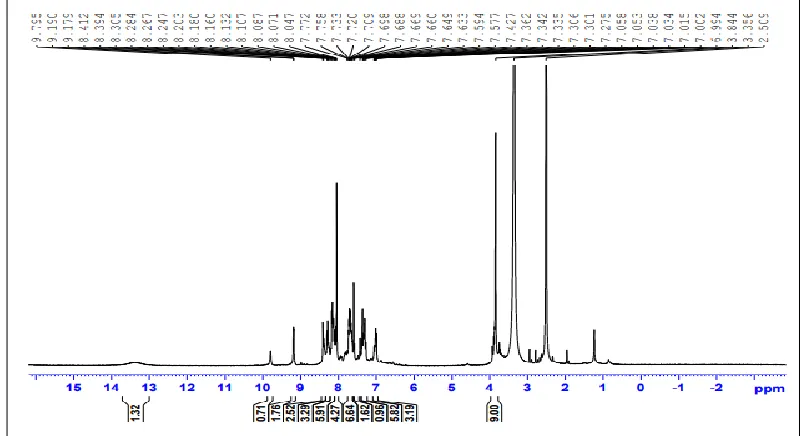
3.4.3 NMR Spectrum
The 1H and 13C NMR spectra of PTSH signals in the range of 170–205ppm and 165–
175ppm in the 13C NMR spectra of the co-polyesters are due to the carbonyl carbon of the α,
β-unsaturated ketone and ester groups respectively, which indicates the formation of the copolyester as shown in Fig.3,4.
1H and 13C NMR spectra of (DMSO-d6 solutions) of PITI are displayed in Fig. 5,6.
The aromatic protons of PITI are observed in the range of 7.2–8.1ppm. The vinylic protons attached to the carbonyl carbon are observed in the range of 6.7–6.9ppm. The methoxy protons in the chalcone moiety are represented in the range of 3.1–3.4 ppm. The methylene protons are observed in the range of 1.3–3.3 ppm with an expected integral value.
Fig. 3. 1H NMR spectra (DMSO-d6 solutions) of PTSH with the assignment of the detected signals (see Figure
Fig. 4. 13C NMR spectra (DMSO-d6 solutions) of PTSH with the assignment of the detected signals (see
Figure for the atom-numbering scheme).
Fig. 5. 1H NMR spectra (DMSO-d6 solutions) of PITI with the assignment of the detected signals (see Figure
Fig. 6. 13C NMR spectra (DMSO-d6 solutions) of PITI with the assignment of the detected signals (see Figure
for the atom-numbering scheme).
3.5. Thermal Stability
3.5.1. Differential Scanning Calorimetry (DSC)
The endothermic peak at 99°C, 103.57°C for PTSH and PITI respectively. They indicate good thermal stability required for their biomedical application. Table 3. Fig. 7 (A),(B).
Table. 3. Both copolyestes PTSH and PITI having transian temperature with melting temperature confirming their stability.
S.No Copolyesters code Glass transition temperature Tg Melting temperature Tm
1. PTSH 52°C 138.19
2. PITI 50°C 103.57
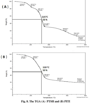
3.5.2. Thermo Gravimetric Analysis (TGA)
The temperature for the 50% weight loss was found to be 325° C, 320°C for PTSH
and PITI respectively in Fig. 8.A & B. This show that the copolyesters were of high thermal stability.
Fig. 8. The TGA (A) PTSH and (B) PITI
3.6. Cytotoxicity effect of PTSH and PITI co-polymers
At higher concentration (100 µg/mL) of the drug PITI, the breast cancer cells were completely disintegrated (figure 1). PIGH treated cells showed a vacuolation in the cell cytoplasm, which might have resulted in autophagy. Interestingly, PITI cells got shrunk, thus denoting cell death. The MTT data substantiates the findings of the morphological studies, as shown in Fig. 10,11.
PSGH PTOI PIOM
PIGI PTSH PITI
Fig. 9. Morphological changes of MDA-MB-231 cells after the exposure of synthetic drugs PSGH, PTOI, PIOM, PIGH, PTSH and PITI.
Fig. 11. The inhibition responses of MDA-MA-231cell lines to 48 h treatments with synthetic drugs such as PSGH, PTOI, PIOM, PIGH, PTSH and PITI at various concentrations (0.1- 100 µg/mL)
The breast cancer cell inhibition is shown in figure 14. The cytotoxicity effect of the synthetic compounds was tested against MDA-MA-231 cells by MTT assay. After 48 hours of synthetic compound treatment, the IC50 dose levels were calculated. Each line graph showed
the percentage of cell inhibition depending on the dose and the inhibition of 50% of the cells is indicated as IC50. The drugs, PSGH, PTOI, PIOM and PIGH showed about 33 % and 43
% inhibition of the cells even at 100 µg/mL. While, the drug, PTSH was effective at 10 µg/mL and 51 % inhibition was observed at 100 µg/mL. Most importantly, the drug, PITI was more significant at 0.1 µg/mL concentration itself and 58 % inhibition was noted at 1 µg/mL, thus signifying the potential of this drug as a promising anti-proliferative agent.
The above synthetic drugs PTSH and PITI primarily involved in the killing mechanism by targeting the Bax proteins, which was stimulated in the late 24-48 h stage leading to apoptosis. This evidence suggested that the induction of other protein, p21 which paves the way to apoptosis through a p53-dependent pathway. Nevertheless, this mechanism needed to be further clarified by immunoblot analysis.
4. CONCLUSIONS
ACKNOWLEDGMENTS
We are also gratefulto FT-IR instrumentation center, Ethiraj College for Women, Chennai for the spectral studies. This study was fully supported by our institute.
AUTHOR CONTRIBUTIONS
S.Kothai and D.Lakshmi Devi, designed the research work and performed the research, analyzed the data and wrote the paper. S.Venkatesh and S.Kothai corrected this manuscript. All authors read and approved the final manuscript.
CONFLICTS OF INTEREST
The authors declare no conflict of interest.
REFERENCES
1. Chen H, Zhang ZW, Guo Y, Wang Y, Liu Y, Luo N, Zhu Y, J buon 17,658-662 (2012).
2. Moore A, N EnglJ Med. 357,1547-1549 (2007).
3. Horwitz SB, Trends Pharmacol Sci, Apr, 13,134-136(1992).
4. Sparano Ja, Wang M, Martino S, Jones V, Perez Ea, Saphner T, Wolff Ac, Sledge Gw Jr, Wood Wc, Davidson Ne, N EnglJ Med, 358,1663-71( 2008).
5. Broxterman Hj, Gotink Kj, Verheul Hm, Drug Resist Updat. 12,114-26 (2009)
Sangrajrang S, Fellous A, Chemotherapy, 46,327-34 (2000).
6. Di Carlo G, Mascolo N, Izzo AA, Capasso F, Life Sci, 65, 337-353 (1999). 7. Nowakowska Z, Eur. J. Med. Chem, 42, 125–137 (2007).
8. Go ML, Wu X, Liu XL, Curr. Med. Chem, 12, 483–499 ( 2005).
9. Sivakumar PM, Ganesan S, Veluchamy P, Doble M, Chem. Biol. Drug. Des, 76, 407–411
(2010).
10. Adibi H, Mojarrad JS, Asgharloo H, Zarrini GSynthesis, Med Chem Res 20, 1318-1324 (2011).
11. Doan TN, Tran DT, Pharmacology &Pharmacy 2, 282-288 (2011).
12. Mizuno CS, Paul S, Suh N, Rimando AM, Bioorg Med Chem Lett 20,7385-7387 (2010).
13. Jasmine Francis S, Reuben Jonathan D, Roop Singh D, Journal of Chemical and
Pharmaceutical Research 6, 1155-1160 (2014).
14. Jasmine Francis S, Roopsingh D, Reuben Jonathan D, Journal of Chemical and