389
*Corresponding author: Manoochehr Makvandi PhD,Health Research Institute, Infectious and Tropical Diseases Research Center; Virology Department, School of Medi-cine, Ahvaz Jundishapur University of Medical Sciences,
Prevalence of Hepatitis C virus Genotype 3a in patients with Hodgkin and
Non-Hodgkin Lymphoma
Hashem Radmehr
1, Manoochehr Makvandi
1, 2*, Alireza Samarbafzadeh
1, 2, Ali Teimoori
2, Niloofar Neisi
2,
Mojtaba Rasti
2, Sara Abasifar
3, Hasan Soltani
2, Samaneh Abbasi
2, Hadis Kiani
2, Hamide
Mehravaran
2, Azarakhsh Azaran
2, Toran Shahani
21
Health Research Institute, Infectious and Tropical Diseases Research Center
;Ahvaz Jundishapur University
of Medical Sciences, Ahvaz, Iran
2
Virology Department, School of Medicine, Ahvaz Jundishapur University of Medical Sciences, Ahvaz, Iran
3Virology Department, School of Medicine, Kerman University of Medical Sciences, Kerman, Iran
Received: July 2016, Accepted: November 2016
ABSTRACT
Background and Objectives: Hepatitis C virus (HCV) is a major public health problem worldwide. Replication and per-sistence of HCV genome have been described in the liver tissue as well as B cells lymphocyte. Several investigations have reported that long-term persistence of HCV in B cells may result in Hodgkin and Non-Hodgkin lymphoma. This study was aimed to determine frequency of HCV RNA in histological tissues obtained from patients suffered from Hodgkin and Non-Hodgkin lymphoma.
Materials and Methods: 52 formalin-fixed paraffin-embedded tissue blocks including 23 (44.3%) Hodgkin and 29 (55.7%) Non-Hodgkin samples were collected and five micrometer sections were prepared. RNA was extracted and cDNA was syn -thesized. Two consecutive Nested RT-PCR assays were carried out for detection of HCV 5’ UTR and core gene. RT-PCR products were sequenced and aligned to construct HCV phylogenic tree to evaluate the homology of sequences in compari-son to the reference sequences retrieved from Genbank.
Results: Overall, 6 Non-Hodgkin (20.6%) and 3 Hodgkin lymphoma (13.04%) samples showed positive PCR results for both 5’ UTR and HCV core RNA via nested PCR (P<0.469). Sequencing results revealed that all detected HCV RNA sam -ples belonged to the genotype 3a.
Conclusion: Despite low prevalence of HCV infection in Iran, high frequency of HCV RNA genotypes 3a (17.3%) has been found in patients with Hodgkin and Non-Hodgkin lymphoma. To improve treatment regimens, screening of HCV RNA in patients suffered from Hodgkin or Non-Hodgkin lymphoma is recommended which can be done through highly sensitive molecular means before and after immunosuppression status.
Keywords: Nested RT-PCR, Hepatitis C virus, Hodgkin lymphoma, Non-Hodgkin lymphoma, Genotype
ORIGINAL
AR
TICLE
Ahvaz, Iran. Tel: +98 9166181683 Fax: +98 61 33738313
INTRODUCTION
Hepatitis C virus (HCV) infection is a major health
problem infecting more than 170-200 million people
worldwide (1). HCV is hepatotropic and
lymphotro-pic and a causative agent for acute, chronic hepatitis.
Persistent chronic HCV may result in cirrhosis and
hepatocellular carcinoma (2). HCV belongs to the
family of flaviviridae and has a single stranded RNA
of 9.7 kb comprises three structural proteins (core,
E1 and E2) and seven non-structural proteins (p7,
NS2, NS3, NS4A, NS4B, NS5A and NS5B) (3). HCV
has been classified into 7 genotypes including over
70 subtypes (4). The rate of HCV infection is varied
from 1.5-3.5% in Eastern and Western Europe and
more than 3.5% in the Middle East (5). Most patients
(80-85%) who become acutely infected cannot clear
the infection and will progress to chronic infection.
The prevalence of HCV in Iranian general population
has been reported less than 1% (6). Different mech
-anisms have been described the implication of HCV
in development of malignant tumors. The expression
of HCV core protein (C) and non-structural protein
3 (NS3) will enhance the generation of nitric oxide
synthase (NOS) and reactive oxygen species (ROS)
and may lead to mutations and abnormalities in DNA
repair system and ultimately in cellular
transforma-tion (7, 8). HCV NS3 can also bind to tumor sup
-pressor p53 molecule and affect DNA repair process
which finally leads to cellular transformation (9).
HCV infected marginal B cells lead to chronic
stim-ulation which is thought to result in accumstim-ulation of
genetic lesions promoting eventually transforming in
diffuse large B-cell lymphoma (DLBCL) (10).
Hodgkin’s disease or Hodgkin lymphoma (HL) is
characterized by multinuclear Reed-Sternberg cells
(RS cells) (11). It has been reported that the role of
Epstein-Barr virus (EBV) may increase risk of HL
even though the exact mechanism remains unknown
(12). Non-Hodgkin’s lymphoma (NHL) is a
lymph-oproliferative disorder which comprises different
hematologic neoplasms that originate from T and B
cells in the lymphatic system (13). The etiology of
NHL is unknown but may be attributed to several
risk factors such as age, sex, genetic and environ
-mental factors (14).
The association of chronic hepatitis C virus (HCV)
infection with Hodgkin and non-Hodgkin lymphoma
has been reported (15-18). Limited data have been
published on the association of HCV infection and
Hodgkin and Non-Hodgkin lymphoma in Iran. Thus,
this study was conducted to determine the
preva-lence of HCV RNA in patients with Hodgkin and
Non Hodgkin lymphoma in Ahvaz, the capital city of
Khuzestan province, located in the south west region
of Iran with about 1.5 million population.
MATERIALS AND METHODS
Patients.
This retrospective study was carried out
on 52 paraffin-embedded tissue blocks including
23/52 Hodgkin (44.23%) and 29/52 Non Hodgkin
(55.77%) lymphomas. Tissue blocks were collected
from archive of Pathology Departments of Shafa and
Imam Khomeini Hospitals of Ahvaz during 2001 to
2011. The diagnosis of Hodgkin and Non-Hodgkin
lymphoma was done by a pathologist. Clinical data
in Table 1 revealed that none of the patients had been
tested for HCV RNA, HBV markers, anti-HCV,
an-ti-HIV-1, 2 and anti-HTLV-1 antibodies.
Deparaffinization of tissue samples. Initially,
sections of 5 µm thickness were prepared from each
tissue block followed by deparaffinization. In brief,
800 µl xylene and 400 µl ethanol was added to each
sample and centrifuged at 13,000 rpm for 2 min. The
supernatant was discarded and 1 ml absolute
etha-nol was added to the pellet. Tubes were incubated at
55°C for 10 min. 100 µl lysate buffer, containing 16
µl 10% SDS and 40 µl proteinase K, was added to
each tube and kept at 55°C overnight (16 hours). The
tubes were centrifuged at 13,000 rpm for 2 min and
the supernatants were collected and used for RNA
extraction.
RNA extraction and cDNA preparation.
Total
RNA was extracted from each sample using High
pure RNA Paraffin kit (Roche, Germany) according
to the manufacturer’s instructions. 10 µl sample was
used for cDNA synthesis using available commercial
kits (Thermo Fisher Scientific, Latvia) according to
the manufacturer’s instructions.
Nested PCR for 5’ UTR.
Detection of HCV RNA
was carried out using cDNA samples via a nested
RT-PCR. The following primers from 5’
untranslat-ed region (UTR) of HCV genome were usuntranslat-ed; for the
first PCR: BKP 7 (outer sense primer) (nucleotides
(out-http://ijm.tums.ac.ir
er anti-sense primer) (nucleotides 319 to 343) (5’-at
-ggtgcacggtctacgagacctcc-3’). 25 microliter reaction
mixture was prepared from 2.5 µl 10X PCR buffer
(Roche), 0.5 µl dNTP mix (0.2 mM), 1 µl each prim
-er (BKP-7 and BKP-8) (0.5 µM), 0.15 µl Taq DNA
polymerase (1 Unit), 7 µl cDNA template and water.
Positive and negative controls were also included in
each PCR. PCR was programmed by initial
denatur-ation at 95°C (5 min) followed by 35 cycles of 94°C (1
min), 60°C (1 min), 72°C (1 min) and final extension
at 72°C (10 min). The second round PCR was per
-formed similar to the first PCR using the same PCR
reaction mixture with inner set of primers (BKP-9
and BKP-10) (BKP 9; nucleotides 63 to 87: 5’-ttcac
-gcagaaagcgtctagccatg-3’; BKP 10; nucleotides 292 to
314: 5’-gcgcactcgcaagcaccctatcagg-3’). PCR condi
-tions were the same as the first round for 30 cycles.
The first PCR product was 306 bp in length whereas
the second product was 252 bp (19) (Fig. 1).
Nested PCR for core of HCV.
Second nested PCR
was fulfilled for detection of core gene of HCV in
tissue samples with the following primers: SC2 (F
primer; 5’-gggaggtctcgtagaccgtgcaccatg-3’), AC2
(R primer; 5’-gagmggkatrtaccccatgagrtcggc-3’),
S7 (F primer; 5’-agaccgtgcaccatgagcac-3’) and 584
(R primer; 5’-cccatgaggtcggcraarc-3’). PCR
reac-tion mix was prepared from 2.5 µl 10X PCR Buffer
(Roche), 0.5 µl dNTP mix (0.2 mM), 1 µl forward
and reverse primers (0.5 µM), 0.15 µl Taq DNA pol
(1 Unit), 7 µl cDNA template and water. PCR condi
-tion was programmed as follows: initial denatura-tion
at 95°C (5 min) and 35 cycles of 95°C (1 min), 55°C
(1 min), 72°C (1 min) and final extension at 72°C (10
min). For the second PCR, the reaction mixture and
PCR temperatures were programmed similar to the
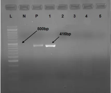
first PCR. 416 bp PCR products on 2% agarose gel
indicated the positive reactions (Fig. 2) (20).
Sequencing.
To determine the genotypes of HCV
Table 1. Profile of patients included in this study
Total %
12(52.17%) 4(17.39%) 7(30.43%)
14(48.28%) 4(13.79%)
1(3.44%) 1(3.44%) 2(6.89%) 4(13.79%)
2(6.89%) 1(3.44%) Female %
8(34.78%) 1(4.34%) 4(17.39%)
10(34.48%) 2(6.89%) 1(3.44%)
0 0 2(6.89%) 1(3.44%)
0 0.5-1±0.25
24.17±7.3 41.02±9.3 Negative Not done Male %
4(17.39%) 3(13.04%) 3(13.04%)
4(13.79%) 2(6.89%)
0 1(3.44%) 2(6.89%) 2(6.89%) 1(3.44%) 1(3.44%) Category
Hodgkin Lymphoma: Number 23 Mixed cellularity (MC)
Lymphocyte Predominant (LP) Nodular Scleorosis (NS)
Non-Hodgkin Lymphoma: Number 29 Diffuse Large cell type
Burkitt Lymphoma
Small lymphocytic lymphoma High grade Immunoblastic lymphoma Mixed Small and Large
Malignant T Cell Malignant B Cell Diffuse Mixed Large Cell Billirubin (mg/dl) AST (U/L) ALT (U/L) HBsAg HCV Ab
Fig. 1. HCV 5’UTR PCR. N: Negative control; L: 100 bp DNA ladder; P: Positive control; #1 to 9: Unknown samples
samples, 9 positive samples of 5’ UTR and core
re-gions were sequenced. The sequences were blasted
using available databases. A phylogenic tree was
constructed with Neighbor joining method using
the partial nucleotide sequences of 5’ UTR region
of HCV positive samples. Reference sequences were
retrieved from GenBank using their accession
num-bers.
Statistics.
Data analysis was done by SPSS (v. 17.0)
using mean, standard deviation (SD), and Chi-square
test. P values < 0.05 were considered as significant.
RESULTS
The age of the patients ranged from 5.48 to 34
years with the mean age of 19.74 ± 14.2 years. In
overall 52 samples, 9 samples (17.3%) represented
positive 5’ UTR and HCV core (P=0.469) including
6 Non-Hodgkin (6 out of 29; 20.68%; 4 males and 2
females) and 3 Hodgkin (3 out of 23; 13.04%; 1 male
and 2 females) lymphomas (P=0.469). Sequence
analysis revealed that all positive HCV RNA samples
belonged to genotype 3a (Fig. 3). Table 2 also
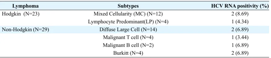
repre-sented the distribution of HCV infection in subtypes
of Hodgkin and Non-Hodgkin lymphomas.
DISCUSSION
In our study 6 Non-Hodgkin (20.6%) and 3 Hod
-gkin (13.04%) lymphoma samples showed positive
PCR results for 5’ UTR and core regions of HCV. The
prevalence of HCV infection in patients with
Hod-gkin lymphoma is controversial. In 2003, Keresztes
et al. have detected HCV RNA in 10 out of 111
Hod-gkin lymphoma patients (9%) in Hungary (21). Dal
Maso et al. have also found HCV RNA in 1.6% of
Hodgkin patients (22). In 2015, Tehseen Iqbal et al.
have observed HCV RNA in 44% of Hodgkin lym
-phoma patients in US (23). We have detected HCV
RNA in 13.04% of Hodgkin lymphoma patients which
indicated that our finding situated between the above
findings. HCV RNA has also been detected in patients
with Non-Hodgkin lymphoma. The rate of HCV RNA
detection among patients with Non-Hodgkin
lympho-ma is varied from 2.4% in Canada to 42.7% in Egypt.
In Egypt, Cowgill et al. have reported a high
preva-lence of HCV RNA (42.7%) in Hodgkin lymphoma
patients (24) while Spinelli et al. have detected 2.4%
HCV RNA positive cases among Canadian
Non-Hod-gkin lymphoma patients (25). John et al. in 2008
re-ported 17.5% HCV RNA positive Non-Hodgkin lym
-phoma cases in Italy (26). We have detected 20.68%
HCV RNA in Non-Hodgkin lymphoma patients. The
overall HCV RNA positivity in the present study was
3 out of 23 Hodgkin lymphoma (13.04%) and 6 out
of 29 (20.6%) Non-Hodgkin Lymphoma (P= 0.469).
The HCV genotypes 1a, 3ad, 1b, 2 and 4 have been
reported in Iran (27, 28).
Fig. 2. HCV core PCR. L: 50 bp DNA ladder; N: Negative control; P: Positive control; #1-5: Unknown samples
http://ijm.tums.ac.ir
We have detected HCV genotype 3a in 2/23 (8.69%)
patients with Hodgkin Mixed Cellularity (MC) sub
-type, and 1/23(4.34%) in patient with Lymphocyte
Predominant (LP) whereas Katalin et al. in Hungary
detected HCV RNA in 10/63(15.87%) patients suf
-fered from Hodgkin mixed cellularity subtype (MC),
7/38(18.42%) nodular scleorosis (NS) and 2/8(25%)
lymphocyte-depleted group (LD) (21).
In the present study, we detected HCV genotype 3a
in Non-Hodgkin subtypes including 2(6.89%) in dif
-fuse large cell group, 2(6.89%) in Burkitt Lymphoma,
1(3.44%) in malignant T-cell group and 1(3.44%) in
malignant B cell group. Visco et al. have detected
HCV RNA in diffuse large B-cell lymphoma
(DLB-CL) subtype of Non-Hodgkin lymphoma (29).
Sever-al studies have been strengthened that antivirSever-al
ther-apy against HCV in patients with low grade B cell
lymphoma was very effective (30, 31).
CONCLUSION
Despite low prevalence of HCV in Iran, high
frequen-cy of HCV RNA genotype 3a(17.3%) has been found
in patients suffered from Hodgkin and Non-Hodgkin
lymphomas. In the light of aforementioned data it is
recommended that for improving treatment regimens,
screening of HCV RNA should be implemented for
Hodgkin and Non-Hodgkin lymphoma patients by
highly sensitive molecular means before and after
im-munosuppression status.
ACKNOWLEDGEMENT
Our great appreciate goes to Farshid Alizadeh for
providing positive control samples. This study was
done by Mr. Hashem Radmehr (MSc,Virology) as a
research project, grant number 92144 in Health Re
-search Institute, Infectious and Tropical Disease
Re-search Center, Ahvaz Jundishapur University of
Med-ical Sciences, Ahvaz, Iran.
REFERENCES
1. Gower E, Estes C, Blach S, Razavi-Shearer K, Razavi H. Global epidemiology and genotype distribution of the hepatitis C virus infection. J Hepatol 2014; 61:S45-57.
2. Perz JF, Armstrong GL, Farrington LA, Hutin YJ, Bell BP. The contributions of hepatitis B virus and hepatitis C virus infections to cirrhosis and primary liver cancer worldwide. J Hepatol 2006; 45:529-538.
3. Halliday J, Klenerman P, Barnes E. Vaccination for hepatitis C virus: closing in on an evasive target. Ex-pert Rev Vaccines 2011; 10:659-672.
4. Smith DB, Bukh J, Kuiken C, Muerhoff AS, Rice CM, Stapleton JT, et al. Expanded classification of hepatitis C virus into 7 genotypes and 67 subtypes: updated cri -teria and genotype assignment web resource. Hepatol-ogy 2014; 59318-327.
5. Mohd Hanafiah K, Groeger J, Flaxman AD, Wiersma ST. Global epidemiology of hepatitis C virus infection: new estimates of age-specific antibody to HCV sero -prevalence. Hepatology 2013; 57:1333-1342.
6. Alavian SM, Adibi P, Zali MR. Hepatitis C virus in Iran: Epidemiology of an emerging infection. Arch Ira-nian Med 2005; 8: 84-90.
7. Machida K, McNamara G, Cheng KT, Huang J, Wang CH, Comai L, et al. Hepatitis C virus inhibits DNA damage repair through reactive oxygen and nitrogen species and by interfering with the ATM-NBS1/Mre11/ Rad50 DNA repair pathway in monocytes and hepato-cytes. J Immunol 2010; 185:6985-6998.
8. Deng L, Nagano-Fujii M, Tanaka M, Nomura-Takiga-wa Y, Ikeda M, Kato N, et al. NS3 protein of Hepatitis C virus associates with the tumour suppressor p53 and inhibits its function in an NS3 sequence-dependent manner. J Gen Virol 2006; 87:1703-1713.
Table 2. Distribution of HCV RNA in Hodgkin and Non-Hogkin Lymphoma subtypes
Lymphoma Hodgkin (N=23)
Non-Hodgkin (N=29)
Subtypes
Mixed Cellularity (MC) (N=12) Lymphocyte Predominant(LP) (N=4)
Diffuse Large Cell (N=14) Malignant T cell (N=4) Malignant B cell (N=2)
Burkitt (N=4)
HCV RNA positivity (%) 2 (8.69) 1 (4.34) 2 (6.89) 1 (3.44) 1 (6.89) 2 (6.89)
9. Canioni D, Michot JM, Rabiega P, Molina TJ, Charlotte F, Lazure T, et al. In Situ Hepatitis C NS3 protein de-tection is associated with high grade features in Hep-atitis C-Associated B-Cell Non-Hodgkin lymphomas.
PLoS One 2016; 11:e0156384.
10. Peveling-Oberhag J, Arcaini L, Hansmann ML, Ze-uzem S. Hepatitis C-associated B-cell non-Hodgkin lymphomas. Epidemiology, molecular signature and clinical management. J Hepatol 2013; 59:169-177. 11. Armitage JO. Early-stage Hodgkin's lymphoma. N
Engl J Med 2010; 363:653-662.
12. Gandhi MK, Tellam JT, Khanna R. Epstein-Barr vi-rus-associated Hodgkin's lymphoma. Br J Haematol
2004; 125:267-281.
13. Bagirath PV, Kumar JV, Arvind UD, Shailaja G. Ag-gressive extranodal peripheral T-cell non-Hodgkin's lymphoma: A rare case report and review. J Oral Max-illofac Pathol 2014; 18:80-83.
14. Morton LM. Dissecting lymphoma incidence to inform epidemiologic and clinical research. Leuk Lymphoma
2013; 54:1575-1576.
15. Yenice N, Gulluk F, Arican N, Turkmen S. HCV preva-lence in Hodg¬kin and non-Hodgkin lymphoma cases.
Turk J Gasteroenterol 2003; 14:173-176.
16. Pozzato G, Mazzaro C, Crovatto M, Modolo ML, Ce-selli S, Mazzi G, et al. Low-grade malignant lympho-ma, hepatitis C virus infection, and mixed cryoglobu -linemia. Blood 1994; 84:3047-3053.
17. Economides MP, Mahale P, Turturro F, Hosry J, Sa -maniego F, Granwehr BP, et al. Development of non-Hodgkin lymphoma as a second primary cancer in hepatitis C virus-infected patients with a different pri-mary malignancy. Leuk Lymphoma 2017; 58:485-488. 18. Michot JM, Canioni D, Driss H, Alric L, Cacoub P,
Suarez F, et al. Antiviral therapy is associated with a better survival in patients with hepatitis C virus and B-cell non-Hodgkin lymphomas, ANRS HC-13 lym-pho-C study. Am J Hematol 2015; 90:197-203. 19. Makvandi M, Khalafkhany D, Rasti M, Neisi N,
Omid-varinia A, Mirghaed A, et al. Detection of Hepatitis C virus RNA in peripheral blood mononuclear cells of patients with abnormal alanine transaminase in Ahvaz.
Indian J Med Microbiol 2014; 32:251-255.
20. Ohno O, Mizokami M, Wu RR, Saleh MG, Ohba K, Orito E, et al. New hepatitis C virus (HCV) genotyping
system that allows for identification of HCV genotypes 1a, 1b, 2a, 2b, 3a, 3b, 4, 5a, and 6a. J Clin Microbiol
1997; 35:201-207.
21. Keresztes K, Takács M, Horányi M, Miltényi Z, Illés Á. HCV and HGV Infection in Hodgkin’s Disease.
Pathol Oncol Res 2003; 9:222-225.
22. Dal Maso L, Talamini R, Montella M, Crovatto M, Franceschi S. Hepatitis B and C viruses and Hodgkin lymphoma: a case-control study from Northern and Southern Italy. Haematologica 2004; 89:ELT17. 23. Iqbal T, Mahale P, Turturro F, Kyvernitakis A, Torres
HA. Prevalence and association of hepatitis C virus in-fection with different types of lymphoma. Int J Cancer
2016;138:1035-1037.
24. Cowgill KD, Loffredo CA, Eissa SA, Mokhtar N, Ab -del-Hamid M, Fahmy A, et al. Case-control study of non-Hodgkin's lymphoma and hepatitis C virus infec-tion in Egypt. Int J Epidemiol 2004; 33:1034-1039. 25. Spinelli JJ, Lai AS, Krajden M, Andonov A, Gascoyne
RD, Connors JM, et al. Hepatitis C virus and risk of non-Hodgkin lymphoma in British Columbia, Canada.
Int J Cancer 2008; 122:630-633.
26. Mele A, Pulsoni A, Bianco E, Musto P, Szklo A, San-paolo MG, et al. Hepatitis C virus and B-cell non-Hod-gkin lymphomas: an Italian multicenter case-control study. Blood 2003; 102:996-999.
27. Khodabandehloo M, Roshani D. Prevalence of hepati -tis C virus genotypes in Iranian patients: a systematic review and meta-analysis. Hepat Mon 2014; 14:e22915. 28. Farshadpour F, Makvandi M, Samarbafzadeh AR,
Jalalifar MA. Determination of hepatitis C virus gen-otypes among blood donors in Ahvaz, Iran. Indian J Med Microbiol 2010; 28:54-56.
29. Visco C, Finotto S. Hepatitis C virus and diffuse large B-cell lymphoma: Pathogenesis, behavior and treat-ment. World J Gastroenterol 2014; 20:11054-11061. 30. Mazzaro C, De Re V, Spina M, Dal Maso L, Festini G,
Comar C, et al. Pegylated-interferon plus ribavirin for HCV-positive indolent non-Hodgkin lymphomas. Br J Haematol 2009; 145:255-257.