Management of Severe Asthma*
WILLIAM B. HUNT, JR., M.D.
Associate Professor of Internal Medicine, Allergy-Pulmonary Division,
University of Virginia School
of
Medicine, Charlottesville, Virginia
In this summary of therapy for severe asthma there is no mention of etiologic factors. The author presumes that if infection is the primary factor in the progression to severe asthma that this will be recognized and appropriately treated. A chest x-ray must be an initial laboratory study for the recognition of pneumonia or complicating pneu-mothorax. However, unlike respiratory failure due to emphysema, here the specific therapy of the altered pulmonary physiology is of paramount importance. Attention to the inciting factors is of minimal therapeutic benefit once infection can be ruled out. After the patient can breathe comfort-ably again the role of allergy can be investigated. Premature investigation of emotional factors can be a lethal therapeutic error in the ill asthmatic.
The treatment of asthma at the University of Virginia Hospital can be divided into three rather specific periods (Table 1).
Mystery Period. Until 1962 arterial blood gas and pH measurement were not routinely avail-able as a clinical tool in our laboratories. Most pa-tients with severe asthma did well with our rou-tine management. Other patients who looked the same might die suddenly with no major change in their clinical course. The mystery revolved around our inability to judge precisely who had life threatening asthma and who did not.
Measurement and Aggression 1962-1968. (Table 2). As soon as arterial puncture became a routine procedure the mystery cleared somewhat. Many patients with severe asthma were found to our surprise to have severe hypoxemia and hy-percarbia. It is presumed that the prior mystery deaths were most likely due to respiratory failure with CO~ narcosis or hypoxemia-induced fatal
ar-* Presented by Dr. Hunt at the Symposium on
Respira-tory Failure, May 26, 1972, at Richmond, Virginia.
MCV QUARTERLY 9(2): 159-163, 1973
rhythmias. These patients were recognized usually late in their disease. Immediate tracheostomy was done; many were hyperventilated with a volume controlled ventilator into frightening states of alkalosis, and the hospitalization was prolonged for two to three weeks. The patients did not die from asthma, however, and survived their respira-tory care (3).
Measurement and Intensive Care. Employ-ing the lessons learned from our middle period, since 1968 our management of severe asthma has changed (Table 3). In the period 1965-1967, 10 patients with severe asthma were ventilated fol-lowing tracheostomy and all survived. From 1968 to 1971 there were 299 admissions of asthmatics to the University of Virginia Hospital. Of these patients, 50 were judged to be in status asthma, and 36 had a P co, greater than 45 mm Hg. Despite the serious nature of their disease, only 8 pa-tients were intubated, 6 were briefly ventilated, and only 1 patient needed a tracheostomy. No deaths occurred ( 4).
Common Errors in Management. (Table 4). A review of patients previously reported with tra-cheostomy and our current series since 1968 re-vealed the same errors in management with the difference being one of degree.
Steroids. In the prior series, two patients re-ceived no steroids at all prior to tracheostomy. In our current patient group, several who were intu-bated received steroids late in their clinical course and in small amounts.
Sedation. The use of sedation continues to be an identifiable factor probably responsible for the deterioration and intubation in at least half of the patients intubated since I 968. Valium® and other tranquilizers are replacing the barbiturates as major offenders. Sedatives have no place in the management of severe asthma.
TABLE I.
MANAGEMENT OF SEVERE ASTHMA University of Virginia Hospital
Mystery Period- Until 1962
Measurement and Aggression 1962-1968 Measurement and Intensive Care 1968-Present
Oxygen. The indiscriminate use of high flow oxygen is not as devastating to the asthmatic in acute respiratory failure as to the patient with chronic bronchitis and emphysema. An attempt should be made to keep the arterial oxygen in the physiologic range (70-110 mm Hg) which gen-erally means the use of no more than 28-35%
oxygen.
Nursing Position. Classically, the patient with asthma is immediately put flat in bed, and, to make sure he stays there, intravenous fluids are begun apparently to facilitate rapid drug admin-istration. Like the patient with emphysema, the asthmatic is much more comfortable breathing in a chair. IV fluids should be replaced with oral medication usually within 24-48 hours except in unusual circumstances. This can be of immense
psychological benefit as well as prevent painful phlebitis.
Tracheostomy. From our current experience and that of others ( 2), tracheostomy should rarely be necessary in the management of asthma. The morbidity associated with the procedure it-self and the prolongation of hospital care, coupled with the short clinical course of aggressively treated asthma, all make nasotracheal tube intubation the procedure of choice when respiratory failure supervenes.
Patients at Risk. Who are the patients who are in the most danger of progression to respiratory fail-ure? The same group (Table 5) appears in every series.
TABLE 2.
MEASUREMENT AND AGGRESSION 1962-1968
Arterial Gas Measurements Begun in 1962 Usually Obtained Late in Course Serious Nature of Disease Recognized Usually Immediate Tracheostomy Frequently Hyperventilated Two to Three Week Hospitalization
TABLE 3.
MEASUREMENT AND INTENSIVE CARE 1968-PRESENT
Arterial Gas Measurement Early in Suspicious Cases Review of Prior Medication Imperative
Uncomplicated Asthma- Low P0,- Lo1v Pco, Severe Asthma-Normal Pco, a Grave Prognostic Sign
How to Evaluate Severity of Asthma.
Prior Medications. A patient who is in marked respiratory distress with asthma, and who has taken only a few tablets in the prior 24 hours, is in a dif-ferent prognostic category than the patient with the same degree of airway obstruction who has tried Tedral®, aminophylline suppositories, and 20 mg of prednisone two hours before. An assess-ment of prior therapy then enables you to plan current therapy and escalate from theophylline, ephedrine and adrenaline, to high dose steroid therapy and hopefully avoid respiratory failure and intubation.
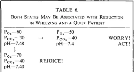
Arterial Blood Gases. The major initial defect in asthma is hypoxemia, as large areas of the lung that have excellent perfusion get progressively less ventilation, shunting poorly oxygenated blood into the arterial circulation. Initially, the asthmatic in respiratory distress can easily overventilate the un-involved areas of the lung lowering arterial C02 (Pac0 ,). The classical arterial gases of the dyspneic, but non-fatigued asthmatic, are a low Pa0 , (50
-60 mm Hg) and a low Paco, (20-30 mm Hg). As the patient tires, is sedated, gets worse, and is re· strained in bed, he will begin to move less air, and underventilation will begin with the Paco, approach-ing normal. This is a grave prognostic sign.
If the arterial oxygen rises as the Pco, rises and the patient is more comfortable, all is well. If the arterial oxygen falls and the Pco, approaches normal, you should immediately escalate your ther-apy (Table 6).
TABLE 4.
COMMON ERRORS IN MANAGEMENT
Too Little Steroids Too Late Sedation
HUNT: SEVERE ASTHMA
Tired
Old Fat
TABLE 5.
PATIENTS AT RISK
Chronic Bronchitis and Emphysema Heart Disease
Escalation of Drug Therapy.
Combination Tablets and lsupre'tJ9. Most pa-tients with asthma who present to the physician with a severe episode have already tried regular therapy with one of the phenobarbital, theophyl-line, or ephedrine combinations such as Tedra!® or Quadrinal®. They have generally used, and at times have overused, an Isuprel® preparation. These drugs then have little place in the manage-ment of the severe asthmatic. It is important, how-ever, to obtain specific historical facts about their use in planning therapy. A patient who has had little or no prior medication can be expected to respond more rapidly to less medication.
One investigator has found that patients with severe asthma are specifically unresponsive to Isuprel®, so that response to Isuprel® was a mea-sure not only of the severity of the disease, but also an index of the patient's response to other therapy, primarily steroids. More and more Isu-prel® is then used by the worsening patient with less and less results. At times the Isuprel'.!ll has
been shown to have been responsible for increas-ing the asthma and may actually cause hypoxemia.
Epinephrine and Aminophyllin. The response to 0.3 cc of epinephrine subcutaneously can at times be magical and should always be tried. The effect occurs within minutes and may last an hour. If no results are obtained after three doses
TABLE 6.
BOTH STATES MAY BE AsSOCIATED WITH REDUCTION IN WHEEZING AND A QUIET PATIENT
Po,-60
Pco,- 30 pH-7.48
! P0, -70 Pco,-40
pH-7.40
P0,-50
-> Pco,- 40 WORRY!
pH- 7.4 ACT!
REJOICE!
161
repeated at 15 minute intervals, a response will usually not be obtained. The response to IV ami -nophyllin may be equally as rapid and last for hours, frequently as long as six. The dose must be large-500 mg for an adult-and must be
given over 15-45 minutes, not over hours in a
large bottle of fluids. Two methods are equally
acceptable:
I. 500 mg ( a 20 cc ampule) given
intra-venously by syringe, over a IO minute
period. This is excellent if you have the time.
2. 500 mg in 250 cc glucose and water given by IV drip over a 30-60 minute period may be equally effective.
Decisions. Only the consultant the next morn-ing has the benefit of the clearly outlined flow
sheet documenting blood gases, clinical state, and
t!me of the therapeutic maneuvers. In actual prac-ttc~, the patient arrives in respiratory distress, ar -terial gases are obtained, and the above medica-tions are given-epinephrine X2 followed by 500 mg of IV aminophyllin. This takes approx -imately one or one and one-half hours. The
ini-tial blood gases become known to the physician ab~ut hal_f way through this therapy. He usually
waits until 15-20 minutes after the aminophyllin
is given and repeats the arterial gases and pH. Initial Gases. If the Paco, is below 50 mm Hg, supplemental oxygen is begun (24 or 28% ventimask).
If the Paco, is greater than 40 mm Hg and the patient has not obtained evident clinical improvement from ?is initial therapy, 250 mg of hydrocortisone is given
mtravenously in addition to the other drugs previously described.
Second Arterial Gases. These results have usually been obtained at a time when full thera-peutic benefits will have occurred from epine-phrine and aminophyllin therapy. A lack of clinical
response by the patient and a lack of significant
reduction in Paco, dictates that aggressive steroid !herapy should be begun. If the Paco, has dropped mto the 30 mm Hg range, therapy with aminophyllin
and epinephrine can be continued and a maintenance drug such as Tedra!® SA begun at an appropriate time.
Steroid Therapy.
General Considerations. There is no reason to give hydrocortisone except for the initial dosage. There is theoretical evidence that the therapeutic
than prednisolone, but there is little data. I am in
agreement with Sheehy et al. that airway
resis-tance is reduced by steroids "in several hours" ( 4). This has been shown to be true by the stud-ies of Pinkerton and Van Meter (1).
This brings up the practical consideration that by the time one decides to escalate to steroid therapy one must wait one to two hours for thera-peutic effect. One must then give steroids one or
two hours before they are needed. This is why 250 mg of hydrocortisone is given if the initial
gases indicate even slight C02 retention instead
of awaiting the second set two hours later. Pinker-ton and Van Meter (1) could show little
differ-ence in the dose response curves of oral and IV
steroids. However, in a seriously ill patient who
may vomit, the intravenous method is preferable.
Dosage Schedule. As long as the patient is in respiratory distress and the Paco, is 44 mm Hg or above, prednisolone 50 mg should be given every two hours. If the Paco, continues to rise, the dosage should be increased to I 00 mg every one to two hours.
Pediatric Dosage. In severe asthma (as de-fined under "Initial Gases") 7 mg/kg of hy-drocortisone should be given as an initial stat dose. Another 7 mg/kg of hydrocortisone may be given during the next 24 hours in divided doses. As is true in adults, there is little specific data to direct prednisone therapy in children within narrow dosage guidelines.
Mucolytic Agents. Hydration with appropriate intravenous fluids and aggressive therapy of
as-thma . are the only two mucolytic techniques of proven benefit. Glyceryl guacolate (Robitussin®) is of proven uselessness and acetylcysteine
(Mu-comyst®) will provoke asthma. Humidified air
can be added to the ventimask if the patient is a mouth breather, but humidity and heat are usually no problem as long as the nose is still in the cir-cuit.
Intubation and Ventilation.
Indications for Intubation. A nsmg Paco,
above 60 mm Hg, a pH below 7 .2, increasing fa-tigue, increasing somnolence with inability to cough despite the therapy as outlined above are indi-cations for intubation and ventilation.
General Considerations. The use of the cuffed
nasotracheal tube has revolutionized the respira-tory care of severe asthma. Only in unusual cir-cumstances should tracheostomy have to be done
for asthma. Once nasotracheal intubation is ac-complished and the patient is being artificially ventilated, aggressive therapy must be continued.
Unlike the patient with chronic bronchitis and
emphysema, the patient with asthma responds
quickly and fairly predictably to therapy (pri-marily corticosteroids) and can be weaned from the ventilator in 24 to 72 hours. The unfortunate tendency is for the physician to relax following in-tubation of the patient. Unless the appropriate ther-apy is continued, weaning from the ventilator can be prolonged by a relapse which increases the pos-sibility that tracheostomy will need to be done.
Plan of Action. The physician should
pre-sume that with appropriate therapy and barring serious complications, the patient can be extubated within 48 hours. The first twelve hours that the patient is on the ventilator should not be
compli-cated by premature weaning procedures. The fa-tigued patient should be allowed to rest. His abil-ity to cycle with the ventilator should be en-hanced with diazepam (Valium®) or if this is not immediately effective, with 5 to 10 mg of
intra-venous morphine (given every one to four hours
as neeqed). Obviously the patient cannot be simul-taneously sedated for control of ventilation and weaned from the ventilator, although this is fre-quently tried with disastrous results. Prednisone
100 mg every two hours should be continued
during this most important phase of therapy. Choice of Ventilator. The tendency of a naso-tracheal tube to leak, coupled with the extremely high airway pressures (3~0 cm H20) necessary to
ventilate asthmatics, makes pressure cycled ventilators (Bird or Bennett) poor choices. A volume controlled
ventilator such as the Bennett MA- I or the Emerson
postoperative ventilator is usually necessary for effective ventilation of these noncompliant lungs. The MA-I enables the respiratory care personnel to
easily monitor tidal volumes and oxygen percentage of
inspired air (F10 .). Both machines will intermittently
and automatically hyperinflate the patient's lungs to
reduce atelectasis. Both are dependable machines.
Complications. All of the legion of complica-tions incident to intubation and mechanical ven-tilation of course can occur. The excellent prog-nosis for return to normal in these patients should make their recognition and reversal all the more important. Several complications however are more peculiar to the asthmatic.
HUNT: SEVERE ASTHMA
extremely high airway pressures needed for venti-lation make this a common complication. Insertion of a chest tube with adequate seal is imperative to prevent a tension pneumothorax. This is a rare complication in adults.
Overventilation with Alkalosis. The very
na-ture of the disease makes this complication
po-tentially common and severe. The patient with
severe, reversible airway obstruction is connected to an efficient ventilator, and aggressive appropriate therapy is given. Success in therapy will almost always result in hyperventilation, unless blood gases are monitored frequently as the patient im-proves and the minute ventilation reduced.
Cardiovascular Collapse as Mechanical
Ven-tilation is Begun. This is more common in the
pa-tient with emphysema since the high airway pres-sures are transmitted more readily to the heart through abnormal lung tissue hindering right heart filling. The asthmatic has some protection due to the normal lung parenchyma. However, the com-bination of high inspiratory airway pressures, hypovolemia, and a short expiration time may rapidly reduce cardiac output even in the asthma-tic. The physician must prolong expiration as much as possible relative to inspiratory time, reduce
tidal volumes even if C02 cannot be lowered
ini-tially, and immediately correct the hypovolemic state.
Weaning. Improvement can be judged by a
return of the Paco, to normal (30-40 mm Hg)
and a reduction in the percentage of oxygen
nec-essary to maintain Pa0 , within a physiologic range
(70-100 mm Hg). Reduction in airway obstruction
can be monitored by following the "effective
com-pliance" (tidal volume divided by airway pressure expressed as liters/cm H20).
If at the end of 12 hours, blood gases and pH are normal and the effective compliance has doubled, sedation should be discontinued and the patient's ventilatory capabilities measured, using
a Wright Respirometer. If it can be determined
that the tidal volume is greater than 5 cc/kg and
the vital capacity greater than 10 cc/kg, a
pe-riod of time off the ventilator should be
moni-tored with blood gas measurements ( arterial
puncture 15-30 minutes off ventilator with
pa-tient receiving humidified 28-30% 02 by means
of a Briggs T-piece). If the patient is able to
maintain a normal or reduced Paco,, extubation should be considered. Alternately this procedure
163
is repeated every 12 hours or at even shorter in-tervals depending upon the observed rate of im-provement.
Preparing for Discharge. The exciting part of the therapy is now over, <1nd the tendency
to-ward management errors increases. If the reason
for the onset of the severe episode of asthma is
known and has been corrected (infection,
ex-posure to irritant fumes, resolved psychic trauma) then the steroid dosage can be quickly reduced
to 100-200 mg of prednisone given as a single
daily dose, then every other day, and finally dis-charge of the patient on 10 or 20 mg of
pred-nisone every other day. If the cause of the asthma
remains unknown, caution will have to be exer-cised in the rapid reduction of steroids.
Two major facts should be borne in mind:
1) Even large amounts of prednisone (50-100 mg)
given as a single early morning dose may be taken by the patient for up to 30 days without significant adrenal suppression. 2) Alternate day prednisone therapy, again a single early morning dose, can be given with little danger of side
ef-fects and may be continued for months.
Summary. The successful management of
se-vere asthma involves recognition and aggressive appropriate therapy, with rapid escalation to ster-oid therapy as soon as indicated. Intubation with a nasotracheal tube and ventilation with a
vol-ume controlled mechanical ventilator should be an
available option in therapy. Tracheostomy need rarely be done since most patients can be
extu-bated within 72 hours.
REFERENCES
1. PINKERTON, HERMAN H., JR. AND VANMETER, THOMAS E., JR. Immediate therapy for the acute attack of asthma.
New Eng. J. Med. 258:363-366, 1958.
2. SHEEHY, A. F., DIBENEDETTO, R., LEFFAK, S., AND LYONS, H. A. Treatment of status asthmaticus. Arch. Int. Med. 130:37-42, 1972.
3. TABB, w.
c.
AND GUERRANT, J. L. Life-threatening asthma. J. Allergy 42:249-260, 1968.4. VIA, C. AND HUNT, W. B., JR. The management of se-vere asthma. (To be published.)