Thallium 201 Myocardial
Imaging
MICHAEL J. COWLEY, M.D.
Assistant Professor of Medicine, Cardiovascular Disease, Medical College of Virginia, Health Sciences Division of Virginia Commonwealth University, Richmond, Virginia
In recent years technological advance-ments in nuclear medicine have resulted in in-creasing interest in the use of radioisotope tech-niques in the evaluation of cardiac disease, and cardiovascular nuclear medicine has developed into a useful noninvasive tool in clinical cardiol-ogy. Myocardial infarct imaging with tech-netium-99m pyrophosphate has been demon-strated to be a reliable method in the diagnosis of suspected myocardial infarction. 1 2
Radio-isotope cardiac flow studies are useful in the di-agnosis and follow-up of congenital heart dis-ease; 3 and gated cardiac blood pool imaging is
emerging as an important technique in the eval-uation of left ventricular function and ejection fraction.4 5 One of the more promising recent
applications of nuclear medicine in cardiology has been the development of myocardial per-fusion imaging in the evaluation of coronary ar-tery disease. Thallium 201 is the major radio-isotope employed in myocardial perfusion imaging and this report will review its basic properties and its use in the diagnosis of is-chemic heart disease.
Myocardial Perfusion Imaging
Myocardial perfusion imaging (MPI) refers to the use of certain radioisotope tracers which are rapidly and selectively concentrated in the heart. It is based on the principle that myo-cardial uptake of these isotopes occurs primar-ily as a function of myocardial blood flow and function. The resultant pattern of activity pro-vides an assessment of regional myocardial blood flow by comparing the amount and
distri-Correspondence and reprint requests to Dr. Michael J Cowley, Box 95, Medical College of Virginia, Richmond, VA 23298.
8 I MCV OUARTERL Y 15(1) 8-15, 1979
bution of activity in different areas of the heart. Regions of normal and abnormal myocardium are thereby distinguished by the presence and location of differences in myocardial radio-activity. Of the various radioisotopes which have been used for MPI, thallium 201 pos-sesses the best radiation characteristics (Table 1) and has been the most widely investigated. Thallium 201 is an analogue of potassium and exhibits similar biologic and physical proper-ties6; it is a monovalent cation with a half-life of 73 hours and is readily concentrated in the myocardium by an active transport process involving membrane sodium-potassium
adenosinetriphosphatase (ATPase). Thallium 201 emits low-energy photons which permit imaging with a scintillation camera and provide for high spatial resolution; it has a high myo-cardial extraction ratio, with 80% to 90% of an injected dose concentrated in the heart within the first few minutes, and exhibits a high myo-cardial-to-background ratio which contributes to improved image resolution. These features al-low for prompt imaging folal-lowing injection and a low patient radiation dose.
Myocardial activity with MPI is dependent on both the initial distribution of the radioisotope and its subsequent redistribution with time. The
distribution phase is most important with early
imaging and is determined both by isotope de-livery to different regions of the heart and by myocardial extraction. Radioisotope delivery is
a flow-dependent process and has been shown experimentally to be proportional to regional myocardial blood flow. 7 Myocardial extraction
of thallium 201 is a cell-dependent process
of myocardium but may be significantly delayed to zones of diminished coronary blood flow and may be absent in regions of abnormal function which are unable to concentrate the isotope. Redistribution is homogeneous in normal hearts but is heterogeneous in zones of significantly
di-minished flow or function. Washout of thallium
201 begins early from normal areas and is de-tectable at one hour; while this is in process from normal areas, continuing uptake is often occurring into flow-limited areas which exhibit initially delayed myocardial uptake. These
re-gions will eventually show normalization of
ac-tivity if viable myocardium is present, and redis-tribution is usually complete within one to four hours. 8 Areas of persistently diminished activity
generally indicate impaired myocardial function
and represent myocardial infarction.
Technique
Thallium 201 in a dose of 1 to 2 mCi is administered intravenously either at rest or at the peak of exercise when MPI is used in con-junction with treadmill exercise testing.
Myo-cardial images are recorded using a stationary
or portable scintillation camera. Early images are obtained 1 0 to 1 5 minutes after injection
and reflect the status of myocardial perfusion at
the time of injection. If the early image is
abnor-mal, delayed imaging three to four hours later is performed to look for redistribution into is-chemic areas. Multiple views are obtained to as-sess the different areas of myocardium with minimal superimposition and overlap. Inter-pretation is done by comparison of regional
myocardial activity; a normal image will
demon-strate a relatively homogeneous distribution of radioisotope within the left ventricle (Figs 1 and 2), and an abnormality is represented by dimin-ished activity, appearing as a "cold" .area (Fig 3) A defect which is persistent on late images indicates damaged myocardium or scar (Fig 4),
and a transient defect which is present on early
images but has resolved with delayed imaging represents an area of reversible ischemia (Fig 5). Difficulty with interpretation may occur when only small differences in regional activity are
present, but this may be improved by use of
computer processing techniques.
Myocardial Infarction
One area of clinical application of thallium 201 MPI is in the diagnosis of myocardial
in-ANT
IVS-LAD/ ( PLW-LC \"-··r /
IW-RCA
LAO
AW~
APEX--/
C-~ JW
LLAH
Fig 1-Normal thallium 201 MPI in the anterior (ANT), left anterior oblique (LAO), and left lateral (LLA TL) projections Myocardial uptake in the left ventricle is intense and
homo-geneous. The diagram to the right of each image indicates
the ventricular and coronary anatomy in each projection.
AW = anterior wall; FW = free wall; IVS = interventricular
septum; IW = left ventricular inferior wall; LAD = left ant
e-rior descending artery distribution; LC = region of left ci
r-cumflex artery; PLW = posterior lateral wall; RCA = right
coronary artery distribution.
farction. Several investigators have demon-strated a high sensitivity for thallium 201 in the detection of acute myocardial infarction. 9 -11 In
these studies sensitivity was greater than 95% in patients with transmural infarction and from 80% to 85% for nontransmural infarction. Sen-sitivity is unaffected by location of infarction but is dependent on infarct size, with decreased
TABLE 1
Myocardial Perfusion Imaging
Radionuclides K-43, Rb-81 , Cs-1 29, Tl-201 • Analogues of K +: physical and biologic
·Tl-201 -best radiation characteristics
• tY2 -73 hours
• low-energy emission characteristics • high myocardial extraction ratio (80%) • high myocardial-to-background ratio
• low patient radiation dose (07 rad/mCi) • usual dose = 1 to 2 mCi
GXT
c
Fig 2 -Normal thallium 201 study with exercise (GXT) in the anterior (left), left anterior oblique (center), and left lateral (right)
views. Activity in the left ventricle is homogeneous and intense, and there is activity seen in the right ventricle in the left and center panels. Right ventricular activity is often detectable with exercise. This patient had typical angina pectoris with normal coronary arteries on angiography and a false-positive exercise electrocardiogram.
THALLIUM - 201
MYOCARDIAL
SCINTIGRAMS
ANT
L
.
A
.
O
L
.
L
.
EE
Fig 3-Abnormal thallium 201 rest image in the anterior (ANT), left anterior oblique (LAO), and left lateral (LL) projections in a patient with acute anterior myocardial infarction. A large area of diminished activity is evident in the anterior and apical regions of the left ventricle in each projection.Fig 4-Thallium 201 exercise MPI with early (left) and delayed ( right) images in the left anterior oblique projection in a patient
with atypical chest pain and history of previous myocardial infarction. The immediate image shows an anteroseptal defect and
the delayed image recorded four hours later shows no redistribution into this area, indicating a fixed or irreversible defect from previous myocardial infarction.
Fig 5-Thallium 201 exercise (GXT) study in a patient with angina pectoris. The image at left was recorded immediately follow-ing completion of exercise and demonstrates a large area of markedly diminished activity in the anteroseptal region of the left ventricle. The image at right was obtained four hours later and shows redistribution of activity into the anteroseptal region with normalization of activity, indicating reversible, exercised-induced ischemia.
Fig 6-Serial thallium 201 MPI in anterior (ANT), left
ante-rior oblique (LAO), and left lateral (LL) projections in a p
a-tient with posterior wall infarction. Arrows indicate a defect at 4.5 hours after onset of chest pain This defect is dimin
-ished on the repeated studies 24 hours and 8 days later,
especially in the LAO view.
sensitivity in small infarctions. Sensitivity is also dependent upon time, with rare false-negative studies being obtained in the first 6 hours after infarction and an increased incidence of false-negative results observed after 24 hours. 9 In ad-dition, serial thallium 201 studies in the same patient have also demonstrated that the size of the defect often changes with time, usually being largest during the first 24 hours and be-coming smaller and more stable in size after 48 hours (Fig 6). 9 This observation probably in -dicates detection of zones of reversible peri-in-farction ischemia in addition to acute infarction in its early course which resolves with time,
TABLE 2
Exercise Thallium Imaging
CLINICAL APPLICATIONS
Improved sensitivity over exercise testing alone
Clarification of equivocal stress test Diagnosis when stress test uninterpretable
Conduction defect (LBBB)
Left ventricular hypertrophy
Resting ST-segment abnormality
Digitalis effect
Identification of false-positive stress test
Evaluation of coronary bypass graft function
1 2 / COWLEY THALLIUM 201 MYOCARDIAL IMAGING
leaving only the residual area of infarction ap-parent on subsequent images. The size of the defect with thallium 201 MPI also correlates well with the size of the infarct as determined by serum enzyme techniques, 12 pathological
anal-ysis, 11 and quantitative left ventricular angiogra-phy. 13 However, application of thallium 201 MPI
in suspected myocardial infarction is limited in patients with previous myocardial infarction be-cause of the isotope's inability to distinguish new infarction from old infarctions or ventricular aneurysm There is also reduced specificity with small infarctions (as with serum enzymes and electrocardiographic diagnosis) because of the limits of resolution of the technique. 14 In
addi-tion, MPI at rest may be abnormal with early
imaging in some patients having unstable
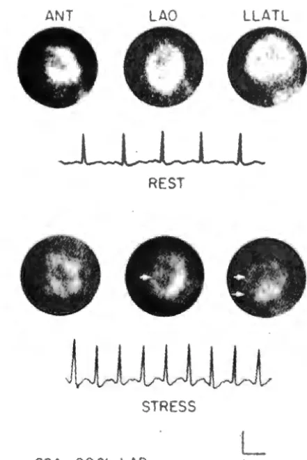
an-LAO
LLATL
REST
ST
R
ESS
I
-SCA -90% LAD
t
e
rn
Fig ?-False-negative stress electrocardiogram. Resting
(top) and stress (bottom) electrocardiogram and thallium
201 MPI in the anterior (ANT), left anterior oblique (LAO),
and left lateral (LLATL) projections in a patient whose
coro-nary angiograms (SCA) demonstrated significant left ante
-rior descending coronary artery stenosis. The resting scinti
-gram reveals homogeneous radioactivity. With stress, the
interventricular septum and anterior left ventricular walls (ar-rows), the regions supplied by the stenotic vessel, are
LVH
SCA-NORMAL
REST
STRESS
L
lcm
Fig 8-Normal thallium 201 MPI and equivocal stress ele c-trocardiogram in a patient with normal coronary arteries on angiography (SCA) Resting (top) and stress (bottom) elec
-trocardiogram and stress imaging in the anterior (left), left
anterior oblique (center) and left lateral (right) projections in
a patient with troublesome atypical chest pain and a history of hypertension. The full resting electrocardiogram revealed left ventricular hypertrophy (L VH) The monitored lead
showed ST-segment depression that deepened with stress
and was difficult to interpret in view of the depression at
rest. The perfusion image was normal and clarified the
stress electrocardiogram
gina witho'ut infarction owing to severely re-duced flow, 15 but this can usually be clarified if
delayed images are also obtained. Thallium
201 studies may also be abnormal in certain
forms of noncoronary heart disease, particularly
cardiomyopathy. 16 For these reasons, thallium
201 MPI appears to have limited general
useful-ness for the diagnosis of acute infarction but
can be of value to confirm infarction in situa-tions when standard criteria are not helpful. It has been useful in screening patients for
coro-nary care unit admission.9
Exercise Myocardial Imaging
Thallium 201 MPI has its greatest
appli-cation when used in combination with treadmill exercise testing for the evaluation of chest pain and transient myocardial ischemia, 11-20 and it
provides advantages over exercise testing alone
ANT LAO LLATL
00
J
1 :1
1)
~J
!
I
t
r-)
('
(
I'
('r
f'r
t''.
/'
~
r
if
(
STRESS
L
SCA-NORMAL lcm
Fig 9-False-positive stress electrocardiogram. Stress thal-lium 201 MPI in the anterior (ANT), left anterior oblique
(LAO), and left lateral (LLA TL) pro1ections ( top) and the rest -ing and stress electrocardiograms ( bottom) in a patient with normal coronary arteries on angiography (SCA), Radi o-nuclide distribution was normal, but the stress elec
-trocardiogram showed distinct and significant horizontal ST -segment depression Scintigraphy proved to be the valid
clinical indicator.
'JP ,
__J
---
1
POST IOP
i
J
[ R[ ST j [ ET T J
Fig 1 0-Preoperative (top) and post-operative (bottom)
thallium 201 MPI in the 45-degree left anterior oblique (LAO) projection in a patient who underwent coronary by-pass surgery The preoperative rest image ( top, left) is nor-mal, but following exercise (ETT) there is an extensive de-fect in the anteroseptal region (top right) preoperatively. Postoperatively both rest (bottom, left) and exercise (bot-tom, right) are normal. The bypass graft was patent on an -giography.
(Table 2). Thallium 201 is administered at the peak of exercise, and early images are o b-tained. If a normal image is recorded, further imaging is unnecessary (Fig 2). If an abnormal -ity is present, delayed images are recorded to assess the redistribution into ischemic areas (Fig 5). When compared with standard exercise electrocardiography, thallium 201 MPI is s ignifi-cantly more sensitive than exercise testing in the detection of coronary artery disease. This improved sensitivity is present in patients with a normal resting electrocardiogram (ECG) in whom exercise testing is most reliable (Fig 7), and is found particularly in patients with an a b-normal resting ECG, in whom ECG changes with exercise are often difficult to interpret or are uninterpretable. This is encountered in patients with intraventricular conduction defects, particu-larly left bundle-branch block; left ventricular hy-pertrophy (Fig 8); nonspecific resting ST-seg-ment abnormalities; and in patients taking digitalis or other medications which may alter the resting and exercise ECG.
Thallium 201 also has a higher specificity in detection of coronary artery disease than standard treadmill exercise testing, as fal se-positive results with thallium 201 are infrequent (Fig 9). 11· 1 9 In contrast, the incidence of
false-positive results with exercise electrocardi o-graphy may be as high as 30% in certain pa-tient populations 21 Thallium 201 exercise im
ag-ing is complementary to exercise testing, and diagnostic accuracy is improved when they are used in combination. In several series, this ac -curacy exceeds 90% in the detection of s ignifi-cant coronary artery disease.17 ·19
MPI with thallium 201 may also be of value in the postoperative evaluation of patients having coronary artery bypass graft surgery, as changes in regional myocardial perfusion with MPI have been shown to correlate well with by-pass graft function, 22 thereby providing a
non-invasive method of determining graft patency or closure (Fig 1 0).
In summary, MPI with thallium 201 is a new and effective noninvasive technique in the diagnosis of ischemic heart disease. It can r e-liably detect myocardial infarction and may be useful when the diagnosis of acute infarction by other means is uncertain. Exercise thallium 201 imaging is the area of greatest clinical appl i-cation of MPI, resulting in improved sensitivity
14 / COWLEY THALLIUM 201 MYOCARDIAL IMAGING
and specificity over exercise testing alone, and in high diagnostic accuracy in the detection of coronary artery disease when used in combina-tion with exercise electrocardiography. Thallium 201 MPI is also of value in the evaluation of cor-onary artery bypass graft function.
Figures 1, 7, 8, and 9 are reproduced with permission
from the American Journal of Cardiology (41 :43-51, 1978)
Figure 6 is reproduced by permission from the New England Journal of Medicine (295 1-5, 1976)
Figure 1 0 is reproduced from Circulation (56 830 -836, 1977) by permission of the American Heart Associ a-tion, Inc.
REFERENCES
1. WILLERSON JT, PARKEY RW, BONTE FJ, ET AL: Tec
h-netium stannous pyrophosphate myocardial scinti
-grams in patients with chest pain of varying etiology. Circulation511046-1052, 1975
2. COWLEY MJ, MANTLE JA, ROGERS WJ, ET AL: Tech
-netium-99m stannous pyrophosphate myocardial sc
in-tigraphy. Reliability and limitations in assessment of acute myocardial infarction. Circulation 56: 192-198,
1977.
3 TREVES S, COLLINS-NAKAI RL: Radioactive tracers in congenital heart disease. Am ..! Cardiol 38:711-721.
1976.
4. STRAUSS HW, ZARET BL, HURLEY PJ, ET AL: A scintiph
o-tographic method for measuring left ventricular ej
ec-tion fraction in man without cardiac catheterization. Am
J Cardiol 28 575-580, 1971.
5. RIGO P, MURRAY M, STRAUSS HW, ET AL: Scintiph
otogra-phic evaluation of patients with suspected left ventricu
-lar aneurysm Circulation 50:985-991, 1974. 6. BRADLEY-MOORE PR, LEBOWITZ E, GREEN MW, ET AL
Thallium-201 for medical use. II: Biologic behavior. J Nucl Med 16 156-160, 1975.
7. STRAUSS HW, HARRISON K, LANGAN JK, ET AL: Thalliu
m-201 for myocardial imaging Relation of thallium-201
to regional myocardial perfusion. Circulation 51 :641
-645, 1975.
8. POHOST GM, ZIR LM, MOORE RH, ET AL: Differentiation
of transiently ischemic from infarcted myocardium by
serial imaging after a single dose of thallium-201. C
ir-culation 55 294-302, 1977.
9. WACKERS FJT, SOKOLE EB, SAMSON G, ET AL Value and
limitations of thallium-201 scintigraphy in the acute
1 0 WACKERS FJT, SOKOLE EB, SAMSON G, ET AL
Myo-cardial imaging in coronary heart disease with
radio-nuclides, with emphasis on thallium-201. Eur J Cardiol
4 273-282, 1976.
11. WACKERS FJT, BECKER AE, SAMSON G, ET AL: Location and size of acute transmural myocardial infarction
esti-mated from Thallium-201 scintiscans. Circulation 56:72-78, 1977
12. WITZTUM K, FLETCHER J, LINDEMAN J, ET AL: Myocardial
infarct size. Thallium-201 perfusion scintigraphy ver
-sus enzymatic (CK-MB) estimates. Circulation 58
(Suppl II) 11-14, 1978
13. NIESS GS, LOGIC JR, RUSSELL RO JR, ET AL Usefulness
and limitations of resting thalium-201 scintigraphy in detecting location and size of myocardial infarction, abstract. AmJCardiol41379, 1978.
14 MUELLER TM, MARCUS ML, EHRHARDT JC, ET AL Limi ta-tions of thallium-201 myocardial scintigrams. Circul
a-tion 54 640-646, 1 976.
1 5. WACKERS FJT, LIE Kl, LIEM KL Thallium-201 scinti
gra-phy in unstable angina pectoris Circulation 5 7 738-742, 1978
1 6. BULKLEY BH, HUTCHINS GM, BAILEY I, ET AL:
Thallium-201 imaging and gated cardiac blood pool scans in
patients with ischemic and idiopathic congestive
car-diomyopathy. Circulation 55 753-760, 1977.
1 7. BAILEY IK, GRIFFITH LSC, ROULEAU J, ET AL
Thallium-201 myocardial perfusion imaging at rest and during exercise. Comparative sensitivity to electrocardio
-graphy in coronary artery disease. Circulation 55:79-87, 1977.
1 8. RITCHIE JL, TROBAUGH GB, HAMIL TON GW, ET AL:
Myo-cardial imaging with thallium-201 at rest and during ex-ercise. Comparison with coronary arteriography and
resting and stress electrocardiography. Circulation
5666-71, 1977.
1 9. HAMIL TON GW, TROBAUGH GB, RITCHIE JL, ET AL:
Myo-cardial imaging with intravenously inJected
thallium-201 in patients with suspected coronary artery
dis-ease. AmJCardiol39347-354, 1977.
20. BOTVINICK EH, TARADASH MR, SHAMES OM, ET AL:
Thal-lium-201 myocardial perfusion scintigraphy for the
clinical clarification of normal, abnormal and equivocal
electrocardiographic stress tests. Am J Cardiol
4143-51, 1978.
21 BORER JS, BRENSIKE JF, REDWOOD DR, ET AL Limit a-tions of the electrocardiographic response to exercise
in predicting coronary-artery disease. N Engl J Med
293.367-371, 1975.