ARTICLE OPEN ACCESS
AP4 deficiency
A novel form of neurodegeneration with brain iron accumulation?
Agathe Roubertie, MD, PhD, Nelson Hieu, MSc,* Charles-Joris Roux, MD,* Nicolas Leboucq, MD, Gael Manes, PhD, Majida Charif, PhD, Bernard Echenne, MD, Cyril Goizet, MD, PhD, Claire Guissart, PhD, Pierre Meyer, MD, Cecilia Marelli, MD, François Rivier, MD, PhD, Lydie Burglen, MD, Rita Horvath, MD, PhD, Christian P. Hamel, MD, PhD, and Guy Lenaers, PhD
Neurol Genet2018;4:e217. doi:10.1212/NXG.0000000000000217
Correspondence Dr. Roubertie
Abstract
ObjectiveTo describe the clinico-radiological phenotype of 3 patients harboring a homozygous novel
AP4M1pathogenic mutation.
Methods
The 3 patients from an inbred family who exhibited early-onset developmental delay, tetra-paresis, juvenile motor function deterioration, and intellectual deficiency were investigated by magnetic brain imaging using T1-weighted, T2-weighted, T2*-weighted,fluid-attenuated in-version recovery, susceptibility weighted imaging (SWI) sequences. Whole-exome sequencing was performed on the 3 patients.
Results
In the 3 patients, brain imaging identified the same pattern of bilateral SWI hyposignal of the globus pallidus, concordant with iron accumulation. A novel homozygous nonsense mutation was identified inAP4M1, segregating with the disease and leading to truncation of half of the
adapdomain of the protein.
Conclusions
Our results suggest thatAP4M1represents a new candidate gene that should be considered in the neurodegeneration with brain iron accumulation (NBIA) spectrum of disorders and highlight the intersections between hereditary spastic paraplegia and NBIA clinical presentations.
*These authors contributed equally to this work.
From the D´epartement de Neurop´ediatrie (A.R., B.E., P.M., F.R.), CHU Gui de Chauliac, Montpellier; Institut des Neurosciences de Montpellier (A.R., N.H., G.M., C.P.H.), INSERM U1051, Universit´e de Montpellier; Service de Neuroradiologie (C.-J.R., N.L.), CHU Gui de Chauliac, Montpellier; Equipe MitoLab (M.C., G.L.), UMR CNRS 6015-INSERM 1083, Institut MitoVasc, University of Angers, France; Department of Medical Genetics (C. Goizet), Hopital Pellegrin, Bordeaux University Hospital; MRGM Laboratory (C. Goizet), INSERM U1211, University of Bordeaux; Laboratoire de G´en´etique Mol´eculaire (C. Guissart), CHU de Montpellier; U1046 INSERM (P.M., F.R.), UMR9214 CNRS, Universit´e de Montpellier; Department of Neurology (C.M.), University Hospital Gui de Chauliac, Montpellier; Centre de R´ef´erence des Malformations et Maladies Cong´enitales du Cervelet (L.B.), Service de G´en´etique, Hˆopital Armand Trousseau, AP-HP, Paris, France; Wellcome Trust Centre for Mitochondrial Research (R.H.), Institute of Genetic Medicine, Newcastle University, United Kingdom; and Centre of Reference for Genetic Sensory Diseases (C.P.H.), Montpellier, France.
Funding information and disclosures are provided at the end of the article. Full disclosure form information provided by the authors is available with the full text of this article at Neurology.org/NG.
The Article Processing charge was funded by the Montpellier Hospital.
Hereditary spastic paraplegias (HSPs) are a heterogeneous group of neurodegenerative diseases clinically characterized by progressive lower extremity weakness and spasticity, which may be isolated (pure HSP) or combined with other neuro-logic or nonneuroneuro-logical signs (complex HSP).1,2More than 70 genes have been implicated, emphasizing diverse molec-ular pathogenic mechanisms.3 In this respect, recessive mutations in genes encoding the different subunits of adaptor protein complex-4, (AP4B1, AP4M1, AP4E1, and AP4S1) have been identified in patients with complex HSP (SPG47, 50, 51, and 52 respectively).4–8The AP4-deficiency syndrome
is characterized by progressive spasticity, microcephaly, in-tellectual deficiency, dysmorphic traits, and growth retardation,4–8 while epilepsy and peripheral neuropathy
might be associated.4,9Brain imaging phenotypes reported up to now are characterized by cerebral atrophy, asymmetric enlargement of lateral ventricles, white matter loss, and thin corpus callosum splenium.8–10Thin and globoid hippocampal cortex9and tortuosity of intraextracranial large vessels were also reported.4
Neurodegeneration with brain iron accumulation (NBIA), which is characterized by dystonia, parkinsonism, spasticity, and brain iron accumulation on MRI, represents another inherited group of neurodegenerative disorders, due to mutations in 10 genes, with molecular overlaps with HSP.11,12
Here, we report 3 patients from the same kindred who harbor a homozygous AP4M1 mutation. They exhibit the typical clinico-radiological phenotype of AP4-deficiency syndrome, but surprisingly associated with bilateral pallidal iron accu-mulation on brain imaging, thus establishing a link between AP4-related complex HSP and NBIA disorders.
Methods
Standard protocol approvals, registrations, and patient consents
The study was conducted in accordance with the Declaration of Helsinki and was approved by the local ethical committee. Written informed consent was obtained from the patients’ legal representatives.
Whole-exome sequencing and brain imaging Whole-exome sequencing (WES) was performed on the DNA from the 3 affected patients by Aros Ltd. Homozygous mutations common to the 3 patients were filtered pro-gressively for their frequency (<1%), alteration of the open reading frame (frameshift, splicing, missense, and nonsense mutations), and ultimately for their localization in the
homozygous regions common to the 3 patients. Sanger se-quencing allowed for their confirmation and segregation study in the family.
CT was performed on a 64-section CT scanner (Discov-ery750 HD; GE Health care, Milwaukee, WI). MRIs were acquired on a 1.5-T system (AVENTO; Siemens medical solutions, Erlangen, Germany) as follows: axial slices T2-weighted, T2*-T2-weighted,fluid-attenuated inversion recovery, susceptibility weighted imaging (SWI) sequences, and sagittal slices T1-weighted sequences.
Results
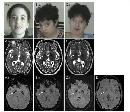
Clinical dataThe clinical features of the 3 patients originating from a large consanguineous Moroccan family (figure 1A) are described in table. Psychomotor retardation with spasticity of the 4 limbs was noticed early in life. Clinical examination from thefirst year showed spastic tetraplegia, with pyramidal tract signs and equinovarus. Patients IV-2 and IV-5 sat unaided at 7 months; patient IV-2 was able to crawl at 2 years but never managed to walk; her sister IV-5 could walk short distances with unsteady spastic gait from the age of 5 years. Patient IV-7 sat unaided at 11 months of age and walked at 3 years, with a broad-based unsteady gait. The patients exhibited stable severe mental deficiency, without behavioral disturbance. Motor achieve-ments progressively deteriorated at adolescence, with loss of the highest motor skills, but without additional cognitive decline; from that time, bradykinesia, hypomimy, drooling, and athetoid movements of the hands were also noticed. Patients IV-5 and IV-7 displayed short stature. Dysmorphic features (figure 2, Aa, Ba, Ca) were also present. The 3 patients needed assistance to most common daily living activities.
The following investigations were normal: electro-myoneurography recording, cardiac ultrasound scan, visual and auditory evoked potentials, fundus examination, karyo-type analysis on lymphocytes (cases IV-2, IV-5 and IV-7),
PANK2andPLA2G6Sanger sequencing (patient IV-7), and analyses of mitochondrial enzymatic activities on a muscle sample (patient IV-2).
Genetic results
Comparison of WES results performed for patients IV-2, IV-5, and IV-7 revealed 3 homozygous regions, 1 on chromosome 7 (5.7 Mb) and 2 on chromosome 9 (2.25 and 1.31 Mb). A total of 14,753 exonic variants were common to the 3 patients, and by progressively filtering them, we identified 4,974
Glossary
homozygous variants, among which 2,546 were non-synonymous, frameshift, splicing, or stop variants. Further filtering for damaging variants with a frequency lower than 1% identified 3 mutations in theAP4M1, HRNR, andNPIPL3
genes, but only the 1 inAP4M1was located in chromosome 7, in 1 of the 3 homozygous regions.
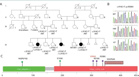
This c.916C>T mutation (rs369459721) is leading to a premature stop codon (p.R306X), truncating the last 147 residues of the protein (figure 1, B and C). It has a global allelic frequency of 2.4 × 10⁻⁵in the ExAC and a frequency of
3.0 × 10⁻⁵ in Non-Finnish European and 9.3 × 10⁻⁵ in African, while it was not encountered in the rest of the world.
Analysis of the homozygous variants located in the 10 known NBIA genes revealed 2 common variants, located in CP
(rs701753) and PANK (rs3737084), but they were not damaging, had a frequency higher than 1%, and were located away from the 3 homozygous regions.
Brain imaging
Brain MRI of the 3 patients showed global cerebral atrophy, white matter loss, asymmetric ventriculomegaly (figure 2, B, E, and H), and thinning of the splenium of the corpus
callosum (data not shown J). T1 sequences showed an iso-intense pattern of the globus pallidus (data not shown). T2 sequences revealed symmetric mild hypointensity of the globus pallidus, which was significantly accentuated on SWI sequences (figure 2, Ab, Ac, Bb, Bc, Cb, Cc, D). Patient IV-7’s CT was normal (data not shown).
Discussion
We identified a homozygous nonsense mutation inAP4M1in 3 women from the same inbred family by WES. This R306X mutation deletes the last 147 residues of the protein, truncating half of theadapdomain, an effect similar to that reported in 2 other families who harbored a stop codon truncating the AP4M1 protein at positions 318 and 338.9Until now, only 5 different AP4M1mutations have been reported in 7 families with a common clinical presentation4,5,9,13–15(figure 1C). The 3 patients from our study share the same clinical phenotype with variable severity, consisting in early-onset developmental delay, tetraparesis, juvenile motor function deterioration, intellectual deficiency, athetoid upper limb movements, bradykinesia, and mild dysmorphism, which fits with the previously de-scribed AP4-deficiency syndrome. Even if the bilateral pallidal
TableClinical features of 3AP4M1individuals
Patient Patient IV-2 Patient IV-5 Patient IV-7
Sex/age at last examination F/25 y F/16 y F/23 y
Perinatal parameter Normal Normal Low birth weight
Seizures No 1 febrile seizure during the second
year of life
1 febrile seizure at 20 m
Age at acquisition of unaided sitting 7 m 7 m 11 m
Highest motor achievements Able to crawl at 2 y Unsteady spastic gait at 5 y Independent broad-based unsteady gait at 3 y
Motor deterioration/age Unable to crawl at 13 y Assisted gait from 12 y Assisted gait from 15 y
Spasticity and pyramidal tract signs Yes Yes Yes
Equinovarus Yes Yes Yes
Bradykinesia and athetoid movements of the hands
From adolescence From adolescence From adolescence
Language Short sentences Short sentences Less than 10 words
Behavior Shy and introverted form
adolescence
Normal Smiley
Intellectual deficiency Severe Moderate Severe
Growth parameter at last follow-up Normal Short stature Short stature
Height 160 cm, weight 68 kg Height 150 cm, weight 55 kg Height 143 cm, weight 44 kg
Microcephaly No No Yes
Head circumference 53 cm 52 cm 48.5 cm
Dysmorphism Yes Yes Yes
hyposignal is mild on T2 sequences and could be interpretated as physiologic iron accumulation at this age, the substantial hyposignal on SWI is totally unusual in patients of the same age. Thesefindings, correlated with the absence of hypersignal on T1-weighted imaging or CT hyperdensities in the patients, are strongly suggestive of brain iron overload.
Iron deposits have not been previously reported in patients with AP4-deficiency syndrome. Nevertheless, magnetic susceptibility sequences, which can confirm the presence of iron, have not been performed in most of the reported cases; therefore, this feature might have been underdiagnosed. A search for homo-zygous mutations common to the 3 patients in the 10 published NBIA genes revealed 2 variants located inPANKandCP, but their frequency and the absence of pathogenicity were somehow incompatible with their involvement as modifier mutations switching HSP clinical presentation to NBIA.
Of interest, a patient withAP4E1mutations, whose brain MRI showed bilateral T2-hypointensity of the globus pallidus, has already been described.8This peculiarfinding, although not discussed in the article, strongly suggests iron accumulation in thisAP4E1patient, as in our 3AP4M1patients.
The pathophysiology of HSP involves many cellular path-ways as cellular transport, nucleotide metabolism, and syn-apse and axon developments, providing a causative link
between HSP and other neurodegenerative diseases.3,16 Overlaps between HSP and NBIA are well known, as already reported for cases with mutations in FA2H and C19orf12
genes (SPG 35 and 43, respectively).11,17The AP-4 complex is a heterotetramer ubiquitously expressed in the CNS early in the embryologic and postnatal development and is im-plicated in vesicle formation, post-Golgi protein trafficking, and sorting processes.18Eventually, AP-4 dysfunction might
affect autophagy by disrupting the early steps of endosomal formation, a process shared with Kufor-Rabeb disease and beta-propeller protein-associated neurodegeneration, 2 forms of NBIA related to ATP13A2 and WDR45 genes, respectively.11,12
Moreover, NBIA disorders are probably underdiagnosed, and the evolution of technologies and practices in radiology leads to the identification of many new candidate genes through the incorporation of susceptibility weighted sequences more frequently in the brain imaging protocols.19 Our study has limitations, especially because of the small sample size.
Nevertheless, according to ourfindings inAP4M1 mutated patients, we recommend that brain MRI with susceptibility weighted sequences be included in the brain imaging protocol for patients with suspected HSP and AP4-deficiency syn-drome to collect a larger group of patients, and we propose
Figure 1Identification of a novelAP4M1mutation
(A) Pedigree showing the segregation of theAP4M1c.916C>T (p.R306X) mutation in the family; black symbols indicate affected patients. (B) Electrophoregrams showing the wild-type (top), the homozygous mutated (middle), and heterozygous (bottom) sequence ofAP4M1. (C) Localization ofAP4M1mutations in the protein: the structure of theAP4M1protein (domain and amino acid positions) is described with all the pathogenic missense mutations5,14(green), nonsense
mutation9,15(violet), frameshift4,13(pink flag) mutations reported to date (in black), and in the present cases (in red). HM = homozygous; HT = heterozygous;
that mutations inAP4genes be considered and screened in a subset of patients with NBIA spectrum disorders.
Author contributions
Design or conceptualization of the study: Agathe Roubertie, Christian P. Hamel, and Guy Lenaers. Analysis or in-terpretation of the data: Charles-Joris Roux, Nicolas Leb-oucq, Gael Manes, Majida Charif, Bernard Echenne, Cyril Goizet, Claire Guissart, Pierre Meyer, Cecilia Marelli, François Rivier, Lydie Burglen, and Rita Horvath. Drafting or revising the manuscript for intellectual content: Agathe Roubertie, Charles-Joris Roux, Cyril Goizet, and Guy Lenaers.
Acknowledgment
The authors acknowledge the patients and their families for participating in the study. They thank La Fonation Maladies Rares et Retina France. They are indebted to the INSERM and CNRS and the Montpellier University for their
institutional supports. They thank the Region Pays de la Loire, Angers Loire-M´etropole, University of Angers, and University Hospital of Angers for their support to the PREMMI project. They are indebted to Dr. Lagavulin for helpful discussions.
Study funding
No targeted funding reported.
Disclosure
A. Roubertie, N. Hieu, C.-J. Roux, N. Leboucq, G. Manes, M. Charif, and B. Echenne report no disclosures. C. Goizet has served on the scientific advisory boards of Amicus Thera-peutics and SanofiGenzyme and has received travel funding/ speaker honoraria from SanofiGenzyme, Amicus Therapeu-tics, and Shire. C. Guissart and P. Meyer report no disclosures. C. Marelli has received travel funding/speaker honoraria from Actelion Pharmaceuticals. F. Rivier, L. Burglen, and
Figure 2Facial appearance and brain imaging
R. Horvath report no disclosures. C.P. Hamel is deceased; disclosures are not included for this author. G. Lenaers reports no disclosures. Full disclosure form information provided by the authors is available with the full text of this article at Neurology.org/NG.
Received September 16, 2017. Accepted infinal form December 10, 2017.
References
1. Finsterer J, L¨oscher W, QuasthoffS, Wanschitz J, Auer-Grumbach M, Stevanin G. Hereditary spastic paraplegias with autosomal dominant, recessive, X-linked, or ma-ternal trait of inheritance. J Neurol Sci 2012;318:1–18.
2. Fink JK. Hereditary spastic paraplegia: clinico-pathologic features and emerging molecular mechanisms. Acta Neuropathol 2013;126:307–328.
3. Kara E, Tucci A, Manzoni C, et al. Genetic and phenotypic characterization of complex hereditary spastic paraplegia. Brain 2016;139:1904–1918.
4. Verkerk AJ, Schot R, Dumee B, et al. AR ticle mutation in the AP4M1 gene provides a model for neuroaxonal injury in cerebral palsy. Am J Hum Genet 2009;85:40–52. 5. Jameel M, Klar J, Tariq M, et al. A novel AP4M1 mutation in autosomal recessive cerebral palsy syndrome and clinical expansion of AP-4 deficiency. BMC Med Genet 2014;15:1–7.
6. Jamra RA, Philippe O, Raas-Rothschild A, et al. Adaptor protein complex 4 deficiency causes severe autosomal-recessive intellectual disability, progressive spastic para-plegia, shy character, and short stature. Am J Hum Genet 2011;88:788–795. 7. Abdollahpour H, Alawi M, Kort¨um F, et al. An AP4B1 frameshift mutation in siblings
with intellectual disability and spastic tetraplegia further delineates the AP-4 de-ficiency syndrome. Eur J Hum Genet 2014;23:256–259.
8. Moreno-De-Luca A, Helmers SL, Mao H, et al. Adaptor protein complex-4 (AP-4) deficiency causes a novel autosomal recessive cerebral palsy syndrome with micro-cephaly and intellectual disability. J Med Genet 2011;48:141–144.
9. T¨uys¨uz B, Bilguvar K, Koçer N, et al. Autosomal recessive spastic tetraplegia caused by AP4M1and AP4B1gene mutation: expansion of the facial and neuroimaging features. Am J Med Genet A 2014;164:1677–1685.
10. Blumkin L, Lerman-Sagie T, Lev D, Yosovich K, Leshinsky-Silver E. A new locus (SPG47) maps to 1p13.2-1p12 in an Arabic family with complicated autosomal recessive hereditary spastic paraplegia and thin corpus callosum. J Neurol Sci 2011;305:67–70. 11. Arber CE, Li A, Houlden H, Wray S. Review: insights into molecular mechanisms of
disease in neurodegeneration with brain iron accumulation: unifying theories. Neu-ropathol Appl Neurobiol 2015;42:220–241.
12. Meyer E, Kurian MA, Hayflick SJ. Neurodegeneration with brain iron accumulation: genetic diversity and pathophysiological mechanisms. Annu Rev Genomics Hum Genet 2015;16:257–279.
13. Langou¨et M, Siquier-Pernet K, Sanquer S, et al. Contiguous mutation syndrome in the era of high-throughput sequencing. Mol Genet Genomic Med 2015;3:215–220. 14. Najmabadi H, Hu H, Garshasbi M, et al. Deep sequencing reveals 50 novel genes for
recessive cognitive disorders. Nature 2011;478:57–63.
15. Duerinckx S, Verhelst H, Perazzolo C, et al. Severe congenital microcephaly with AP4M1 mutation, a case report. BMC Med Genet 2017;18:48.
16. Novarino G, Fenstermaker AG, Zaki MS, et al. Exome sequencing links corticospinal motor neuron disease to common neurodegenerative disorders. Science 2014;343: 506–511.
17. Landour´e G, Zhu PP, Lourenco CM, et al. Hereditary spastic paraplegia type 43 (SPG43) is caused by mutation in C19orf12. Hum Mutat 2013;34:1357–1360. 18. Simmen T, H¨oning S, Icking A, Tikkanen R, Hunziker W. AP-4 binds basolateral
signals and participates in basolateral sorting in epithelial MDCK cells. Nat Cell Biol 2002;4:154–159.
DOI 10.1212/NXG.0000000000000217
2018;4;
Neurol Genet
Agathe Roubertie, Nelson Hieu, Charles-Joris Roux, et al.
AP4 deficiency: A novel form of neurodegeneration with brain iron accumulation?
This information is current as of January 24, 2018
reserved. Online ISSN: 2376-7839.
Published by Wolters Kluwer Health, Inc. on behalf of the American Academy of Neurology.. All rights an open-access, online-only, continuous publication journal. Copyright Copyright © 2018 The Author(s).
Services
Updated Information &
http://ng.neurology.org/content/4/1/e217.full.html
including high resolution figures, can be found at:
References
http://ng.neurology.org/content/4/1/e217.full.html##ref-list-1
This article cites 19 articles, 2 of which you can access for free at:
Citations
http://ng.neurology.org/content/4/1/e217.full.html##otherarticles
This article has been cited by 1 HighWire-hosted articles:
Subspecialty Collections
http://ng.neurology.org//cgi/collection/spastic_paraplegia
Spastic paraplegia
http://ng.neurology.org//cgi/collection/mri
MRI
http://ng.neurology.org//cgi/collection/developmental_disorders
Developmental disorders
http://ng.neurology.org//cgi/collection/all_genetics
All Genetics
following collection(s):
This article, along with others on similar topics, appears in the
Permissions & Licensing
http://ng.neurology.org/misc/about.xhtml#permissions
its entirety can be found online at:
Information about reproducing this article in parts (figures,tables) or in
Reprints
http://ng.neurology.org/misc/addir.xhtml#reprintsus
Information about ordering reprints can be found online:
reserved. Online ISSN: 2376-7839.
Published by Wolters Kluwer Health, Inc. on behalf of the American Academy of Neurology.. All rights an open-access, online-only, continuous publication journal. Copyright Copyright © 2018 The Author(s).