Comparison of the Efficacy of Oral Simvastatin and Topical Simvastatin
Solution in Decreasing Post-Laparotomy Adhesions in Rats
Mojtaba Javaherzadeh1, Babak Sabet1, Mohammad Nejati2, Ali Shekarchizadeh3,
Abass Mirafsharieh4, Mohammad Bayat5
1
Associate Professor, Department of Surgery, School of Medicine, Shahid Beheshti University of Medical Sciences, Tehran, Iran 2 Resident, Department of Surgery, School of Medicine, Shahid Beheshti University of Medical Sciences, Tehran, Iran
3 Researcher, Poursina Hakim Gastrointestinal Research Center, Poursina Hakim Research Institution, Isfahan, Iran
4
Associate Professor, Department of Pathology, School of Medicine, Shahid Beheshti University of Medical Sciences, Tehran, Iran 5 Professor, Department of Anatomical Sciences, School of Medicine, Shahid Beheshti University of Medical Sciences, Tehran, Iran
Received: 05 Feb. 2018; Received in revised form: 10 Apr. 2018; Accepted: 18 May 2018
Abstract
Background: Intra-abdominal adhesions and their complications occur frequently after laparotomy. The aim of this study was to compare oral versus intraperitoneal administration of simvastatin in decreasing post-laparotomy adhesions in rat.
Methods: Thirty male Wistar albino rats were divided into three groups of ten. All the rats underwent laparotomy and induction of adhesions using the method of Meso-Stitch approximation of injured cecum and abdominal wall. One group received oral simvastatin (40 mg/kg) daily during two weeks before the laparotomy. In the two other groups, 2 ml of solution of simvastatin (40 mg/kg) or distilled water (as placebo) was spilled into abdomen before closing abdominal wall, respectively. After 14 days, all the rats were put under laparotomy again to be compared. Rates and grades of adhesions were assessed using Hoffman et al. and Lauder et al. Scale and histopathological reports.
Results: In placebo group, the grade II and III adhesion was seen in 2 and 8 rats, respectively. In local simvastatin group, there was no adhesion in 5 rats, and grade I and II adhesion was seen in 3 and 2 rats, respectively. In oral simvastatin group, 6 rats were without adhesion, and 3 cases with grade I and 1 case with grade II adhesion. The frequency and grade of adhesion were statistically different in simvastatin groups compared to the placebo group (P < 0.001), but not with each other.
Conclusions: Oral simvastatin for two weeks before the laparotomy can reduce post-laparotomy adhesion bands as well as local administration of simvastatin solution.
© 2018 Tehran University of Medical Sciences. All rights reserved.
Citation: Javaherzadeh M, Sabet B, Nejati M, Shekarchizadeh A, Mirafsharieh A, Bayat M. Comparison of
the Efficacy of Oral Simvastatin and Topical Simvastatin Solution in Decreasing Post-Laparotomy Adhesions in Rats. Acad J Surg, 2018; 5(1-2): 2-6.
Keywords: Abdomen; Surgical adhesions; Simvastatin; Laparotomy
Introduction
Annually, over 300000 patients undergo surgery for the treatment of intestinal obstruction due to adhesions in the United States (1) and 20-years analysis (1988 to 2007) did not show any reduction in this amount (2).
Although all the patients with adhesion do not show clinical signs, but early or late intestinal obstruction (49-79 percent), chronic abdominal pain (20-70 percent) and infertility (15-20 percent) are considered as the major surgical problems (1).
The studies using serial laparoscopies showed that
at the 8th day after the surgery, 55% of the adhesions
are appeared and 100% of them were visible between
the 4th to 8th weeks (3,4). Although laparoscopy and
microsurgery techniques have reduced the formation of adhesions, but the benefits of adjuvant use of different substances, despite their widespread use, is still questionable (5).
Many drugs, such as anti-inflammatory or
fibrinolytic agents and physical barriers are
recommended; but there is still no ideal substance to
prevent adhesions. Hyaluronic acid, Intergel
anastomoses (1). Therefore, efforts are ongoing to examine other factors in reducing adhesions. Among these factors, are the statins that have anti-inflammatory and fibrinolytic effects (9).
According to the our previous study and the positive effect of local simvastatin at a dose of 30 mg per kg of body weight in preventing the formation of adhesion bands in rats (6), and based on similar studies and the significant effect of oral statins prior to laparotomy in this animal model (8), this study aimed to investigate the anti-adhesion effect of oral and intraperitoneal simvastatin, compared to placebo (distilled water) in vivo conditions.
Materials and Methods
In this experimental study, 30 adult male Wistar albino rats weighing 250 to 300 g with the age of 4-6 months and the same race were selected and divided into three groups of ten. In the first group, during two weeks before the study, oral simvastatin HEXAL (Germany) with the dose of 40 mg/kg dissolved in water was administrated daily.
This study was done under the regarding the Guide for the Care and Use of Laboratory Animal provided by Shahid Beheshti Laboratory Animals Center in accordance with global standards for laboratory biosafety guidelines (10). The rats were housed in the animal nest of the Department of Anatomy in School of Medicine. The study was approved by the institutional review board and the Ethics Committee of Shahid Beheshti University of Medical Sciences, Iran.
The rats were intramuscularly anesthetized with ketamine 60 mg/kg ROTEXMEDICA (Germany) and then, underwent midline laparotomy incision with the length of 4 cm. In all three groups, powder-free gloves were used during the surgery. To create adhesion, according to Daniel study, the method of Meso-Stitch approximation of injured cecum and abdominal wall was used in this way: scratching with a blade on the cecum and creating one-square-centimeter defects on the abdominal wall and creating sutures for abdominal wall and the skin with 3-0 nylon (11). All the rats underwent the surgery in one day.
At the end of the surgery, in the second and groups, before closing the abdomen, 2 cc of simvastatin solution (40 mg/kg) and 2 cc distilled water as placebo was spilled into abdomen, respectively, and then the abdomen was closed. For the first group, that had
received drug for two weeks, nothing was
administrated in this step.
After two weeks, the rats were reevaluated in terms of adhesions. They were anesthetized using ketamine and then euthanized using chloroform gas; re-laparotomy was performed and adhesions in the three groups were compared via observation during the surgery based on the quantitative questionnaire of
Hoffman et al. use by Poehnert et al. (11), observation
during surgery based on the qualitative questionnaire of Lauder et al. (12), and histological reports (biopsy).
The degree of clinical adhesion was scaled as: 0: No adhesion, 1: One adhesion band, no vessel, easily separated, 2: Two thin adhesion bands, no vessel, easily separated, 3: Three thin adhesion bands, no vessel, easily separated, and 4: More than three thin adhesion bands, easily separated with no vessel or diffuse adhesion bands with vessels (13).
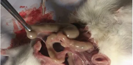
The degree of pathological adhesion was scaled as: 0: No adhesion, 1: Fat (Figure 1), 2: Fat and fibrosis, and 3: Fibrosis (Figure 2).
Figure 1. Fourth-degree intra-abdominal adhesion in a rat receiving normal saline (control group). Note the intraperitoneal adhesion between the colon and the mesothelium of small intestine. More than three thin adhesion bands, easily separated with no vessel or diffuse
adhesion bands with vessels
The obtained data were analyzed using Kruskal-Wallis and Mann-Whitney U evaluated using SPSS software (version 20, IBM Corporation, Armonk, NY) at the significant level of P < 0.050.
Figure 2. First-degree intra-abdominal adhesion in a rat receiving normal saline (local simvastatin group). Note the
intraperitoneal loose adhesion between the colon and the mesothelium of small intestine. One adhesion band, no
vessel, easily separated
Results
Table 1. Comparison of the degree of clinical adhesion between the study groups
The degree of clinical adhesion Local simvastatin
[n (%)]
Oral simvastatin [n (%)]
Placebo [n (%)]
0: No adhesion 5 (50) 6 (60) 0 (0)
1: One adhesion band, no vessel, easily separated 3 (30) 1 (10) 0 (0) 2: Two thin adhesion bands, no vessel, easily separated 2 (20) 3 (30) 0 (0) 3: Three thin adhesion bands, no vessel, easily separated 0 (0) 0 (0) 2 (20) 4: More than three thin adhesion bands, easily separated with no vessel
or diffuse adhesion bands with vessels
0 (0) 0 (0) 8 (80)
P < 0.001 between the both simvastatin groups and placebo group, P = 0.910 between the simvastatin groups
Clinical finding showed that in placebo group, 8 cases had grade 4 and 2 rats had grade 3 adhesions; while in intra-abdominal and oral simvastatin groups, 5 and 6 cases did not have adhesion, respectively. Based on the clinical findings, the formation of severe adhesions in the oral and topical simvastatin groups was significantly less than the placebo group (P < 0.001); but no significant difference was observed between the two simvastatin groups (P = 0.910) (Table 1).
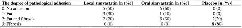
Based on the pathological findings, the formation of fibrosis in the oral and topical simvastatin groups was significantly less than the placebo group (P < 0.001); there were no significant difference between the two simvastatin groups (P = 0.907) (Table 2).
Discussion
The findings of this study showed that oral and local administration of simvastatin can reduce the formation of intra-abdominal adhesions following laparotomy. Arons et al., in an experimental study on rats, examined the effect of statins in the prevention of adhesion and its effect on wound healing. Oral and topical atorvastatin (30 mg/kg) were administrated into two groups of 15 rats. After 7 days, repeated laparotomy showed that only in the topical type, adhesion formation reduced (3). This is not consistent with the findings of this investigation in oral form. In this study, simvastatin with a dose of 40 mg/kg was used and in both oral and topical groups, decreased clinical and pathological adhesion after 14 days.
Hoşcan et al. study was conducted on 40 rats with fluvastatin. In the group of intra-abdominal fluvastatin with dosage of 10 mg/kg, all the rats died after one day and this group was excluded from the study. But in the oral group with the same dosage, reduction in adhesion was observed after two weeks (9). In our study, simvastatin was used and none of the rats died. Perhaps, because of the influence of different groups of different
classes of statins, all the rats which received topical fluvastatin, died Actachir et al. study. This is the reason that simvastatin was used in the present study.
Kucuk et al. by administering 0.57 mg/kg intraperitoneal simvastatin and measurement of tissue-type plasminogen activator (t-PA) in the abdomen in 30 rats, after a week, found that reducing adhesions in the abdominal cavity is associated with increased levels of t-PA (13). In the present study, the adhesion criteria was the clinical and histological observation and laboratory criteria were not used.
Jahovic et al. showed that the two drugs of simvastatin and fluvastatin were not similar in decreasing inflammatory parameters in colitis in two groups of 10 rats. Fluvastatin decreased the colon tissue collagen content and the level of tumor necrosis factor (TNF) more (14). In the case of simvastatin, the capacity of hydroxyl radicalism is more, while fluvastatin had higher antioxidants for taking the anti-peroxyl radicals (15). Fluvastatin, decreased the monocyte CD11b expression and endothelial adhesion, too (16).
In Hoşcan et al. study, the effect of fluvastatin on
wound healing was measured by wound
hydroxyproline content which did not differ with the control sample and the effect of simvastatin on the restoration of tissues was not studied (9). This is one of the issues that must be evaluated for assessment of the effects of drug on the anastomosis to determine whether topical or oral simvastatin impairs healing or not.
Yildiz et al. showed that oral simvastatin had no effect in reducing adhesions (8); their findings are not consistent with this study. In the Yildiz et al. study, simvastatin was given just 48 hours before the laparotomy, whereas in the present study, the medication was given 14 days prior to surgery (8). Another difference is that they performed another laparotomy 7 days later, while this was done after 14 days in the present study.
Table 2. Comparison of the degree of pathological adhesion between the study groups
The degree of pathological adhesion Local simvastatin [n (%)] Oral simvastatin [n (%)] Placebo [n (%)]
0: No adhesion 5 (50) 6 (60) 0 (0)
1: Fat 3 (30) 1 (10) 0 (0)
2: Fat and fibrosis 2 (20) 3 (30) 2(20)
3: Fibrosis 0 (0) 0 (0) 8 (80)
The adhesion criterion in that study was the activity of t-PA, which did not increase in oral form. Although the dose of simvastatin was similar to the present study, but perhaps with 48 hours of oral medication, the appropriate level of the drug was not created in mesothelial cells.
In a pilot study on human model, analyzing the data of 46 patients on oral simvastatin for 3-7 days before the surgery did not show any significant difference on the level of t-PA and plasminogen activator inhibitor-1 (PAI-1) compared to control group at the first day after the surgery (17). The shortness of sample size as well as the low duration of drug administration, were the limitations of that pilot study.
In another human study, oral administration of 40 mg of simvastatin from 3-7 days before the surgery to 4 days after it, tended to significant decrease of inflammatory factors (18).
Haslinger et al. in in-vitro study on mesothelial cells showed that simvastatin stimulated fibrinolytic activity with increasing levels of t-PA and decreasing PAI-1 (19). In another study, Bea et al. showed that simvastatin was the stimulant of fibrinolytic system in mesothelial cells by reducing TNF-a (20). We showed the effect of local simvastatin (30 mg/kg body weight) in reducing adhesion, with a significant difference compared to the control group in a sample of 32 rats with clinical and pathological criteria (6).
Conclusion
The present study showed that oral simvastatin similar to topical simvastatin was significantly effective in reducing adhesion compared to the control group in rats. More studies are also needed on human subjects to examine the effects on healing of anastomosis.
Conflict of Interests
Authors have no conflict of interests.
Acknowledgments
This article has been extracted from the thesis written by Mohammad Nejati (Registration No.: 589) Department of surgery, School of Medicine, Shahid Beheshti University of Medical Sciences, Tehran, Iran.
References
1. Brunicardi F, Andersen D, Billiar T, Dunn D, Hunter J, Matthews J, et al. Schwartz’s Principles of Surgery. 10th
ed. New York, NY: McGraw-Hill Education; 2015. p. 1146-52.
2. Scott FI, Osterman MT, Mahmoud NN, Lewis JD. Secular trends in small-bowel obstruction and adhesiolysis in the United States: 1988-2007. Am J Surg
2012; 204(3): 315-20.
3. Aarons CB, Cohen PA, Gower A, Reed KL, Leeman SE, Stucchi AF, et al. Statins (HMG-CoA reductase inhibitors) decrease postoperative adhesions by increasing peritoneal fibrinolytic activity. Ann Surg 2007; 245(2): 176-84.
4. Lalountas MA, Ballas KD, Skouras C, Asteriou C, Kontoulis T, Pissas D, et al. Preventing intraperitoneal adhesions with atorvastatin and sodium hyaluronate/carboxymethylcellulose: a comparative study in rats. Am J Surg 2010; 200(1): 118-23.
5. Schönbeck U, Libby P. Inflammation, immunity, and HMG-CoA reductase inhibitors: statins as antiinflammatory agents? Circulation 2004; 109(21 Suppl 1): II18-26.
6. Javaherzadeh M, Shekarchizadeh A, Kafaei M, Mirafshrieh A, Mosaffa N, Sabet B. Effects of Intraperitoneal Administration of Simvastatin in Prevention of Postoperative Intra-Abdominal Adhesion Formation in Animal Model of Rat. Bull Emerg Trauma 2016; 4(3): 156-60.
7. Menzies D, Ellis H. Intestinal obstruction from adhesions--how big is the problem? Ann R Coll Surg Engl 1990; 72(1): 60-3.
8. Yildiz MK, Okan I, Dursun N, Bas G, Alimoglu O, Kaya B, et al. Effect of orally administered simvastatin on prevention of postoperative adhesion in rats. Int J Clin Exp Med 2014; 7(2): 405-10.
9. Hoşcan Y1, Karabulut Z, Hoşcan MB, Arikan S, Oğüş E, Müderrisoğlu H. Oral fluvastatin reduces the severity of peritoneal adhesions in rats. Acta Chir Belg 2010; 110(1): 66-70.
10.National Research Council (US) Committee for the Update of the Guide for the Care and Use of Laboratory Animals. Guide for the Care and Use of Laboratory Animals. 8th ed. Washington, DC: National Academies Press (US); 2011.
11.Poehnert D, Abbas M, Kreipe HH, Klempnauer J, Winny M. High reproducibility of adhesion formation in rat with meso-stitch approximation of injured cecum and abdominal wall. Int J Med Sci 2015; 12(1): 1-6.
12.Lauder CI, Garcea G, Strickland A, Maddern GJ. Use of a modified chitosan-dextran gel to prevent peritoneal adhesions in a rat model. J Surg Res 2011; 171(2): 877-82. 13.Kucuk HF, Kaptanoglu L, Kurt N, Uzun H, Eser M,
Bingul S, et al. The role of simvastatin on postoperative peritoneal adhesion formation in an animal model. Eur Surg Res 2007; 39(2): 98-102.
14.Jahovic N, Gedik N, Ercan F, Sirvanci S, Yüksel M, Sener G, et al. Effects of statins on experimental colitis in normocholesterolemic rats. Scand J Gastroenterol 2006; 41(8): 954-62.
15.Franzoni F, Quiñones-Galvan A, Regoli F, Ferrannini E, Galetta F. A comparative study of the in vitro antioxidant activity of statins. Int J Cardiol 2003; 90(2-3): 317-21. 16.Weber C, Erl W, Weber KS, Weber PC. HMG-CoA
reductase inhibitors decrease CD11b expression and CD11b-dependent adhesion of monocytes to endothelium and reduce increased adhesiveness of monocytes isolated from patients with hypercholesterolemia. J Am Coll Cardiol 1997; 30(5): 1212-7.
activity after colorectal surgery-a pilot study. J Surg Res 2016; 205(1): 28-32.
18.Singh PP, Lemanu DP, Soop M, Bissett IP, HarrisonJ, Hill AG. Perioperative Simvastatin Therapy in Major Colorectal Surgery: A Prospective, Double-Blind Randomized Controlled Trial. J Am Coll Surg 2016; 223(2): 308-20.
19.Haslinger B, Goedde MF, Toet KH, Kooistra T. Simvastatin increases fibrinolytic activity in human
peritoneal mesothelial cells independent of cholesterol lowering. Kidney Int 2002; 62(5): 1611-9.