Iranian Rehabilitation Journal, Vol. 13, Issue 3, Autumn 2015
Original Article
Long Term Effects of Volar-Dorsal Wrist/ Hand Immobilization
Splint on Motor Components and Function of Stroke Patients
Malek Amini
Iran University of medical sciences
Aryan shamili
Tabriz University of medical sciences
Marzieh Pshmdarfard
*Occupational therapist, Zanjan, Iran
Objectives: This study was designed to determine the effects of Volar-Dorsal Wrist/Hand Immobilization
Splint on upper extremity motor components and function of stroke patients.
Methods: fourteen patients were participated in this study. The patients were selected based on the
inclusion and exclusion criteria, and were given the splint after a primary evaluation. They were re-evaluated after one, two and three months. In order to assess spasticity, the range of motion, and upper extremity function, the Modified Ashworth Scale (MAS), the goniometry, and fugl-meyer assessment were used respectively. The splints were used 2 hours during the day and the whole night in three months.
Results: The amount of the upper extremity function changes (P (v) =0.07) was not significant: and the
amount of the ranges of the motion changes (P (v) =0.02) were statistically significant. The changes in range of motion of other joints and spasticity were not significant (P (v)>.05).
Discussion: The results of the present study indicated that, volar-dorsal wrist/hand immobilization splints
can improve the AROM of metha-carpophalengeal joints. Although spasticity, the range of motion of other joints, and the upper extremity function went through some changes due to splinting (3month),these changes were not significant, which requires further research.
.Keywords: Stroke, Splint, Spasticity, Range of motion, Function
Submitted :27 June 2015 Accepted: 25 August 2015
Introduction
The most common problem that happens after stroke is spasticity or the increase in muscle tone (1). Because of the increase in muscle tone (the increase in the reflexive pattern and the reduction in the inhibitive control), patient will have a lot of problems in motor control. This problem leads to deformity, shortness, contractures, and problems in activities of daily living. Neurolysis, denervation with chemical materials, and serial casting are common methods utilized for the prevention of spasticity's problems (1). In this case, splinting is a usual method for the prevention of shortness and the increase in muscle tone; however, if the splints are used incorrectly, they bring about adverse results (2, 3). Gracies et al, concluded that short-term use of Lycra dynamic splints improves the position of wrist
range of motion exercises, static stretching, and splinting (6). However, in another study, Lannin concluded that casting is not a suitable method for reducing spasticity since casting leads to hand immobilization, which prevents the hand motion, and brings about weakness in hands (7). Therefore, owing to the fact that there is not any study on the
function of Volar-Dorsal Wrist/Hand Immobilization, This study was designed to determine the effects of Volar-Dorsal Wrist/Hand Immobilization Splint on upper extremity motor components and function of stroke patients (Figure 1).
Fig 1. posterior and anterior view of Volar-Dorsal Wrist/Hand Immobilization
Methods
This study was an interventional study and had a pretest-posttest design. This survey was undertaken in the occupational therapy clinics of rehabilitation faculty and Occupational Therapy clinic of Firoozgar hospital. In this study, 14 chronic stroke patients were selected via non-probability sampling. These patients were 6 women and 8 men with the average cognitive score of 27.55. The inclusion criteria of this study were: experiencing the stroke at least a year ago, aged between 20 to 64 years, gaining a score above 22 on the cognitive test of Mini Mental Status Exam (MMSE), having no other neurological diseases, having a maximum spasticity score of 3 on the Modified Ashworth scale (MAS), having the ability to sit at least 10 minutes independently on the edge of the bed and not receiving Botulinum toxin or similar splints while entering the study. Any of the patients who had the following conditions, were excluded from the research: having orthopedic lesions in the upper extremity, having any other neurological disease, or being absence in the post-test evaluation.
To construct the target splint, initially positive patterns were made in two sizes of men and women for their left/right hand. Then, all splints were fabricated based on the patterns. Splints immobilized the wrist, thumb, and fingers in 10 degrees of extension, in hyper-abduction, and in zero respectively, so the angles of the splint were the same for all patients. Before the initiation of the
interventions [splinting], active and passive ranges of the motion of the elbow, wrist, and meta carpophalengeal joints were gauged. Other outcome measures were the elbow and wrist spasticity and the upper extremity function. The method used to measure the range of motion and the spasticity was Goniometry and Modified Ashworth scale respectively. The upper extremity function was scored based on Fugl-Meyer assessment. The whole initial data were gathered and recorded.
Results
The results obtained for 11 patients at the end of the first month were as follows: the difference in the upper extremity functions before and after the splint
using was significant, and regarding the other variables, the changes caused by using the splint wasn’t significant (P (v)> ٠.05) (Table 1).
Table1. Quantitative analysis of variables at the end of first month
Changes score Standard deviation t P value Variables 9.09 42.06 -0.71 0.49
Active range of motion of elbow (degree)
2.27 6.84
-1.1 0.29
Passive range of motion of elbow( degree)
4.8 8.9
-1.78 0.1
Active range of motion of wrist(degree)
14 30.48
-1.53 0.15
Passive range of motion of wrist
3 6
-1.46 0.12
Active range of motion of methacarpophalngeal (degree)
4.09 9.17
-1.48 -.17
passive range of motion of methacarpophalngeal (degree)
2.09 2.89
-2.23 0.04
Function of hand based on fugel- Mayer scale
The differences between the spasticity of the elbow and that of the wrist with the p value of P (v)> 0.05 weren’t significant (Table 2) at the end of the second
month; however, due to the absence of most patients, the statistical analysis was not applied.
Table 2. changes in spasticity over 1 month
Mod Z P value Variables +1 -0.81 0.41 Elbow spasticity ( MAS)
2 -0.81
0.41 Wrist spasticity (MAS)
The results obtained for 9 patients at the end of the third month indicated that the change, caused by the use of splinting, in the active range of the motion of
meta carpophalangial joint was significant, however, regarding other variables, the change wasn’t significant (P (v)>0.05) (Table 3).
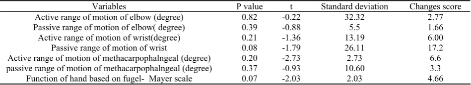
Table 3. Quantitative statistical analysis of variable for usage of the splint for 3 month
Changes score Standard deviation t P value Variables 2.77 32.32 -0.22 0.82
Active range of motion of elbow (degree)
1.66 5.5
-0.88 0.39
Passive range of motion of elbow( degree)
6.00 13.19
-1.36 0.21
Active range of motion of wrist(degree)
17.2 26.11
-1.79 0.08
Passive range of motion of wrist
6.6 2.73
-2.73 0.20
Active range of motion of methacarpophalngeal (degree)
3.3 10.60
-0.93 0.37
passive range of motion of methacarpophalngeal (degree)
4.66 2.03
-2.03 0.07
Function of hand based on fugel- Mayer scale
The changes in spasticity of the elbow and wrist, based on MAS, with the p value of P (v)> 0.5 wasn’t significant (Table 4).
Table 4. Spasticity changes over 3 month for usage of splint
Mod Z P value Variables +1 -2.64 0.08 Elbow spasticity (MAS)
2 -1.41
0.12 Wrist spasticity (MAS)
Discussion
In this study, based on MAS, the results showed that the elbow spasticity and the wrist spasticity after a month and after three months of using the splint did not display any significant differences. In this study, the sample size was low, and in order to obtain better results, it is required to use bigger sample sizes. Another reason for achieving these results
other scales such as MAS. In 2005, Pizzi et al observed that, after 3 months of the use of volar splint, the spasticity decreased, This decrease was significant by using electrophysiological parameters, while it wasn’t significant by using the MAS test (8). Pizzi et al observed that the spasticity of both flexor groups of the wrist and elbow declined, and the reason of this decrease was the inhibitation of biceps via stretching. By stretching, the biceps muscle of the afferent II of flexors was activated and led to the inhibitation of biceps. This mechanism was also reported by Ushiba et al, investigating on 17 chronic patients, (this mechanism happens in lower extremity too) (9). Although in our study, the statistical analysis did not yield significant changes, the chart changes and the percent changes indicated some decrease in the spasticity which may augment via increasing the sample size and using neurophysiological tests. In this study, it was found that the range of motion of the MP joints was changed, and this change was owing to the increase in the extension of the MP joint. This change was due to the structure of the splint which allowed the MP joint to be free from posterior and have active extension. In this study, it was found that the changes in the passive range of motion of the MP and the active and passive ranges of motion of the elbow weren't significant. But the chart changes and the coefficient of variations showed some changes in the range of motion. Pizzi et al concluded passive range of motion of the wrist increased in the range of extension movement (8). The passive range of motion of the wrist in the chronic patients increased significantly more than that in the sub-acute patients, however, the passive range of motion of the elbow in the sub-acute patients increased significantly. The passive range of motion of the wrist extension was changed more than that of the flexion, maybe owing to the decrease in the flexor Carpi radialis spasticity and other wrist flexors spasticity. The results obtained for the chronic patients were greater than those achieved for the sub-acute patients, perhaps because the hand fingers of the chronic patients were more flex than those of the sub-acute patients (9). In 2000, Gracies et al showed that by using the Lycra dynamic splint for a short time, the active range of motion of the finger flexion decreased maybe because of the hand fingers’ stretching (4). The results of the chart changes and coefficient of variations indicated some changes in the range of motion, which we declared that maybe future studies
would obtain the results achieved by other researchers. In this study, it was demonstrated that after using the splint for one month, 11 patients could significantly improve their range of motion, but after the usage of the splint for 3 months, the results weren’t significant (0.07 =P(v) ), so it can be declared that the sample size is one of the most important parameter for achieving better results, in this study it was also shown that the results obtained for 9 patients using the splint for one month wasn’t significant, which can be due to the insufficient sample size, not the duration of the usage, because in this duration, 11 patients could achieve better and significant results. Gracies et al. showed that, the upper extremity functions improved in some assignments (p (V)= 0.07 and the reasons were the better sensory perception in the hemiplegic part, the decrease in the spasticity, and the improvement of the range of motion in some parts because of the usage of the splint (4). Kinghorn et al. reported that the muscle tone of the patients decreased using an inhibitory weight bearing splint, and the changes of fine motors function in some assignments were variable, and the position of the hand and elbow improved (10). Katz et al observed that there was a significant correlation between the hand spasticity and its function (11). Although some clinical scales like MAS can be used in such studies, because of their low sensitivity, it's better to use neurophysiologic tests (8). In the present study, the spasticity, upper extremity function and range of motion improved but they were not statically significant using the clinical tests, which shows that more studies are required.
Conclusion
The results didn’t show any significant improvement in the spasticity, upper extremity function, and range of motion after the use of the splint for 3 months. But the changes in the active range of motion of the MP joint showed some improvement, which needs more study. On the whole, we suggest that it is better to undertake this study via applying case control, and using more sample size and other neuro-physiological tests.
Acknowledgment
Researchers are grateful to Dr. Hassanbeygi and other professors, as well as all of the patients and their families that have participated in this survey for their valuable cooperation.
References
1. Andrew J SM, Samir G. Reliability of Biomechanical
Spasticity Measurements at the Elbow of People post Stoke. Arch Phys Med Rehabil. 2005;86:1648-54.
2. D. J. Atlas of orthosis and Assistive Devises. 4.
Philadelphia. Mosby. Inc. /an affiliate of Elsevier Inc.
2008:191-200.
3. Lannin N HR. Is hand splinting effective for adults
following stroke? A systemic review and methodological critique of published research. Clin rehabil. 2003;17(8):807-16.
4. Gracies JM MJ, Renton R, Sandanam J, Gandevia SC,
Burke D. Short term effect of dynamic lycra splints on
upper limb in hemiplegic Patients. Arch Phys Med Rehabil. 2000;81:1547-55.
5. Gossman M SS. Review of length-associated change in
muscle. Physical Therapy. 1982;62:1799-808.
6. J H. the effects of casting on upper extremity motor
disorders after brain injury. Am J occup ther. 1994;48(3):219-24.
7. Lannin N NI, Cusick A. the effects of splinting on wrist
contracture after stroke. Stroke. 2007;38:111-6.
8. Pizzi A CG, Falsini C, Verdesca S, Grippo A. Application of
a Volar static splint in Post stroke spastisity of upper
limb. Arch phys Med Rehabil. 2005;86:1855-9.
9. Ushiba J MY, Komune Y, Muraoka Y, Chino N, Tomita Y.
Changes of reflex size in upper limbs using wrist splint
in hemiplegic Patients. Electromyography clin Neurophysiology 2004;44:175-82.
10.Kinghorn J RG. The effect of on Inhibitive weight – bearing
splint on tone and function: a single case study. Am J occup ther. 1996;50:807-14.
11.Katz T RG. Objective quantification of spastic hypertonia: