Published online 2016 May 23. Review Article
Metacarpal Neck Fractures: A Review of Surgical Indications and
Techniques
Eric M. Padegimas,
1,*William J. Warrender,
1Christopher M. Jones,
2and Asif M. Ilyas
21Department of Orthopedic Surgery, Thomas Jefferson University Hospital, Philadelphia, USA
2Department of Orthopedic Surgery, The Rothman Institute, Thomas Jefferson University Hospital, Philadelphia, USA
*Corresponding author: Eric M. Padegimas, Department of Orthopedic Surgery, Thomas Jefferson University Hospital, 1025 Walnut Street, Room 516 College, Philadelphia, USA. Tel: +1-2159551500; +1-8606040902, Fax: +1-2155030530, E-mail: padegimase@gmail.com
Received2015 September 04;Accepted2016 January 26.
Abstract
Context:Hand injuries are a common emergency department presentation. Metacarpal fractures account for 40% of all hand frac-tures and can be seen in the setting of low or high energy trauma. The most common injury pattern is a metacarpal neck fracture. In this study, the authors aim to review the surgical indications for metacarpal neck fractures, the fixation options available along with the risk and benefits of each.
Evidence Acquisition:Literature review of the different treatment modalities for metacarpal neck fractures. Review focuses on surgical indications and the risks and benefits of different operative techniques.
Results:The indications for surgery are based on the amount of dorsal angulation of the distal fragment. The ulnar digits can toler-ate gretoler-ater angulation as the radial digits more easily lose grip strength. The most widely utilized fixation techniques are pinning with k-wires, dorsal plating, or intramedullary fixation. There is currently no consensus on an optimal fixation technique as sur-gical management has been found to have a complication rate up to 36%. Plate and screw fixation demonstrated especially high complication rates.
Conclusions:Metacarpal neck fractures are a common injury in young and active patients that results in substantial missed time from work. While the surgical indications are well-described, there is no consensus on the optimal treatment modality because of high complication rates. Dorsal plating has higher complication rates than closed reduction and percutaneous pinning, but is necessary in comminuted fractures. The lack of an ideal fixation construct suggests that further study of the commonly utilized techniques as well as novel techniques is necessary.
Keywords:Metacarpal Bone, Neck Fractures, Hand, Fracture Fixation, Internal, Bone Wires
1. Context
Hand injuries are a common presentation to an emer-gency department (1). Fractures involving the hand ac-count for up to 28% of all fractures enac-countered (2). While the most common fractures of the hand are of the distal phalanx (3), metacarpal fractures alone account for about 40% of all hand fractures with 1.5 million injuries occur-ring annually (4,5). They are the second most common fracture to present to an orthopaedic surgeon (after distal radius fractures) with an incidence of 130.3 per 100,000 pa-tients annually (6). The economic burden is particularly high in these patients as they typically affect young and healthy men (7) leading to missed time from work. Isolated metacarpal injuries can result in up to 3 - 6 weeks of missed time with non-operative management alone (8-10). There-fore, optimizing treatment of these injuries may have sig-nificant benefits in both quality of life and return to work for these patients.
Among metacarpal fractures, the metacarpal neck is the most common site of injury (11). Metacarpal neck frac-tures typically result from a patient striking a solid surface with a clenched fist causing volar comminution and dor-sal apex angulation (12,13). The most frequent scenario encountered is a fracture of fifth metacarpal neck other-wise known as the “boxer’s fracture.” Metacarpal neck frac-tures provide a particular dilemma to orthopaedic sur-geons as there is no consensus for their management (14,
15). Non-operative management typically delivers an ac-ceptable outcome, but can be associated with a poor cos-metic result, weakness, extensor lag, and palmar promi-nence (16). Conversely, with surgical intervention there have been reports of high complication rates, up to 36% (17-19). The controversy surrounding the management of metacarpal neck fractures, prompted review of the treat-ment options available.
2. Evidence Acquisition
This article will provide an evidence-based review of the presentation and different surgical management op-tions for metacarpal neck fractures. This is intended to clarify the surgical options for these injuries with the spe-cific risks and benefits for each technique. We will re-view the typical presentation of metacarpal neck fractures, radiographic findings, surgical indications, and fixation techniques available to the surgeon.
PubMed was used as an electronic search engine in this review. The search terms used to identify studies were “metacarpal fractures” and “metacarpal neck fractures”. All studies were scrutinized to reflect a wide spectrum of treatment options for metacarpal neck fractures. Addi-tionally, two widely circulated orthopaedic textbooks were utilized for general information on the presentation, ra-diographic features, surgical indications and fixation tech-niques.
3. Results
3.1. History and Physical Examination
A thorough history and physical exam must be done for evaluation of metacarpal neck fractures. Key aspects of the patient history include age, hand dominance and occu-pation as they can significantly affect management. Mech-anism of injury is an important variable for early manage-ment and outcome of metacarpal neck fractures. The clas-sic presentation of a metacarpal neck fracture is a patient that strikes a solid surface with a closed fist (12,13). How-ever, these injuries can also occur in multiply injured poly-trauma patients. Metacarpal neck fractures can be subtle, as can most hand fractures, in the setting of a multiply injured patient which can lead to delays in diagnosis and treatment (20).
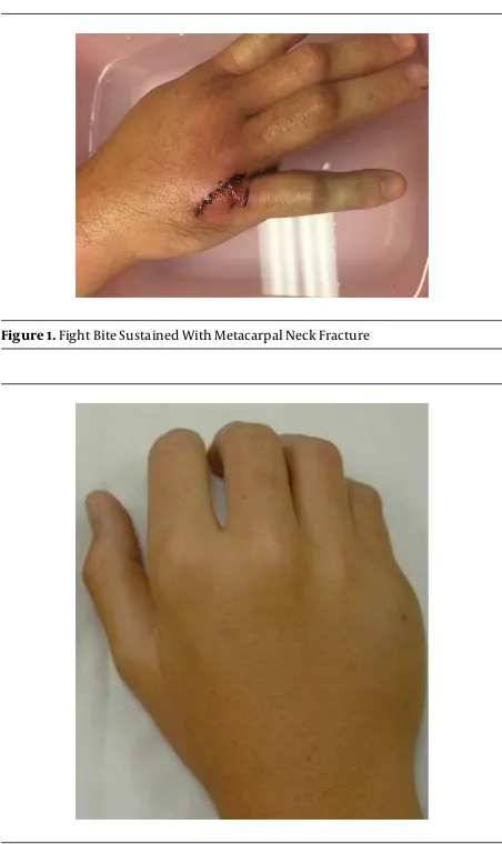
The first component of physical examination is care-ful inspection of the skin and soft tissue. The clinician must be careful to fully examine for lacerations to rule out an open fracture. Wounds over the metacarpophalangeal (MCP) joints should be considered to communicate with the fracture or the MCP joint until proven otherwise ( Fig-ure 1). For less obvious injuries, localized swelling, ecchy-mosis and tenderness to palpation can be helpful isolating the fracture. MCP joint depression or loss of normal joint contour may be present (Figure 2). If there is a soft tissue defect, the patient should receive both tetanus prophylaxis as well as antibiotic prophylaxis that covers common oral flora (amoxicillin/clavulanic acid), presuming the lacera-tion was from a tooth sustained from punching someone in the mouth, also known as a “fight bite” (21,22).
Figure 1.Fight Bite Sustained With Metacarpal Neck Fracture
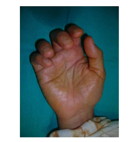
Figure 2.Note the Altered Contour and Deformity of the Fifth Small Finger ray Along the Border due to an Underlying Angulated Metacarpal Neck Fracture
Additionally, the specifics of the deformity can be noted. Shortening of the ray can be assessed by compar-ison to the contralateral hand. The rotational alignment should also be evaluated. A rotational deformity may not be appreciated with the fingers in extension, however test-ing them in flexion may reveal subtle deformity. Normally in flexion, the digits will point to the distal pole of the scaphoid, but in metacarpal fractures with rotational de-formity, there will be some degree of external rotation of the affected finger (Figure 3). If the patient is unable to ac-tively flex his or her fingers due to pain, this deformity will also be present with passive flexion.
dor-Figure 3.Note the Rotational Deformity due to an Underlying Spiral Metacarpal Fracture
Rotational deformity is most accentuated with active or passive finger flexion, while angular deformity is most prominent with finger extension.
sal sensory branches of the ulnar or radial nerves. Sim-ilarly, volar wounds are commonly associated with digi-tal nerve injury. Motor function is checked in the finger flexors and extensors. Extensor tendons can be lacerated and retracted especially when dorsal wounds are present. “Pseudo-clawing” (hyperextension) of the ray may result after repeated attempts to extend the finger if the angula-tion of the metacarpal neck fracture is advanced (23,24). Evaluation of finger extension and grip strength in the sub-acute or chronic setting (not sub-acutely) should also be noted.
3.2. Imaging
Imaging and subsequent diagnosis of metacarpal neck fractures can be readily achieved with plain radiographs. Anteroposterior, lateral and oblique radiographs of the af-fected digit should preferably be obtained. A “skyline” view of the metacarpophalangeal joint has been described and can show a vertical impaction defect in the metacarpal head which is typically sustained as a result of tooth pen-etration through the extensor hood into the joint (25). In-jured digits should be viewed individually to minimize overlap of other digits. Additionally, every attempt at a perfect lateral should be made as it has been shown that the obliquity of the hand at the time of radiograph can effect the angulation measurement (26). Advanced imag-ing is typically not necessary unless there is concern for intra-articular extension of the fracture to the metacarpal
head. Articular involvement would best be evaluated by computed tomography (CT).
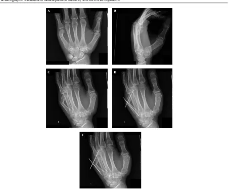
Imaging should be surveyed for comminution, dis-placement, angulation, shortening and fractures to sur-rounding bones of the hand and wrist (Figure 4A - C). There are two different techniques for measuring angula-tion on the lateral radiograph. In one, the angle is mea-sured between the line along the longitudinal axis of the metacarpal shaft (medullary canal) and the line from the center of the metacarpal head to the fracture site (Figure 4D). In the other, the angle is measured after drawing two lines tangential to the dorsal cortices of the proximal and distal fragments using the intersecting angle between the two lines as the measure of the fracture angulation (this method may underestimate angulation) (Figure 4E). Nor-mal angulation of the 5th metacarpal neck is fifteen de-grees using the first of the aforementioned methods (12). The measurement of fracture angulation of small finger metacarpal neck fractures is subject to a high degree of inter- and intraobserver variability (27). Reliability and va-lidity can be adequate only when the degree of small fin-ger metacarpal neck fracture angulation is measured af-ter drawing lines on laaf-teral radiographs. Oblique radio-graph measurements consistently produce higher read-ings (28). Finally, one must account for the fact that the normal metacarpal head-to-neck angle is 15 degrees so any fracture angulation is equal to the measured angle on the lateral radiograph minus 15 degrees.
3.3. Surgical Indications
There are a number of surgical indications for metacarpal neck fractures. Firstly, open fractures are an absolute indication for irrigation, debridement, and fracture reduction (29). Secondly, if there is extension of the fracture into the metacarpal head with greater than 1 mm of displacement or any intra-articular fracture fragment blocking joint motion, surgery is recommended (29). Finally, and most commonly, the degree of volar an-gulation and displacement is used to determine operative versus nonoperative management. If volar angulation is not corrected, patients can have a prominent dorsal deformity, decreased grip strength, a prominent palmar metacarpal head and pseudo clawing (Figure 2) (23,24). The degrees of volar angulation for each digit that may be considered for surgical fixation are (29):
- 150 for the index finger - 250 for the long finger - 350 for the ring finger - 450 for the small finger
Figure 4.Radiographic Assessment of Metacarpal Neck Fractures, Note the Dorsal Angulation
A, B and C, PA, lateral, and oblique X-rays demonstrating displaced, dorsally angulated fifth metacarpal neck fracture; D, measurement of dorsal angulation between the line along the longitudinal axis of the metacarpal shaft (medullary canal) and the line from the center of the metacarpal head to the fracture site; E, measurement of dorsal angulation between the two lines tangential to the dorsal cortices of the proximal and distal fragments using the intersecting angle between the two lines as the measure of the fracture angulation.
of angulation increases for the more ulnar digits owing to the compensatory movement of the 4th and 5th car-pometacarpal joints. Less angulation of the second and third metacarpals can be tolerated due to the fact that their CMC joints are much less mobile. When considering oper-ative indications, it is important to note that in the small finger, greater than 30 degrees of angulation is associated with a decrease in flexor digiti minimi grip strength and range of motion (30).
3.4. Surgical Technique
3.4.1. Closed Reduction and Pinning
Metacarpal neck fractures are reduced by maximal flex-ion of the metcarpophalangeal (MCP) joint with an axial
load directed dorsally across the flexed proximal interpha-langeal (PIP) joint. The fracture site can then be pinned by three different techniques.
3.4.1.1. Cross Pinning
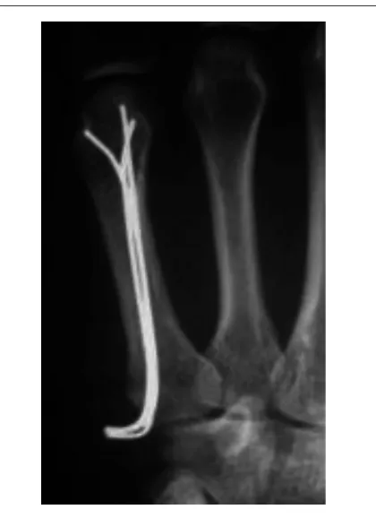
This technique utilizes 0.9 or 1.1 mm K-wires placed dis-tal to the fracture site at the flare of the metacarpal neck or in the collateral recess of the metacarpal head. The pins are advanced in a retrograde fashion bicortically without crossing at the fracture site to control rotation (Figure 5).
3.4.1.2. Crucifix Pinning
Figure 5.Radiographic Assessment of Metacarpal Neck Fracture After Reduction and Cross-pinning
A and B, PA and lateral X-rays demonstrating cross-pinning construct showing reduction of the metacarpal neck fracture.
canal, and then placing a 0.045 K-wire laterally through the metacarpal head and advancing it bicortically into the ad-jacent metacarpal head to hold the rotation (Figure 6).
Figure 6.Crucifix Pinning Showing Reduction of the Metacarpal Neck Fracture by Two-crossed Pins
3.4.1.3. Bouquet Pinning
This technique utilizes a “bouquet” of typically three K-wires placed for intramedullary fixation in antegrade fash-ion. These have a dorsal bend to preferentially support the dorsal aspect of the metacarpal head to hold reduction (Figure 7) (31,32).
Benefits of the technique include: - Minimally invasive
- Good post-operative range of motion (97.7% com-pared to contralateral side) (33)
Complications/Drawbacks of the technique include: - Less stable with extensive comminution (29) - Pinsite infection (34)
- Nonunion/malunion (35)
- Tendon adhesion and joint contracture from pro-longed immobilization
- Need to protect exposed pins
3.4.2. Plate and Screw Fixation
Figure 7.Bouquet Pinning Showing Reduction of the Metacarpal Neck Fracture by Three Antegrade Pins
placing the distal screws obliquely in different planes or by utilizing unicortical locking screws (Figure 8) (29).
Figure 8.Plate and Screw Fixation of Metacarpal Neck Fracture
Benefits of the technique:
- Most biomechanically stable construct (36)
- Can be used to treat multiple concurrent metacarpal neck fractures (29)
- Can be used when significant comminution precludes closed reduction and percutaneous pinning (29)
Complications of the technique:
- Higher rates of stiffness/poor post-operative range of motion (58.7% compared to contralateral side) (33,37)
- Metacarpal head avascular necrosis (33) - Extensor tendon injury (17)
- Nonunion/malunion (37) - Hardware irritation
3.4.3. Intramedullary Fixation
Distal fragment reduced by maximal flexion of the MCP joint with an axial load directed dorsally across the flexed PIP joint. Intramedullary fixation of the fracture can be accomplished with antegrade placement of pre-fabricated intramedullary nails (Figure 9) (29) or retrograde fixation with a headless compression screw placed through the metacarpal head (Figure 10).
Benefits of the techniques:
- Improved range of motion compared to k-wire cross-pinning (38)
- Lower incidence of shortening compared to k-wire cross-pinning (38)
Complications/Drawbacks of the techniques: - Nonunion/malunion (35)
- Limited rotational stability
4. Conclusions
Figure 9.Intramedullary Fixation of Metacarpal Fractures With Commercially Available Nails Placed Antegrade
Later removal is required, (Courtesy Jorge Orbay, MD).
Figure 10.Fluoroscopic Assessment of Metacarpal Neck Fracture Treated by Reduction and Intramedullary Fixation With a Headless Compression Screw
References
1. Hile D, Hile L. The emergent evaluation and treatment of hand injuries. Emerg Med Clin North Am. 2015;33(2):397–408. doi:
10.1016/j.emc.2014.12.009. [PubMed:25892728].
2. Meals C, Meals R. Hand fractures: a review of current treatment strate-gies.J Hand Surg Am.2013;38(5):1021–31. doi:10.1016/j.jhsa.2013.02.017. [PubMed:23618458] quiz 1031.
3. Day C, Stern PJ. Fractures of the Metacarpals and Phalanges. 6th ed. Philadelphia: Elsevier Churchill Livingstone; 2011.
4. Ashkenaze DM, Ruby LK. Metacarpal fractures and dislocations. Or-thop Clin North Am.1992;23(1):19–33. [PubMed:1729666].
5. Diao E. Metacarpal fixation.Hand Clin. 1997;13(4):557–71. [PubMed:
9403293].
6. Court-Brown CM, Caesar B. Epidemiology of adult fractures: A review. Injury. 2006;37(8):691–7. doi: 10.1016/j.injury.2006.04.130. [PubMed:
16814787].
7. Nakashian MN, Pointer L, Owens BD, Wolf JM. Incidence of metacarpal fractures in the US population.Hand (N Y). 2012;7(4):426–30. doi:
10.1007/s11552-012-9442-0. [PubMed:24294164].
8. Statius Muller MG, Poolman RW, van Hoogstraten MJ, Steller EP. Im-mediate mobilization gives good results in boxer’s fractures with volar angulation up to 70 degrees: a prospective randomized trial comparing immediate mobilization with cast immobilization.Arch Orthop Trauma Surg. 2003;123(10):534–7. doi:
10.1007/s00402-003-0580-2. [PubMed:14639483].
9. Harding IJ, Parry D, Barrington RL. The use of a moulded metacarpal brace versus neighbour strapping for fractures of the little finger metacarpal neck. J Hand Surg Br. 2001;26(3):261–3. doi:
10.1054/jhsb.2000.0509. [PubMed:11386781].
10. van Aaken J, Kampfen S, Berli M, Fritschy D, Della Santa D, Fusetti C. Outcome of boxer’s fractures treated by a soft wrap and buddy taping: a prospective study.Hand (N Y). 2007;2(4):212–7. doi:
10.1007/s11552-007-9054-2. [PubMed:18780055].
11. Jupiter JB, Goldfarb CA, Nagy L, Boyer MI. Posttraumatic reconstruc-tion in the hand.J Bone Joint Surg Am. 2007;89(2):428–35. [PubMed:
17326313].
12. Henry M. Hand fractures and dislocations. Philadelphia: Lippincott Williams & Wilkins; 2010.
13. Stanton JS, Dias JJ, Burke FD. Fractures of the tubular bones of the hand. J Hand Surg Eur Vol. 2007;32(6):626–36. doi:
10.1016/j.jhse.2007.06.017. [PubMed:17993422].
14. Tosti R, Ilyas AM, Mellema JJ, Guitton TG, Ring D, Science of Vari-ation G. Interobserver variability in the treatment of little finger metacarpal neck fractures.J Hand Surg Am. 2014;39(9):1722–7. doi:
10.1016/j.jhsa.2014.05.023. [PubMed:25034789].
15. Beredjiklian PK. Small finger metacarpal neck fractures. J Hand Surg Am.2009;34(8):1524–6. doi:10.1016/j.jhsa.2009.06.015. [PubMed:
19729252].
16. Strub B, Schindele S, Sonderegger J, Sproedt J, von Campe A, Gruenert JG. Intramedullary splinting or conservative treatment for displaced fractures of the little finger metacarpal neck? A prospective study. J Hand Surg Eur Vol. 2010;35(9):725–9. doi: 10.1177/1753193410377845. [PubMed:20659966].
17. Page SM, Stern PJ. Complications and range of motion following plate fixation of metacarpal and phalangeal fractures.J Hand Surg Am. 1998;23(5):827–32. doi:10.1016/S0363-5023(98)80157-3. [PubMed:
9763256].
18. Kollitz KM, Hammert WC, Vedder NB, Huang JI. Metacarpal frac-tures: treatment and complications.Hand (N Y). 2014;9(1):16–23. doi:
10.1007/s11552-013-9562-1. [PubMed:24570632].
19. Creighton Jr JJ, Steichen JB. Complications in phalangeal and metacarpal fracture management. Results of extensor tenolysis.
Hand Clin.1994;10(1):111–6. [PubMed:8188771].
20. Adkinson JM, Shafqat MS, Eid SM, Miles MG. Delayed diagnosis of hand injuries in polytrauma patients.Ann Plast Surg.2012;69(4):442– 5. doi:10.1097/SAP.0b013e31824b26e7. [PubMed:22868310]. 21. Perron AD, Miller MD, Brady WJ. Orthopedic pitfalls in the ED: fight
bite.Am J Emerg Med.2002;20(2):114–7. [PubMed:11880877]. 22. Shoji K, Cavanaugh Z, Rodner CM. Acute fight bite.J Hand Surg Am.
2013;38(8):1612–4. doi:10.1016/j.jhsa.2013.03.002. [PubMed:23660199]. 23. Kamath JB, Harshvardhan , Naik DM, Bansal A. Current concepts in managing fractures of metacarpal and phalangess.Indian J Plast Surg. 2011;44(2):203–11. doi:10.4103/0970-0358.85341. [PubMed:22022030]. 24. Thurston AJ. Pivot osteotomy for the correction of malunion of
metacarpal neck fractures.J Hand Surg Br.1992;17(5):580–2. [PubMed:
1479255].
25. Eyres KS, Allen TR. Skyline view of the metacarpal head in the as-sessment of human fight-bite injuries.J Hand Surg Br.1993;18(1):43–4. [PubMed:8436860].
26. Lane CS, Kennedy JF, Kuschner SH. The reverse oblique x-ray film: metacarpal fractures revealed.J Hand Surg Am. 1992;17(3):504–6. [PubMed:1613232].
27. Leung YL, Beredjiklian PK, Monaghan BA, Bozentka DJ. Radiographic assessment of small finger metacarpal neck fractures.J Hand Surg Am. 2002;27(3):443–8. [PubMed:12015718].
28. Lamraski G, Monsaert A, De Maeseneer M, Haentjens P. Reliability and validity of plain radiographs to assess angulation of small fin-ger metacarpal neck fractures: human cadaveric study.J Orthop Res. 2006;24(1):37–45. doi:10.1002/jor.20025. [PubMed:16419967]. 29. Leinberry C, Ukomadu U, Ilyas A. Metacarpal Fractures and
Car-pometacarpal Fracture-Dislocations. 1st ed. New Delhi, India: Jaypee Brothers Medical Publishers Ltd; 2013.
30. Ali A, Hamman J, Mass DP. The biomechanical effects of angu-lated boxer’s fractures.J Hand Surg Am.1999;24(4):835–44. [PubMed:
10447177].
31. Foucher G, Chemorin C, Sibilly A. [A new technic of osteosynthesis in fractures of the distal 3d of the 5th metacarpus].Nouv Presse Med. 1976;5(17):1139–40. [PubMed:934828].
32. Winter M, Balaguer T, Bessiere C, Carles M, Lebreton E. Surgical treatment of the boxer’s fracture: transverse pinning versus in-tramedullary pinning.J Hand Surg Eur Vol. 2007;32(6):709–13. doi:
10.1016/j.jhse.2007.07.011. [PubMed:17993437].
33. Facca S, Ramdhian R, Pelissier A, Diaconu M, Liverneaux P. Fifth metacarpal neck fracture fixation: Locking plate ver-sus K-wire?. Orthop Traumatol Surg Res. 2010;96(5):506–12. doi:
10.1016/j.otsr.2010.02.009. [PubMed:20580630].
34. Botte MJ, Davis JL, Rose BA, von Schroeder HP, Gellman H, Zinberg EM, et al. Complications of smooth pin fixation of fractures and disloca-tions in the hand and wrist.Clin Orthop Relat Res. 1992(276):194–201. [PubMed:1537152].
35. Balaram AK, Bednar MS. Complications after the fractures of metacarpal and phalanges. Hand Clin. 2010;26(2):169–77. doi:
10.1016/j.hcl.2010.01.005. [PubMed:20494743].
36. Jones WW. Biomechanics of small bone fixation.Clin Orthop Relat Res. 1987(214):11–8. [PubMed:3539435].
37. Fusetti C, Meyer H, Borisch N, Stern R, Santa DD, Papaloizos M. Complications of plate fixation in metacarpal fractures.J Trauma. 2002;52(3):535–9. [PubMed:11901331].