2017
FCA Part II
Anaesthetic Refresher
Course
Department of Anaesthesia
University of the
Contents
Contents
South Afr J Anaesth Analg • 2017 • Volume 23 • Number 2 • Supplement 1
• Peribulbar (extraconal) eye block and its complications
KK Purbhoo ... S3 • Molar pregnancies with a focus on the thyroid
F Bham ... S7 • Cerebral perfusion monitoring during non-neurosurgical procedures:
The practicalities of Near Infrared Spectroscopy (NIRS)/Bispectral Index (BIS) M Mphomane ...S12 • Anaesthesia for related living donor liver transplantation
M Hatchett ...S15 • The bleeding parturient: anaesthetic perspective
D Nel ...S18 • Carcinoid syndrome
MA Eagar ...S26 • Analgesia for thoracic surgery
NT Hlongwane-Gukuta ...S31 • Extracorporeal Membrane Oxygenation (ECMO)
A Alli ...S36 • Anaesthetic considerations for inhaled and ingested foreign bodies in children:
What? Where? When?
AZ Bhettay ...S42 • Anaesthesia for audiology brainstem response testing in children with autism
spectrum disorders
MR Correia ...S46 • Anaesthesia for craniosynotosis surgery
D Lines ...S53 • Anaesthesia for thoracoscopy in paediatric patients
S Spijkerman ...S59 • Mediastinoscopy in paediatric patients
S Spijkerman ...S65 • The obese parturient
E Mostert ...S70 • Laryngospasm in anaesthesia
S Spijkerman ...S74 • Epilepsy and anaesthesia
C Hosking ...S81 • Neonatal eye surgery
KL Borrill ...S84 • Conventional cardiopulmonary bypass and mini-cardiopulmonary bypass
MEA Kemp ...S87 • Posterior of fossa surgery
MMA Mashinini ...S92
FCA Part II
Anaesthetic
Refresher
Course 2017
2017
FCA Part II
Anaesthetic Refresher
Course
Department of Anaesthesia
University of the
FCA Part II
Anaesthetic
Refresher
Course 2017
Contents
Contents
© 2017 SASA. Unless otherwise stated, opinions expressed in the editorial columns of SAJAA should not be taken as reflecting official SASA policy. The appearance of advertising in this publication does not denote a guarantee or an endorsement by SASA of the
• The anaesthetist’s role in the endoscopy suite
C Quan ...S98 • Inadvertent burns in the theatre
EE Oosthuizen ...S103 • The postoperative management of a supraventricular tachyarrhythmia
HJ Moutlana ...S106 • A study of perioperative deaths in Charlotte Maxeke Johannesburg Academic
Hospital, a South African level four central hospital
C Lundgren, P Cleaton-Jones ...S110 • Anaesthetic considerations for children with malignancies
C Lee ...S114 • Can you sedate a child for a dental procedure?
BM Gardner ...S119 • Anaesthesia in pregnancy for non-obstetric surgery
NR Madima ...S123 • Anaesthesia for cleft lip and palate surgery
A Oosthuizen ...S128 • New concepts in anaesthetic emergence
AW Moodley ...S134 • Three-stage oesophagectomy
ME Sethusa ...S138 • Managing an acute pulmonary embolism on the table
E Welch ...S140 • Clinical use of nerve stimulators
E Welch ...S143 • Anaesthesia for a patient with Achondroplasia presenting for Caesarean section
T Kleyenstuber ...S147 • Anaesthetic management in paediatric burns
© 2017 The Author(s) FCA 2 REFRESHER COURSE Open Access article distributed under the terms of the
Creative Commons License [CC BY-NC-ND 4.0] http://creativecommons.org/licenses/by-nc-nd/4.0
Peribulbar (extraconal) eye block and its complications
K K PurbhooDepartment of Anaesthesia, Helen Joseph Hospital, University of the Witwatersrand Correspondence to: kashmirapurbhoo@gmail.com
Local anaesthesia is preferred over general anaesthesia by many ophthalmic surgeons with the main reasons being lower perioperative morbidity with quicker patient recovery and fewer complications.1–3
Anaesthetists have become increasingly involved in eye blocks that were previously performed by surgeons. Good anaesthesia is essential for the performance of safe intraocular surgery and may contribute to the success or failure of the surgery.4,5
Goals of anaesthetic care during elective eye surgery
1. Anaesthesia of the globe, lids and adnexa ensuring pain-freesurgery
2. Akinesia of the globe and lids facilitating the surgical procedure, and
3. Rapid recovery with adequate postoperative analgesia4,5
Anaesthetic techniques available:
Type of anaesthesia chosen
This would depend on: 1. The type of surgery 2. The surgeon’s preference 3. Patient’s suitability, and 4. Patient cooperation
Preoperative considerations
A routine preoperative assessment has to be done to ensure that co-morbid conditions are reasonably well controlled.
A focused medical history needs to be taken and should include a history determining if the patient is a suitable candidate for same day surgery.6 A history of anticoagulant or antithrombotic therapies is vital. Patients having cataract surgery that are at a high risk of clotting and embolic complications due to cardiac or vascular pathology should continue therapeutic doses of aspirin and warfarin throughout the perioperative period.7,8 There is,
Anaesthesia for eye surgery
Peribulbar (extraconal) block
Topical
Local anaesthesia
Retrobulbar (intraconal) block
Regional
General anaesthesia
Southern African Journal of Anaesthesia and Analgesia 2017; 23(2)(Supplement 1) 4
however, insufficient data available on dual antiplatelet therapy for stents and eye surgery. Eye surgery, if possible, should be postponed until after the minimum period recommended for daily administration of dual antiplatelet therapy.8,9
A full physical examination needs to be done. It is important to determine if the patient will be able to lie supine comfortably under drapes. Conditions that preclude this, e.g. congestive cardiac failure, claustrophobia or back pain need to be excluded. The patient’s communication and cooperation skills also need to be established. During regional anaesthesia you have to be able to communicate with the patient and the patient has to be able to follow commands such as avoiding all movement.
Monitoring
Standard ASA monitoring, which includes electrocardiogram, pulse oximetry and non-invasive blood pressure monitoring together with capnography is essential. Patients need to be monitored because they can get an occulocardiac reflex or an accidental injection into a blood vessel or the brain.10
Supplemental oxygen should be given to decrease the risk of hypoxaemia during sedation and to have a fresh gas flow to prevent the patient rebreathing clinically significant amounts of carbon dioxide that may occur as a result of completely covering the face.
Vacuum suctioning under the drapes can be used to lower the oxygen concentration and remove expired carbon dioxide.11,12 Before the block is performed one needs to establish good intravenous access.
Sedation and analgesia
Short-acting agents can be given immediately before the block to decrease or eliminate pain of needle insertion and injection of local anaesthetic. Minimal sedation is preferable during the procedure because the surgery requires an awake and cooperative patient. Excess sedation can lead to a restless, confused or unresponsive patient who may have respiratory depression with airway obstruction. This can pose a significant intraoperative challenge. Even minor movement (highly magnified under a microscope) may result in severe eye injury.4 If the patient has pain intraoperatively, a supplemental block with local anaesthesia should be given. Inadequate analgesia should not be substituted with heavy sedation.13
Anatomy of the eye
The orbit is pyramidal in shape with its base at the orbital opening and its apex pointing to the optic foramen. The orbit is occupied by the globe, its muscle cone, loose connective tissue and fat. The globe lies in the anterior part of the orbit and sits high and lateral, therefore it is nearer the roof than the floor and nearer the lateral than medial wall (important for needle insertion points). There are four recti muscles and two oblique muscles that control eye movement. The recti muscles form the muscle ‘cone’ around the globe. The muscle cone encloses the sensory and motor nerves, ciliary ganglion, optic nerve, ophthalmic artery and vein.14 Retrobulbar anaesthesia is injected in the intraconal compartment, whereas peribulbar anaesthesia is injected in the
extraconal compartment and hence considered safer since it avoids potential damage to the intraconal structures.
The motor supply to the eye is from the oculomotor nerve (CN III), the trochlear nerve (CN IV) and the abducens nerve (CN VI). CN III supplies all the extraocular muscles except superior oblique and lateral rectus muscles. CN III also supplies levator palpebrae superioris. CN IV supplies the superior oblique muscle and CN VI supplies the lateral rectus muscle. CN IV lies outside the muscle cone and hence is not usually blocked.5
The sensory supply is from the trigeminal nerve (CN V). CN V is divided into the ophthalmic branch which is further divided into nasociliary, lacrimal and frontal branches .The nasociliary branch supplies the cornea, perilimbal conjunctiva and the superonasal quadrant of the bulbar conjunctiva. The rest of the conjunctiva is supplied by the lacrimal, infraorbital and frontal nerves.5 The ciliary ganglion lying within the cone relays sensory fibres from the globe to the ophthalmic division of CN V. It receives a parasympathetic branch from CN III and sympathetic fibres from the carotid plexus.14
Peribulbar (extraconal) block
The peribulbar block (PBB) was first described in 1986 and was developed as a safer alternative to the retrobulbar (intraconal) block (RBB) for providing anaesthesia and akinesia of the eye. Similar clinical efficacy of the PBB and the RBB, as evidenced by similar spreading of local anaesthetic injected, but with a higher risk of complications associated with introducing the needle into the cone, makes the PBB a better alternative to the RBB.15 With PBB the needle is placed less deeply and at a different angle compared to the RBB. The local anaesthetic (larger volume than with RBB) is injected outside the muscle cone and spreads by way of diffusion to block the orbital nerves including the ciliary ganglion, ciliary nerves, CNs II, III, VI and even CN IV. The modifications with needle placement make the PBB less likely to result in perforation of the globe posteriorly, injury to the optic nerve or injection into the brain with resultant brainstem anaesthesia.15,16
Technique
The classic technique involves two injections. An initial inferolateral injection supplemented with a medial injection. A single inferolateral injection is often adequate for anaesthesia but may not be predictable for akinesia.5,17
After establishing intravenous access, connecting monitors and giving minimal sedation, the patient is asked to lie supine and look straight ahead.
The first injection is inferior and temporal. The junction of the medial two-thirds and lateral one-third of the inferior orbital rim is palpated, where a groove is felt at the junction of the maxilla and zygoma. Slightly lateral to this point a 2.5 cm needle is introduced through the skin and passed slowly backwards perpendicular to all planes. If the needle tip contacts bone it is redirected slightly superomedially to follow the orbital floor once more. The needle is advanced until its tip is about level with the posterior pole of the globe (until the hub reaches the plane of the iris). The globe should be observed closely for any signs of rotation during insertion, indicating scleral contact. After aspiration 6–10 mls of local anaesthetic is injected slowly.5,14 A larger volume is needed to spread into the whole corpus adiposum of the orbit including the intraconal space. The larger volume also allows anterior spread of the local anaesthetic to the lids to provide a block of the orbicularis muscles and to avoid the need for an additional lid block.5 PBB produces more reliable akinesia of the orbicularis oculi as compared to a RBB.22,23 If the globe becomes tense or proptosed or if the upper eyelid falls during injection then you should stop immediately as this may indicate a RBB which requires a smaller volume of local anaesthesia.14
Compression to the eye can be done after injection to lower intraocular pressure, which increases after injection. Applying a pressure of 30 mmHg for 5–10 minutes is usually sufficient. Assess the block after 5–10 minutes and if a greater degree of akinesia or additional anaesthesia is required a second injection should be performed. In the classic technique the second injection is superior and nasal between the medial one-third and the lateral two-thirds of the orbital roof edge.5 It is important to remember that at the superior nasal site, the distance between the orbital roof and the globe is reduced, theoretically increasing the risk of globe perforation. Additionally the superior oblique muscle can be injured.14
If both medial and lateral injections are planned, the volume of the solution is 4–5 mls for each. It is important to remember that the first injection may increase the risk of complications associated with consecutive injections, as it may distort the anatomy.24
Alternate techniques
Numerous alternative techniques and different sites for needle insertion for a PBB have been described.5 The most common alternative is the medial canthus peribulbar block. Here the needle is introduced at the medial junction of the lids, nasal to the lacrimal caruncle, in a strictly posterior direction to a depth of 15 mm or less. At this site the space between the orbital wall and the globe is similar in size to that of the inferolateral approach and is free from blood vessels. This site can be used as the second injection point when the first inferolateral injection needs to be supplemented.5,12
Complications
The main reason for complications is needle misplacement.
1. Globe perforation
This can occur as a result of direct trauma. More common in patients with myopia (axial length > 26 mm), posterior staphyloma, posterior scleral buckling and severe exophthalmos.25 The symptoms of globe perforation are variable, ranging from intense ocular pain with abrupt loss of vision and hypotonus, to no signs or symptoms. Globe perforation and rupture is the most devastating complication of eye blocks and has a poor prognosis especially when the diagnosis is delayed.5
2. Intravascular injection
Inadvertent intra-arterial injection may reverse the blood flow in the ophthalmic artery up to the anterior cerebral or the internal carotid artery. It can result in convulsions, loss of consciousness or, rarely, cardiopulmonary arrest. Treatment is symptomatic.26
Inadvertent intravascular injection can lead to systemic toxicity but is unlikely to because of the small amount of local anaesthetic given. Treatment would be symptomatic and intravenous administration of lipid emulsion.4
3. Retrobulbar haemorrhage
This occurs as a result of direct trauma to the artery or vein. It may lead to a compressive haematoma (artery injury) which can threaten retinal perfusion. It is very important that at the time of the haemorrhage an ophthalmologist be present to monitor intraocular pressure and treat appropriately. Surgical decompression may be required, but in most cases surgery has only to be postponed.5
Venous puncture may occur and leads to a noncompressive haematoma, the consequence of which is less severe and in most cases surgery can be continued.5
4. Optic nerve damage
Direct optic nerve trauma by the needle is rare but can cause blindness. Optic nerve damage may also occur as a result of vascular occlusion.5
5. Occulocardiac reflex
This occurs as a result of stimulation of the trigeminal nerve (afferent limb) and leads to a response via the vagus nerve (efferent limb). The patient may develop a bradycardia, an arrhythmia or cardiac asystole. It is usually self-limiting but can be treated with atropine.14
6. Optic nerve sheath injection
This can result in a subdural or subarachnoid injection. It can cause partial or total brainstem anaesthesia. Depending on the concentration and volume of the local anaesthetic, a bilateral block, cranial nerve palsy with sympathetic activation, confusion, restlessness or total spinal anaesthesia with quadriparesis, arterial hypotension, bradycardia and eventually respiratory arrest can occur. Treatment is symptomatic until the block resolves.27,28
7. Chemosis (subconjunctival oedema)
This is usually of minimal concern and disappears with pressure.5
8. Decreased visual acuity
Southern African Journal of Anaesthesia and Analgesia 2017; 23(2)(Supplement 1) 6
9. Myotoxicity
This can occur with a high concentration of local anaesthetic or direct injection into the muscle and can result in muscle palsy.29
Myotoxicity may progress in three steps; firstly the muscle is paralysed, secondly it seems to recover and thirdly a retractile scar develops.5
Agents used
All available local anaesthetics have been used for eye blocks, either alone or as a mixture of two different agents. The choice and concentration of local anaesthetic used depends on: 1. Speed of onset required (lignocaine for quicker onset of
action)
2. Duration of block desired or need for postoperative analgesia (ropivicaine or bupivacaine for longer duration)
3. Availability, and
4. Need for akinesia (higher concentration)
Additives
1. Hyaluronidase
This is an enzyme that helps facilitate spread of anaesthetic through tissues by increasing permeability of fibrous septa. It improves the speed of onset and quality of the nerve block. It also reduces the increase in intraocular pressure and the risk of injury to extraocular muscles.4 Concentrations between 1 and 7.5 units per ml are commonly used, but concentrations as low as 0.75 units per ml may be effective.30 2. Clonidine
It enhances intra- and postoperative analgesia when added to the local anaesthetic. At a dose of 1 microgram per kilogram it does not increase the incidence of systemic adverse events such as hypotension or excessive sedation.5 3. Epinephrine
It is sometimes used to increase the duration of action of the local anaesthetic. Its use has decreased with the availability of longer acting agents and also because of fear of vasospasm and subsequent retinal ischaemia.5
4. Sodium bicarbonate
Alkalinisation of the local anaesthetic solution has been proposed for decreasing pain during injection and accelerating the block onset. The efficacy of this has not been proven.5
References
1. Eke T, Thompson JR. The national survey of local anaesthesia for ocular surgery. II. Safety profiles of local anaesthesia techniques. Eye. 1999;13(2):196-204. 2. Hamilton RC, Gimbel HV, Strunin L. Regional anaesthesia for 12,000 cataract
extraction and intraocular lens implantation procedures. Canadian Journal of Anaesthesia. 1988;35(6):615-23.
3. Hodgkins P, Luff A, Morrell A, et al. Current practice of cataract extraction and anaesthesia. British Journal of Ophthalmology. 1992;76(6):323-6.
4. Macias AA, Bayes J, McGoldrick KE. Anesthesia for elective eye surgery: Up to Date; 2016. Available from: http://www.uptodate.com/contents/ anesthesia-for-elective-eye-surgery.
5. Ripart J, Mehrige K, Robert Della Rocca M, Blocks MCOE. Local and regional anesthesia for eye surgery: NYSORA; 2009 [00]. Available from: http://www.
nysora.com/mobile/regional-anesthesia/sub-specialties/3029-local-regional-anesthesia-for-eye-surgery.html.
6. Lawrence D, Fedorowicz Z, van Zuuren EJ. Day care versus in‐patient surgery for age‐related cataract. The Cochrane Library. 2015.
7. Douketis JD, Berger PB, Dunn AS, et al. The perioperative management of antithrombotic therapy: American College of Chest Physicians evidence-based clinical practice guidelines. CHEST Journal. 2008;133(6_suppl):299S-339S. 8. Fleisher LA, Fleischmann KE, Auerbach AD, et al. 2014 ACC/AHA guideline
on perioperative cardiovascular evaluation and management of patients undergoing noncardiac surgery. Circulation. 2014:CIR. 0000000000000106. 9. American Society of Anesthesiologists Committee on Standards and practice
parameters. Practice alert for the perioperative management of patients with coronary artery stents. Anesthesiology; 2009.
10. Committee of Origin: Standards and Practice Parameters. Standards for basic anesthetic monitoring. 2011. Available from: https://www.asahq.org/ For-Members/Standards-Guidelines-and-Statements.aspx
11. Apfelbaum JL, Caplan RA, Barker SJ, et al. Practice Advisory for the Prevention and Management of Operating Room Fires. An Updated Report by the American Society of Anesthesiologists Task Force on Operating Room Fires. The Journal of the American Society of Anesthesiologists. 2013;118(2):271-90.
12. Kung TA, Kong SW, Aliu O, et al. Effects of vacuum suctioning and strategic drape tenting on oxygen concentration in a simulated surgical field. Journal of Clinical Anesthesia. 2016;28:56-61.
13. American Society of Anesthesiologists. Continuum of depth of sedation: Definition of general anesthesia and levels of sedation/analgesia. 2009. Available from: https://www.asahq.org/~/media/For%20Members/Standards%20and%20 Guidelines/2012/CONTINUUM%20OF%20DEPTH%20OF%20SEDATION%20 442012.pdf
14. Farmery A. Opthalmic Surgery. In: Allman KG, Wilson IH, editors. Oxford Handbook of Anaesthesia. Oxford: Oxford University Press inc.; 2011. p. 558-67. 15. Ripart J, Lefrant J-Y, de La Coussaye J-E, et al. Peribulbar versus Retrobulbar
Anesthesia for Ophthalmic Surgery, An Anatomical Comparison of Extraconal and Intraconal Injections 2001 [56-62].
16. Davis DB, Mandel MR. Posterior peribulbar anesthesia: an alternative to retrobulbar anesthesia. Journal of Cataract and Refractive Surgery. 1986;12(2):182-4.
17. Demirok A, Simsek S, Çinal A, Yasar T. Peribulbar anesthesia: one versus two injections. Ophthalmic Surgery, Lasers and Imaging Retina. 1997;28(12):998-9. 18. Sarvela J, Nikki P. Comparison of two needle lengths in regional ophthalmic
anesthesia with etidocaine and hyaluronidase. Ophthalmic Surgery, Lasers and Imaging Retina. 1992;23(11):742-5.
19. Karampatakis V, Natsis K, Gigis P, Stangos N. The risk of optic nerve injury in retrobulbar anesthesia: a comparative study of 35 and 40 mm retrobulbar needles in 12 cadavers. European Journal of Ophthalmology. 1997;8(3):184-7. 20. Katsev DA, Drews RC, Rose BT. An anatomic study of retrobulbar needle path
length. Ophthalmology. 1989;96(8):1221-4.
21. Waller SG, Taboada J, O'Connor P. Retrobulbar anesthesia risk: do sharp needles really perforate the eye more easily than blunt needles? Ophthalmology. 1993;100(4):506-10.
22. Hessemer V. Peribulbäranästhesie versus Retrobulbäranästhesie mit Fazialisblock-Techniken, Lokalanästhetika und Zusätze, Akinesie und sensible Blockade, Komplikationen. Klinische Monatsblätter für Augenheilkunde. 1994;204(02):75-89.
23. McGoldrick K, Gayer S. Anesthesia for Opthalmologic Surgery. In: Barash P, editor. Clinical Anesthesia. Philadelphia: Lippincott Williams and Wilkins; 2013. p. 1373. 24. Ball J, Woon W, Smith S. Globe perforation by the second peribulbar injection.
Eye. 2002;16(5):663-5.
25. Edge R, Navon S. Scleral perforation during retrobulbar and peribulbar anesthesia: risk factors and outcome in 50 000 consecutive injections. Journal of Cataract and Refractive Surgery. 1999;25(9):1237-44.
26. Aldrete JA, Romo-salas F, Arora S, et al. Reverse arterial blood flow as a pathway for central nervous system toxic responses following injection of local anesthetics. Anesthesia and Analgesia. 1978;57(4):428-33.
27. Nicoll JMV, Acharya PA, Ahlen K, et al. Central nervous system complications after 6000 retrobulbar blocks. Anesthesia and Analgesia. 1987;66(12):1298-302. 28. Singer S, Preston R, Hodge W. Respiratory arrest following peribulbar anesthesia
for cataract surgery: case report and review of the literature. Canadian Journal of Ophthalmology Journal Canadien d'Ophtalmologie. 1997;32(7):450-4. 29. Rainin EA, Carlson BM. Postoperative diplopia and ptosis: a clinical hypothesis
based on the myotoxicity of local anesthetics. Archives of Ophthalmology. 1985;103(9):1337-9.
© 2017 The Author(s) FCA 2 REFRESHER COURSE Open Access article distributed under the terms of the
Creative Commons License [CC BY-NC-ND 4.0] http://creativecommons.org/licenses/by-nc-nd/4.0
Molar pregnancies with a focus on the thyroid
F BhamDepartment of Anaesthesia, Charlotte Maxeke Johannesburg Academic Hospital, University of the Witwatersrand Correspondence to: faizalbham@gmail.com
Gestational trophoblastic disease (GTD) represents a spectrum of conditions that arise from abnormal cellular proliferation of trophoblastic tissue of the developing placenta. It encompasses the following main forms:
1. Complete and partial hydatiform mole (non-invasive molar pregnancy)
2. Invasive hydatiform mole 3. Choriocarcinoma
4. Placental site trophoblastic tumour1,2
The latter three forms can invade the myometrium and metastasise, and are collectively termed gestational trophoblastic neoplasia (GTN). Since the introduction of chemotherapy into their management, they are amongst the most curable of all solid tumours, with cure rates of more than 90% even in the presence of metastatic disease.1 A detailed discussion of GTN is beyond the scope of this review.
There are wide regional variations in the incidence of GTD. The incidence of hydatiform mole ranges from 0.57 to 2.00 per 1 000 pregnancies.1 One South African study from a single tertiary referral hospital estimates the incidence of molar pregnancy at 1.2 per 1 000 deliveries.3
Pathophysiology
The placental trophoblast is composed of a cytotrophoblast, intermediate trophoblast and syncitiotrophoblast, all of which may result in GTD when they proliferate. The syncitiotrophoblast produces human chorionic gonadotropin (hCG).1
Hydatiform mole is characterised by varying degrees of trophoblastic hyperplasia and vesicles of swollen hydropic villi, and is associated with absent (complete mole) or abnormal (partial mole) foetal tissue. GTN can develop after 15–20% of complete moles and 1–5% of partial moles, as well as after a non-molar gestation.1,4
Clinical presentation
Molar pregnancies usually present clinically in the second trimester with:
• Vaginal bleeding (most common) • Anaemia
• Excessive uterine enlargement • Theca lutein ovarian cysts • Hyperemesis gravidarum • Passing vesicles (grape-like tissue) • Features of pre-eclampsia • Abdominal pain
• Hyperthyroidism • Respiratory distress1,2,5
Diagnosis
• Ultrasonography: A vesicular pattern consisting of multiple echoes (holes) within the placental mass (“snowstorm” or “bunch of grapes” appearance) together with absent foetal tissue is suggestive of a complete mole. A partial mole demonstrates a more focal distribution of placental cystic spaces and the presence of foetal tissue.1,4–6
• β-hCG: The β-subunit of hCG can be measured quantitatively in urine and blood. Hydatiform moles are associated with markedly elevated levels. A single β-hCG measurement may not differentiate a molar pregnancy from a normal intrauterine pregnancy, a multiple gestation, a pregnancy complicated by erythroblastocis foetalis or intrauterine infections associated with an enlarged placenta. Stable or rising β-hCG levels following evacuation of a molar pregnancy is suggestive of GTN.1,2
• Pathology: Examination of curettage, biopsy, placenta or hysterectomy specimens.
Treatment
Southern African Journal of Anaesthesia and Analgesia 2017; 23(2)(Supplement 1) 8
preservation of fertility is not required. Medical induction of labour is not recommended as it increases maternal morbidity, such as blood loss and trophoblastic dissemination. Chemotherapy is the principal therapy for GTN with or without adjuvant surgery or radiation in high risk disease.1
The remainder of this review will focus mainly on hyperthyroidism. Other perioperative considerations in patients presenting for evacuation of a molar pregnancy include the risk of heavy bleeding, pre-eclampsia, trophoblastic embolisation, respiratory insufficiency, cardiac failure, disseminated intravascular coagulopathy and hyperemesis gravidarum with dehydration, ketosis and electrolyte abnormalities.1
FOCUS ON HYPERTHYROIDISM
Hyperthyroidism: Thyroid gland overactivity leading to excessive thyroid hormone production.
Thyrotoxicosis: Clinical syndrome caused by the circulation of excess thyroid hormones.7
HCG biochemistry and physiology
HCG is a placental glycoprotein hormone that consists of two subunits: a β-subunit, which is unique to hCG, and an α-subunit, which is identical to the α-subunit in thyroid stimulating hormone (TSH) and pituitary gonadotropins.1,4
HCG has a plasma half-life of about 24 hours.1 Normal secretion begins in early pregnancy and peaks at 8–12 weeks gestation with levels of 30–100 U/ml.4,6,8 This gradually declines to 5–10 U/ ml during the second trimester.1 HCG stimulates TSH-receptors on the thyroid follicular cell.9 Peak hCG secretion results in an increase in total thyroxine (T4) and triiodothyronine (T3) levels, and suppression of TSH, which reflects a mirror image to the hCG peak.8 As a result, up to 20% of pregnant women have subnormal TSH levels in the first half of pregnancy, with 1.4% experiencing transient gestational thyrotoxicosis.8,10 The thyroid gland releases more T4 than the more potent T3. Most of the circulating T3 is formed by peripheral partial deiodination of T4.11
Biological activity is influenced by variations in the structure of the hormone. Normal hCG has weak thyrotropic activity, whereas hCG extracted from molar tissue contains less sialic acid, which confers greater thyrotropic potency.1,4,12 Furthermore, the addition of desalyliated hCG to normal TSH has synergistic effects.12
The main biological function of hCG is the maintenance of corpus luteum function, allowing continued progesterone production. HCG also promotes male sexual differentiation. Hyperemesis gravidarum is associated with increased serum β-hCG levels.1
Causes of hyperthyroidism in pregnancy
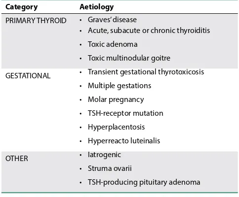
Graves’ disease is the most common differential diagnosis of non-transient thyrotoxicosis in pregnancy. Other causes are listed in Table I.
Placental tumours and hyperthyroidism
The association between hyperthyroidism and molar pregnancy was first reported in 1955.6 Hydatiform moles secrete large
amounts of hCG, proportional to the mass of the tumour. Measured β-hCG level, the amount of desialylation, and the duration of the molar pregnancy correlate with the severity of hyperthyroidism.4,6,12 The prevalence of hyperthyroidism in patients with hydatiform mole has been reported as 25–64%, with about 5% of patients presenting with thyrotoxicosis.4 Various studies have shown a paucity of clinical features of thyrotoxicosis, despite elevated levels of free T4 in many molar pregnancies.6 Hyperthyroidism usually becomes clinically evident when β-hCG levels exceed 300 U/ml.13 In a developing country like South Africa, the lack of ultrasound and quantitative β-hCG screening may lead to a higher incidence due to delayed presentation with higher hCG levels. There have also been case reports of hyperthyroidism in patients with choriocarcinoma.4,6
Clinical features
• Sinus tachycardia • Palpitations
• Hypertension • Atrial fibrillation
• Cardiac failure • Weight loss
• Hyperactivity • Tremors
• Insomnia • Heat intolerance
• Diaphoresis • Diarrhoea
• Thrombocytopaenia • Muscle weakness8,11,14
The clinical diagnosis of thyrotoxicosis may present difficulties as many hypermetabolic signs and symptoms are seen during normal pregnancy, i.e. palpitations, heart rate 90 to 100 beats/min, mild heat intolerance, warm skin and decreased effort tolerance.7
Diagnosis
• Elevated serum free T3 and T4 concentrations • Suppressed serum TSH level
• Greatly increased thyroid radioiodine uptake
• Lower T3:T4 ratio compared to Graves’ disease and multinodular goitre in which the ratio is > 20.4,13
Treatment
Evacuation of the mole or effective chemotherapy for choriocarcinoma rapidly achieves clinical and biochemical euthyroidism.4,6,9
Table I. Causes of hyperthyroidism in pregnancy7,8
Category Aetiology
PRIMARY THYROID • Graves’ disease
• Acute, subacute or chronic thyroiditis • Toxic adenoma
• Toxic multinodular goitre
GESTATIONAL • Transient gestational thyrotoxicosis • Multiple gestations
• Molar pregnancy • TSH-receptor mutation • Hyperplacentosis • Hyperreacto luteinalis
OTHER • Iatrogenic
• Struma ovarii
Perioperative anaesthetic management
Patients present to theatre for suction curettage or, less commonly, hysterectomy. A multidisciplinary team approach involving the anaesthesiologist, gynaecologist, endocrinologist and intensivist is essential.
History and examination: Includes identification of thyroid dysfunction and its complications, as well as other sequelae of a molar pregnancy.
Investigations: FBC, U&E, thyroid hormone levels, ECG, crossmatch – blood immediately available due to risk of major bleeding.
An ABG, INR/PTT, LFT, CXR and/or echocardiogram may be necessary depending on clinical presentation and baseline investigations.
Preoperative optimisation – treatment of hyperthyroidism: Evacuation of the molar pregnancy is the only definitive management of thyrotoxicosis. Antithyroid drugs usually lead to some clinical improvement after one week of therapy, but may need 4–6 weeks for full effect.15 The combined use of propylthiouracil (PTU), iodide and dexamethasone has been shown to restore serum T3 concentration to normal within
48 hours.13 Delaying surgery increases the risk of
complications such as haemorrhage, pre-eclampsia and pulmonary embolisation.10,13 Untreated or poorly controlled hyperthyroidism on the other hand increases the risk of life-threatening perioperative complications, including a thyroid storm. There is no consensus on the optimum time needed for medical stabilisation of the hypermetabolic state.13 Patients require rapid hormonal improvement and should be clinically euthyroid before surgery. A normal serum TSH is not the primary management target, as it remains suppressed beyond normalisation of free T4.7
• Antithyroid drugs (thioamides): PTU; carbimazole; methimazole. Inhibit thyroxine synthesis.14 PTU also inhibits peripheral conversion of T4 to T3.15
• Beta-blockers: Propranolol. Thyroid hormones sensitise adrenergic receptors to catecholamines. Beta-blockers control the hyperadrenergic effects of thyrotoxicosis (β1) and decrease the peripheral conversion of T4 to T3 (β2).5,14,15 Target maintaining a heart rate < 100 beats/min.13
• Iodides: Sodium iodide; potassium iodide; Lugol’s solution. Inhibit thyroid hormone release. Start at least one hour after administration of antithyroid drugs since iodide may cause a reflex release of thyroid hormone.7,8,13
The use of plasmapheresis has been reported to decrease thyroid hormone levels more rapidly in preparation for suction curettage. This is however an invasive procedure and patients should be monitored carefully for coagulopathy.9
In a bleeding patient it will not be possible to delay emergency surgery for optimisation of thyroid status. These patients are at a particularly increased risk of developing a thyroid storm.14 In addition to the above-mentioned drugs, they should receive a glucocorticoid and proceed for surgery with perioperative optimisation. (Beta-blockers are important – however exercise caution in the hypovolaemic hypotensive patient.)
Anaesthetic technique: Safe use of general anaesthesia (GA), both volatile- and total intravenous anaesthesia (TIVA) based techniques, and spinal anaesthesia have been reported.5
Evacuation poses a considerable risk of heavy bleeding. GA provides better haemodynamic stability in these patients and is a definitive indication in unstable patients.
Hyperthyroid patients are more sensitive to sympathetic nervous system (SNS) activation, which may lead to tachycardia, hypertension and ventricular arrhythmias. Laryngoscopy and intubation, surgical stimulation and extubation are vulnerable periods. Ensure an adequate depth of anaesthesia and maintain normovolaemia and normothermia.
A short-acting opioid, esmolol, lignocaine, or magnesium sulphate may be used to suppress the response to intubation. Depending on the uterine size, patients will probably require a rapid sequence induction. Incompletely treated hyperthyroid patients can be chronically hypovolaemic, and are prone to an exaggerated hypotensive response during induction of anaesthesia,11 particularly if bleeding as well.
Successful use of spinal anaesthesia for evacuation of molar pregnancies with thyrotoxicosis has been reported in both bleeding (NOT hypovolaemic) and non-bleeding patients.13,16,17 Advantages of spinal anaesthesia include the ease of the technique, providing a sympathetic block, earlier detection of uterine perforation and thyroid storm in an awake patient, and avoiding airway manipulation, the tocolytic effects of volatile agents and the effects of ventilation on the pulmonary system.5,16,17
TIVA using propofol and remifentanil with the concurrent administration of an esmolol infusion has also been used successfully. This technique avoids the tocolytic effects of volatile agents. It decreases the stress hormone release and haemodynamic response to stimulation. It also allows for a dose-dependent decrease in blood pressure and heart rate, which is advantageous in thyrotoxic patients and may decrease blood loss during surgery.18
An oxytocin infusion should be started after dilatation of the cervix and application of suction.2 Oxytocin decreases the risk of bleeding but early administration increases the risk of trophoblastic embolisation and metastases.5
Anaesthetic drugs: Hyperthyroidism and its hypermetabolic state can affect the pharmacokinetics of anaesthetic drugs. Propofol or thiopentone may be used for induction of anaesthesia. The latter possesses antithyroid activity.5,18 The volume of distribution and clearance of propofol is increased in hyperthyroid patients, increasing requirements if TIVA/TCI is used.14 Alternatively etomidate can be used in haemodynamically unstable patients. Ketamine, indirect acting adrenergic agonists, and other drugs that activate the SNS or are unpredictable muscarinic antagonists are best avoided in patients with current or recently treated thyrotoxicosis, as they may lead to exaggerated elevations in heart rate and blood pressure.11 Agents causing histamine release (atracurium, morphine) and its associated tachycardia should also be avoided.5,18
amounts of hCG, proportional to the mass of the tumour. Measured β-hCG level, the amount of desialylation, and the duration of the molar pregnancy correlate with the severity of hyperthyroidism.4,6,12 The prevalence of hyperthyroidism in patients with hydatiform mole has been reported as 25–64%, with about 5% of patients presenting with thyrotoxicosis.4 Various studies have shown a paucity of clinical features of thyrotoxicosis, despite elevated levels of free T4 in many molar pregnancies.6 Hyperthyroidism usually becomes clinically evident when β-hCG levels exceed 300 U/ml.13 In a developing country like South Africa, the lack of ultrasound and quantitative β-hCG screening may lead to a higher incidence due to delayed presentation with higher hCG levels. There have also been case reports of hyperthyroidism in patients with choriocarcinoma.4,6
Clinical features
• Sinus tachycardia • Palpitations
• Hypertension • Atrial fibrillation
• Cardiac failure • Weight loss
• Hyperactivity • Tremors
• Insomnia • Heat intolerance
• Diaphoresis • Diarrhoea
• Thrombocytopaenia • Muscle weakness8,11,14
The clinical diagnosis of thyrotoxicosis may present difficulties as many hypermetabolic signs and symptoms are seen during normal pregnancy, i.e. palpitations, heart rate 90 to 100 beats/min, mild heat intolerance, warm skin and decreased effort tolerance.7
Diagnosis
• Elevated serum free T3 and T4 concentrations • Suppressed serum TSH level
• Greatly increased thyroid radioiodine uptake
• Lower T3:T4 ratio compared to Graves’ disease and multinodular goitre in which the ratio is > 20.4,13
Treatment
Southern African Journal of Anaesthesia and Analgesia 2017; 23(2)(Supplement 1) 10
Hyperthyroidism does not increase the minimum alveolar concentration of volatile agents11 and excessive concentrations should be avoided to minimise the associated uterine tocolytic effects, which may increase blood loss. A combination of fentanyl or sufentanil and paracetamol provide suitable analgesia.
Monitoring and vascular access: Standard monitoring; large bore IV access; arterial line (+/- CVP) depending on the severity of hyperthyroidism; close temperature monitoring.
Postoperative care: Hyperthyroid patients should be managed in a high care or ICU. There remains a risk of developing a thyroid storm postoperatively,11 despite preoperative medical optimisation.13 Hyperthyroidism resolves rapidly and antithyroid medication can be weaned.
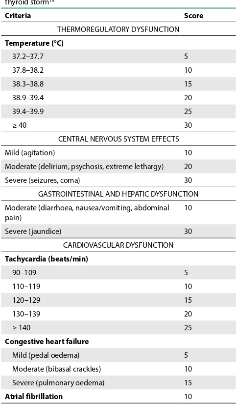
Thyroid storm
A life-threatening hypermetabolic crisis precipitated by infection, trauma, surgery, and labour, with mortality rates as high as 15%.7,8,18 It is a clinical diagnosis with accentuated signs and symptoms of thyrotoxicosis, and dysfunction in one or more organ systems. Table II lists the main clinical features. Free T4 levels do not correlate with the severity of the symptoms.7
Table II. Burch and Wartofsky scoring system for thyroid storm19
Criteria Score
THERMOREGULATORY DYSFUNCTION Temperature (°C)
37.2–37.7 5
37.8–38.2 10
38.3–38.8 15
38.9–39.4 20
39.4–39.9 25
≥ 40 30
CENTRAL NERVOUS SYSTEM EFFECTS
Mild (agitation) 10
Moderate (delirium, psychosis, extreme lethargy) 20
Severe (seizures, coma) 30
GASTROINTESTINAL AND HEPATIC DYSFUNCTION Moderate (diarrhoea, nausea/vomiting, abdominal pain)
10
Severe (jaundice) 30
CARDIOVASCULAR DYSFUNCTION Tachycardia (beats/min)
90–109 5
110–119 10
120–129 15
130–139 20
≥ 140 25
Congestive heart failure
Mild (pedal oedema) 5
Moderate (bibasal crackles) 10
Severe (pulmonary oedema) 15
Atrial fibrillation 10
PRECIPITANT HISTORY (TRIGGERING FACTOR) Present
< 25 unlikely thyroid storm 25–44 suggestive of impending storm ≥ 45 highly suggestive of thyroid storm
10
Management
• Supportive measures:▫ Adequate oxygenation and ventilation ▫ Intravenous fluids
▫ Inotropes/vasopressors
▫ Correction of glucose and electrolyte abnormalities ▫ Cooling measures (paracetamol; tepid sponging. NOT
Aspirin or NSAIDs – may increase free thyroid hormones). • Management of congestive cardiac failure if present • Appropriate antibiotics if precipitated by infection
• Drugs: Beta-blockade and antithyroid drugs are used as the first-line treatment.14
Beta-blockers:
▫ Esmolol 250–500 µg/kg IV loading dose followed by 50–200 µg/kg/min infusion OR
▫ Propranolol 1 mg/min IV OR 60–80 mg PO 4–6 hourly, OR ▫ Labetalol
Glucocorticoids: Inhibit peripheral conversion of T4 to T3. ▫ Hydrocortisone 50–100 mg IV 8 hourly OR ▫ Dexamethasone 2–4 mg IV 6 hourly Antithyroid drugs:
▫ Carbimazole 40–60 mg PO/NGT daily, OR
▫ PTU 1g PO/NGT loading dose then 200–300 mg 6 hourly, OR
▫ Methimazole 30 mg PO/NGT 6 hourly
Iodides: Start at least one hour after antithyroid drugs. ▫ Sodium iodide 500 mg IV 8 hourly, OR
▫ Lugol’s Solution 10 drops PO/NGT 8 hourly, OR ▫ Potassium iodide 5 drops PO/NGT 6 hourly7,8,13 Magnesium sulphate: Decreases adrenergic receptor sensitivity to endogenous catecholamines and associated arrhythmias.14
• ICU postoperatively with frequent evaluation of cardiac and respiratory status.
Conclusion
References
1. Lurain JR. Gestational trophoblastic disease I: epidemiology, pathology, clinical presentation and diagnosis of gestational trophoblastic disease, and management of hydatidiform mole. American Journal of Obstetrics and Gynecology. 2010;203(6):531-9. Epub 2010/08/24.
2. Snyman LC. Gestational trophoblastic disease: An overview. SA Journal of Gynaecological Oncology. 2009;1(1):32-7.
3. Moodley M, Tunkyi K, Moodley J. Gestational trophoblastic syndrome: an audit of 112 patients. A South African experience. International Journal of Gynecological Cancer: Official Journal of the International Gynecological Cancer Society. 2003;13(2):234-9. Epub 2003/03/27.
4. Hershman JM. Physiological and pathological aspects of the effect of human chorionic gonadotropin on the thyroid. Best Practice and Research: Clinical Endocrinology and Metabolism. 2004;18(2):249-65. Epub 2004/05/26.
5. Biyani G, Mohammed S, Bhatia P. Anaesthetic Challenges in Molar Pregnancy. The Indian Anaesthetists’ Forum. 2013;14(8):1-9.
6. Hershman JM. Human chorionic gonadotropin and the thyroid: hyperemesis gravidarum and trophoblastic tumors. Thyroid: Official Journal of the American Thyroid Association. 1999;9(7):653-7. Epub 1999/08/14.
7. Mestman JH. Hyperthyroidism in pregnancy. Best Practice and Research: Clinical Endocrinology and Metabolism. 2004;18(2):267-88. Epub 2004/05/26.
8. King JR, Lachica R, Lee RH, et al. Diagnosis and Management of Hyperthyroidism in Pregnancy: A Review. Obstetrical and Gynecological Survey. 2016;71(11):675-85. Epub 2016/12/03.
9. Azezli A, Bayraktaroglu T, Topuz S, Kalayoglu-Besisik S. Hyperthyroidism in molar pregnancy: rapid preoperative preparation by plasmapheresis and complete improvement after evacuation. Transfusion and Apheresis Science: Official Journal of the World Apheresis Association: Official Journal of the European Society for Haemapheresis. 2007;36(1):87-9. Epub 2007/03/10.
10. Chiniwala NU, Woolf PD, Bruno CP, et al. Thyroid storm caused by a partial hydatidiform mole. Thyroid: Official Journal of the American Thyroid Association. 2008;18(4):479-81. Epub 2008/03/21.
11. Butterworth JF, Mackey DC, Wasnick JD. Morgan and Mikhail's Clinical Anesthesiology. 5th ed. New York: McGraw-Hill Education; 2013:733-5.
12. Moskovitz JB, Bond MC. Molar pregnancy-induced thyroid storm. The Journal of Emergency Medicine. 2010;38(5):e71-6. Epub 2009/12/05.
13. Samra T, Kaur R, Sharma N, Chaudhary L. Peri-operative concerns in a patient with thyroid storm secondary to molar pregnancy. Indian Journal of Anaesthesia. 2015;59(11):739-42. Epub 2016/01/13.
14. Farling PA. Thyroid disease. British Journal of Anaesthesia. 2000;85(1):15-28. Epub 2000/08/06.
15. Marx H, Amin P, Lazarus JH. Hyperthyroidism and pregnancy. British Medical Journal. 2008;336(7645):663-7. Epub 2008/03/22.
16. Bhatia N, Mitharwal SM. Hydatidiform mole with uncontrolled hyperthyroidism: An anesthetic challenge. Journal of Anaesthesiology Clinical Pharmacology. 2016;32(4):537-8. Epub 2017/01/18.
17. Solak M, Akturk G. Spinal anesthesia in a patient with hyperthyroidism due to hydatidiform mole. Anesthesia and Analgesia. 1993;77(4):851-2. Epub 1993/10/01.
18. Erturk E, Bostan H, Geze S, et al. Total intravenous anesthesia for evacuation of a hydatidiform mole and termination of pregnancy in a patient with thyrotoxicosis. International Journal of Obstetric Anesthesia. 2007;16(4):363-6. Epub 2007/04/27.
South Afr J Anaesth Analg
ISSN 2220-1181 EISSN 2220-1173© 2017 The Author(s) FCA 2 REFRESHER COURSE Southern African Journal of Anaesthesia and Analgesia 2017; 23(2)(Supplement 1)
Open Access article distributed under the terms of the Creative Commons License [CC BY-NC-ND 4.0] http://creativecommons.org/licenses/by-nc-nd/4.0
Keywords: neuro-monitoring, cerebral perfusion, Near Infrared Spectroscopy (NIRS), Bispectral (BIS), Transcranial Doppler (TCD), monitoring in non-neurosurgical procedures
Cerebral perfusion monitoring during non-neurosurgical procedures: The
practicalities of Near Infrared Spectroscopy (NIRS)/Bispectral Index (BIS)
M MphomaneDepartment of Anaesthesia, Chris Hani Baragwanath Academic Hospital, University of Witwatersrand Correspondence to: m_mphomane@yahoo.com
Introduction
Complex non-neurosurgical procedures such as cardiac surgery, major vascular procedures (including carotid endarterectomy), liver transplantation, orthopaedic procedures in beach chair position, etc. are commonly associated with devastating neurologic complications and poor patient outcome. Neuromonitoring in these procedures assumes a pivotal role in ensuring timely detection and prevention of permanent brain and spinal cord dysfunction.
The most common aetiology of intraoperative neurologic injury that can potentially be detected by neuro-physiologic monitoring is cerebral ischaemia, which may occur as a consequence of hypoperfusion, embolic stroke, or malperfusion.1
Clinically, the major neurologic deficits most commonly observed have been cognitive impairment, seizures, choreoathetosis, bilateral motor deficits and hemiparesis.2
Multimodal integrated techniques are used to monitor cerebral perfusion and thus avoid the above-mentioned complications. There is no one modality that has shown to be superior to the other and in practice monitors are used to complement each other.
Cerebral perfusion monitoring modalities
Cerebral perfusion monitors seek to ensure that adequate perfusion to the brain is maintained at all times. In order to perfuse the brain adequately cerebral perfusion pressure (CPP) has to be maintained, and CPP is defined by the following equation:
CPP = MAP – ICP (or CVP)3.4 (MAP – mean arterial pressure, ICP – intracranial pressure, CVP – cerebral venous pressure)
Adequate perfusion of the brain and prevention of ischaemia can be achieved by measuring the following parameters:
Cerebral oxygenation and cerebral blood flow (CBF)
Cerebral oxygenation monitoring
Near Infrared Spectroscopy (NIRS) monitor
This is a non-invasive, continuous monitor that measures regional cerebral oxygen saturation (rSO2), usually of the frontal area. The values reflect primarily venous saturation as opposed to arterial pulsatile component. Added advantages of NIRS include: that it can identify both hypoperfusion and hyperperfusion; and there is no need for blood flow.5
A number of drawbacks are recognised however, and these include: that it is affected by extracranial blood flow and ambient light; there are no recognised standards or normal values (as variations between individual patients do occur). This means that every patient’s baseline is needed, absolute values are not useful, and trends are more important.
Electroencephalogram (EEG) monitor
This is a measure of spontaneous electrical activity generated by the brain, specifically the cortex; therefore the deeper structures are not assessed. It requires a robust machine with multiple electrodes ranging from 8 to 16. Measurements of CBF are implied from changes in the amplitude, frequency and the patterns of the EEG bands (α, β, δ).3
The added limitations are that the raw data produced from the EEG is quite complex, and needs interpretation by experienced personnel and therefore limiting its use. Anaesthetic agents and hypothermia also extensively affect it.
Bispectral Index (BIS) monitor
This is a processed EEG, originally designed to assess the hypnotic level of a patient. But it can now be used to detect cerebral ischaemia in the intraoperative period. The monitor gives a value, called a BIS value ranging from 0 to 100. Reduction in BIS value by more than 10 or by 30–40% may correlate with ischaemia.4
raw EEG data. BIS also does not differentiate between localised and global ischaemia.
Cerebral Blood Flow (CBF) monitoring
CBF monitoring offers a rational approach to detect and prevent ischaemic insults to the brain and improve outcome in patients undergoing major non-neurologic procedures. Normal value of CBF in young adults is 50 ml/100 g brain tissue/min. The ischaemic threshold is 18 ml/100 g/min and below 10 ml/ 100 g/min, irreversible brain damage ensues.3
The CBF monitors may be classified into two broad categories: direct and indirect measurements.
Indirect monitoring
Transcranial Doppler (TCD)
This is a non-invasive, bedside technique that measures local blood flow velocity. It presents these data graphically as velocity against time. The changes in velocity indicate pathology, e.g. hypoperfusion due to stenosis, thrombosis or hyperperfusion.
The limitations of TCD are that it is operator-dependent, there is signal interference with numerous electromagnetic apparatus in the operating room. In 10–15% of patients, there is a lack of acoustic transtemporal window, and therefore the test cannot be carried out.
Stump pressure
During carotid endarterectomy, when the proximal common carotid and external carotid arteries are clamped, the pressure measured in the internal carotid craniad to the clamp site is the stump pressure. It reflects the back pressure on the internal carotid artery, supplied by the collaterals from the circle of Willis. It is considered to be a surrogate marker of perfusion in the ipsilateral cerebral hemisphere from the contralateral side. A pressure below 40–55 mm Hg is often used as a threshold for intervention.4
Direct monitoring
Positron Emission Tomography (PET)
This mode is an invasive, quantitative measure of CBF, cerebral metabolic rate of oxygen (CMRO2), cerebral blood volume (CBV), and oxygen extraction fraction. It uses a radioactive compound, and requires patients to be transported to a radiology facility, therefore impractical for intraoperative use. Although it is considered a gold standard, it is expensive, not universally available and gives a short single assessment.
Practical use of NIRS and BIS during
non-neurosurgical procedures
Near Infrared Spectroscopy (NIRS)
6NIRS is a measure of cerebral perfusion that relies on the difference of absorption spectra between oxyhaemoglobin, deoxyhaemoglobin and the total haemoglobin for calculating estimates of regional cerebral oxygen saturation (rSO2).2 (Beer Lambert law).
The technique requires the use of two sensors separated by a fixed distance. The proximal sensor records infrared light reflected from superficial tissue while the distal signal represents the brain tissue saturation. The subtraction between these two signals represents a venous weighted estimate of rSO2.
Usefulness of cerebral NIRS
It is a non-invasive technology that provides continuous monitoring of regional cerebral oxygenation. The machine is portable, sensors are easy to apply, and interpretation of results needs no specialised expertise.6
The monitor can identify both hypoperfusion and hyperperfusion. Due to the strategic placement of the electrodes, the watershed area between the anterior and the middle cerebral artery territories are included in the NIRS measurements.
NIRS is relatively resistant to the effects of anaesthetic agents, but can be affected by the indirect changes in the global blood flow due to physiological changes due to drugs. Therefore, to allow meaningful interpretation of neuromonitoring results, steady state anaesthetic depth, physiological variables, such as blood pressure, temperature, and patient position should be kept stable.4
In non-neurological procedures NIRS has been found very useful in determining the risk of significant cerebral ischaemia, and therefore implementing appropriate treatment algorithms are the following: procedures in beach chair position, repair of aortic coarctation, aortic aneurysm repair, cardiac surgery, carotid endarterectomy, etc.
Spinal NIRS monitoring looks to be an attractive alternative monitoring during procedures such as thoracic and abdominal aorta repair.6,7 The same advantages as mentioned above apply here. The problematic issues are also as depicted above. For instance, the interference of the surrounding paraspinal space tissues.
Limitations of NIRS
NIRS reading is affected by the scalp tissue composition in different individuals, and therefore it is difficult to compare to ‘standards’/‘normals’. Ambient light also affects NIRS reading, necessitating proper padding of the electrodes.
The monitor can only assess regional oxymetry, usually frontal, so clinically relevant focal cerebral ischaemia outside the monitored area may easily go unnoticed. Patients with previous infarcts may produce erroneous measurements.
Bispectral monitor (BIS)
Southern African Journal of Anaesthesia and Analgesia 2017; 23(2)(Supplement 1) 14
a set of three disposable electrodes attached to the patient’s frontal and temporal regions. The machine is portable, and in fact some anaesthetic machines are fitted with cartridges for BIS monitoring. The accuracy of the BIS to monitor hypnotic state has been questioned due to inconsistencies associated with it in many studies.4
The recent use of BIS has been to detect cerebral ischaemia during non-neurosurgical procedures. Reduction in BIS value may correlate with ischaemia.4
Limitations of BIS monitor for use in detecting cerebral ischaemia intraoperatively are numerous, and include:
• Delay in reporting owing to the time needed for processing of EEG data.
• Lack of differentiation between global and focal ischaemia. • Inhalational and intravenous anaesthetic agents decrease
BIS value in a dose-dependent manner, ultimately resulting in burst suppression and electrical silence similar to severe hypoperfusion and ischaemia.4,9 Table I gives comparison amongst different types of cerebral perfusion monitors.9
Conclusions
Numerous modalities for measurement of cerebral perfusion during high-risk non-neurosurgical procedures are available. The aim of monitoring cerebral perfusion in these procedures is to detect cerebral hypoperfusion, malperfusion, and embolic complications early and therefore avoid cerebral ischaemia.
Studies have shown that no single monitor is capable of exclusively and conclusively detecting cerebral malperfusion. A multimodal, complementary approach is recommended.
NIRS promises to be quite a valuable technique to use in non-neurosurgical procedures to avoid neurologic complications associated with these high-risk procedures. On the other hand, BIS has been found to be disappointing in terms of accurately determining depth of anaesthesia as well as detecting cerebral ischaemia.
References
1. Cheung AT. Neuromonitoring in cardiac surgery or deep hypothermic circulatory arrest. Optional or imperative? Scahq.org 2014.
2. Rodriguez RA. Intraoperative brain monitoring: www.anaesthesia.org/ winterlude/w197/l_neuro.html/
3. Mahajan C, Rath GP, Bithal PK. Advances in neuro-monitoring. Anesthesia essays and researches. 2013; 7(3) 31 -318.
4. So V C, Poon CCM. Intraoperative neuromonitoring in major vascular surgery. Br J Anaesth (2016) 117 (suppl_2): ii 13-ii 25.
5. Mėrat S, Levecque JP, Gulluche Y, et al. Monitoring may allow the detection of severe cerebral ischaemia. Can J Anaesth. 2001; 48:1066-1069.
6. Moerman A. Clinical application of near-infrared spectroscopy in perioperative. biblio.urgent.be/publication/
7. Kawano H, Matsumoto T. Anaesthesia for arthroscopic shoulder surgery in the beach chair position: monitoring cerebral oxygenation using bispectral index and near-infrared spectroscopy. Middle East J Anaesthesiol. 2014; 22 (6):613-617. 8. William M, Lee JK. Intraoperative blood pressure and cerebral perfusion:
strategies to clarify haemodynamic goals: Paediatr Anaesth. 2014; 24 (7): 657-667.
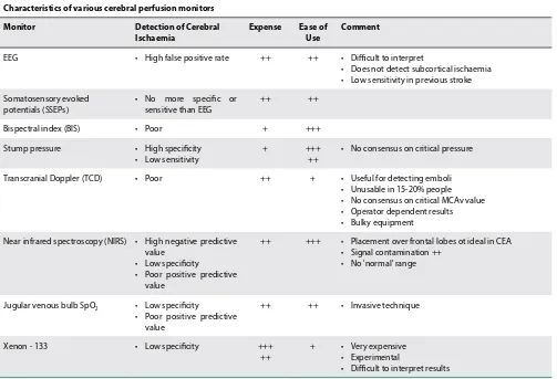
9. Whiten C, Gunning P. Carotid endarterectomy: intraoperative monitoring of cerebral perfusion: current anaesthesia and critical care 20 (2009) 42-45 Table I. Characteristics of various cerebral perfusion monitors9
Characteristics of various cerebral perfusion monitors
Monitor Detection of Cerebral
Ischaemia Expense Ease of Use Comment
EEG • High false positive rate ++ ++ • Difficult to interpret
• Does not detect subcortical ischaemia • Low sensitivity in previous stroke
Somatosensory evoked potentials (SSEPs)
• No more specific or sensitive than EEG
++ ++
Bispectral index (BIS) • Poor + +++
Stump pressure • High specificity
• Low sensitivity
+ +++
++
• No consensus on critical pressure
Transcranial Doppler (TCD) • Poor ++ + • Useful for detecting emboli
• Unusable in 15-20% people • No consensus on critical MCAv value • Operator dependent results • Bulky equipment
Near infrared spectroscopy (NIRS) • High negative predictive value
• Low specificity
• Poor positive predictive value
++ +++ • Placement over frontal lobes ot ideal in CEA • Signal contamination ++
• No 'normal' range
Jugular venous bulb SpO2 • Low specificity
• Poor positive predictive value
++ ++ • Invasive technique
Xenon - 133 • Low specificity +++
++
+ • Very expensive • Experimental
© 2017 The Author(s) FCA 2 REFRESHER COURSE Open Access article distributed under the terms of the
Creative Commons License [CC BY-NC-ND 4.0] http://creativecommons.org/licenses/by-nc-nd/4.0
Anaesthesia for related living donor liver transplantation
M Hatchett
Wits Donald Gordon Medical Centre Transplant Unit
Dunkeld Anaesthetic Practice
Introduction
The first liver transplant was performed in 1963 in Colorado, USA by Dr Thomas Starzl, a man who is acclaimed world-wide as the father of organ transplantation. His career and list of accomplishments can, and has been, the individual subject of lectures by themselves, but suffice to say that it is difficult to foresee many people surpassing his achievements in medicine. At one stage in his career it was calculated he was producing a scientific paper for publication every seven days. He died in March this year at the age of 90.
As we are all aware, liver transplantation is the only real hope for end-stage liver disease. In kidney disease we have dialysis as a method of prolonging life, albeit very restrictive for the patient and certainly not ideal. For progressive liver disease we have liver transplantation or death. The results and outcomes of liver transplantation have improved exponentially since 1963, particularly as a result of advances in immunosuppression and decreased rejection of transplanted organs. The main problem that still remains is the perennial shortage of donor organs for the huge number of recipients requiring them.
Deceased vs living donors
Traditionally, particularly in South Africa, donor organs have come from brain-dead patients with functional cardiac systems on ventilators. Elsewhere organs have also been procured from controlled non-heart-beating donors (often for religious and cultural reasons), or patients on ECMO support after cardiac death. The huge step forward has been the successful introduction of removal of a portion of a healthy donor’s liver so that both donor and recipient survive thereafter. There is a significant difference in transplant patterns between Western and Eastern countries. The Western countries continue to have higher numbers of liver transplants where the donor organ came from deceased donors, whereas in the Eastern countries, because of religious, cultural and social differences, living donor liver transplants (LDLT) are the commonest means of a patient receiving a transplant.
Critical to the concept of LDLT, specifically for adult-to-adult procedures, is the fact that the liver can regenerate its tissue extremely rapidly. Within two to three months of 40% of a liver having been removed as a donor graft, the liver has regenerated that volume so that it has regained full function. As regards the South African situation with LDLT, most of our cases are those where a parent is donating a portion of their liver to a child. This is usually the left lateral lobe and the volume of liver lost is small relative to the liver’s original volume. The situation is very different where a large proportion of the liver has to be donated because the recipient is an adult. There can then be a problem having sufficient functioning liver tissue for the donor and/or the recipient. This phenomenon is known as ‘small for size’ and can be extremely serious, if not life-threatening.
Advantages of LDLT
1. The LDLT is done as an elective procedure. This means everything is preplanned and controlled. As it is invariably scheduled for a morning start, staff are fresh and all support staff should be readily available. This is in stark contrast to cadaver transplants where there is little, if any, control over when the procedure will go ahead, and often occurs in the middle of the night with on-call staff that may be tired. Ancillary discipline staff may also not be readily available and have to be roused from bed and brought in at short notice. 2. Donors are thoroughly investigated with multiple
examinations and investigations over a period of time before being passed fit to donate a liver segment. Any deviations from the norm in the donor’s health usually means they are turned down and other potential related donors are sought. This also translates into a good quality donated organ being recovered. This luxury of extensive preoperative assessment of the donor is not always present with cadaver-derived organs.