Determination of Monosomy 7 among In Vitro
Generated CD
1
a Positive Cells in Patients with
Juvenile Myelomonocytic Leukemia
Shayan P
1, 2*, Izadyar M
3, Brigitte Eckert
41- Department of Pathobiology, University of Tehran
2- Devision of Pediatric Hematology and Oncology, Department of Pediatrics and Adolescent Medicine, University of Freiburg, Mathildenstrasse, Germany
3- Children’s Medical Center, Tehran University of Medical Sciences
4- Molecular Biological System Transfer Research Institute, Tehran, Iran
*Corresponding Author: Shayan P, Email: jpshayan@ut.ac.ir Submitted: 30-08-2011, Accepted: 20-03-2012
Abstract
Objective: To study whether the generated CD1a positive cells belong to the leukemic cells among patients with juvenile myelomonocytic leukemia.
Materials and Methods: We used mononuclear cells from 3 patients with juvenile myelomonocytic leukemia, from which two had monosomy 7. The mononuclear cells from these patients were cultured in RPMI/10%FCS without adding exogeneous growth factors for 7 days. At day 7 the cultured cells were harvested and analyzed using antibodies against Ki 67, CD20 and CD1a. Additionally the cultured cells were analyzed using antibodies against CD1a and CD20 and chromosome 7 specific DNA probe, using combined fluorescence immunophenotyping and interphase cytogenetic techniques.
Results: The immunocytochemistry assay demonstrated that a high number of cells were in proliferation status, which was determined by antibody against proliferation associated nuclear protein ki 67. The percentage of Ki 67 positive cells was between 24% and 38% respectively. The percentage of CD1a positive cells was between 8% and 31% and the percentage of CD20 positive cells was between 5% and 12% respectively. The fluorescence immunophenotyping and interphase cytogenetic analysis showed that nearly all CD1a positive cells of one patient with monosomy 7 had one chromosome 7, whereas in other patient with monosomy 7, the amount of CD1a positive cells having only one chromosome 7 was approximately 11%. Furthermore, the combined immunophenotyping and cytogenetic analysis showed that the CD20 positive cells in all patients had normal karyotype.
Conclusion: Our results suggest that CD1a positive cells generated by mononuclear cells from patients with juvenile myelomonocytic leukemia in vitro most probably belong to the leukemic cells. Since monosomy 7 could not be detected in all CD1a positive cells in juvenile myelomonocytic leukemia patients with monosomy 7, it is to assume that monosomy 7 is a secondary event in the pathophysiology of juvenile myelomonocytic leukemia.
Keywords: Leukemia, mononuclear cells, CD1a antigen, chromosome 7, monosomy, cell proliferation, myelomonocytic,
juvenile
Introduction
Juvenile myelomonocytic leukemia (JMML) is a rare, unique clonal myeloproliferative disorder of early childhood. JMML is characterized by massive infilteration of various organs with monocytic and geanulocytic cell lineages resulting in hepatosplenomegaly, lymphadenopathy, skin rash and pulmonary infiltrates1, 2, 3. It has been shown that the blood mononuclear cells isolated from JMML patients can grow spontaneously in
semi solid or liquid medium4, 5. The proliferation
of mononuclear cells (MNC) seems to be GM-CSF dependent, which is produced by macrophages. The monoclonal antibody against GM-CSF leads to the dose depended inhibition of cell growth in vitro culture5. One interesting characteristic among
JMML patients is that a population within the MNC cells differentiates in the in vitro culture to the CD1a positive cells 5. This cells harbor multilamellar
ORIGINAL ARTICLE
IJBC 2012;3: 111-118
structures resembling major histocompatibility complex (MHC) class II compartiments (MCIIC) 5 .
Interestingly, monoclonal antibody against human GM-CSF inhibits, in a dose dependent manner, the generation of CD1a positive cells in JMML patients7. It has also been demonstrated thatribozyme, a catalytic RNA molecule down regulating the expression of GM-CSF, decreases colony formation and viability of JMML cells in culture 8. Although
the molecular mechanisms of pathophysiology of JMML is not well characterized, but there are various data relating the GM-CSF hypersensitivity and abnormal regulation of RAS-RAF-MEK-ERK signal transduction pathway with the pathogenesis of JMML 9, 10.
Another interesting aspect of JMML patients is that not only mutations at gene level but also chromosomal aberrations occur among some patients. Monosomy 7 could be detected in about 25% of the patients with JMML 11. The monosomy 7 is ussualy determined by chromosomal staining and banding in good spreading metaphases or by fluorescence in situ hybridization of interphase cells. It is of gross importance to be able to analyze the karyotype of phenotype defined cells. Weber-Matthiesen et al. (1992) 12 developed a method for the simultaneous determination of karyotype and phenotype of the cells using immunophenotyping followed by in sito hybridization. Since a population of MNC from patients with JMML differentiates to CD1a positive cells, it will be interesting to know whether the generated CD1a positive cells in JMML patients with monosomy 7 have normal karyotype or monosomy 7.
In the present study we analyzed chromosome 7 in the in vitro generated CD1a positive cells and CD20 positive cells among JMML patients using FICION technique (Fluorescence Immunophenotyping and Interphase Cytogenetics as a Tool for Investigation of Neoplasms) 12.
Materials and Methods
Isolation of mononuclear cells
Mononuclear cells (MNC) were isolated by Ficoll-Paque (Pharmacia, Uppsala, Sweden) density gradient centrifugation. Spleen samples from three patients with JMML, two with monosomy 7 and one with normal karyotype (table 1), were cut into small pieces and placed in phosphate-buffered saline (PBS). The cells were isolated by scraping,
filtering through a cell strainer (70 μm Nylon, Becton Dickinson) and by Ficoll-Paque. MNC from an individeum with normal karyotype was used for additional determination of cutoff level. All tissues were obtained with consent given according to institutional guidelines.
Cell culture
The isolated MNC from spleen were cultured at a concentration of 1 x 10 6 /ml in RPMI medium with 10% FCS and 1% penicillin-streptomycin. Cultures were incubated for 7 days at 37º C in atmosphere containing 5% CO2. At day 7 cells in suspension were used for cytospin preparation and analysed using immuncytochemistry and combined immunophenotyping and fluorescence in situ hybridization technique known as Fluoresence Immunophenotyping and Interphase Cytogenetics as Tool for Investigation of Neoplasms (FICTION) 12.
Immunocytochemistry
For immunostaining of cells we used an immunoperoxidase system from DAKO Hamburg, Germany. The used antibodies were CD1a, Ki-67 and CD20 (DAKO, Hamburg, Germany). The cytospin slides were first fixed in 4% formalin for 10 minutes. The slides were then washed three times in Millipore-purified water and incubated for 10 min in 0.6%H2O2/methanol to inhibit the endogenuous peroxidase activity. After washing the slides in TBS buffer (20 mM Tris, 0.15 mM NaCl), the cell region on the slides were separately covered with corresponding diluted (1:100) antibody (CD1a, CD20 or Ki 67) and incubated for 1 hour on ice. Afetr that the slides were washed three times with TBS and the cells were covered with the biotinulated anti mouse antibodies supplied by universal SterptABComplex/HRP Duet, mouse/Rabbbit kit (DAKO) and incubated for 30 minets on ice. After incubation and washing the slides, the cells were covered with streptavidin-peroxidase (supplied with kit) and incubated for 30 minets on ice. After that the cells were covered with substrate (H2O2/ Diaminobenzidine, supplied with kit) for one to five minutes. After washing the slides three times in water, the cells were analysed by light microscopy. The number of positive reaction was determined in at least 1000 cells.
Table 1: Patients’ characteristics, Data obtained from European Working Group on Child
-hood MDS (EWOG-MDS)
Abbreviations: Age at diag. (mon.): Age at diagnosis (months); (cm*): cm below costal margin; WBC: white blood cells; Mono.: monocytes; -7: monosomy 7.
Fluoresence Immunophenotyping and Interphase Cytogenetics as Tool for Investigation of Neoplasms (FICTION)
The method of FICTION has been previously described by Weber Matthiesen et al. (1992) 12. Briefly, slides were fixed in fresh acetone for 10 min, and then they were airdryed. One hundred microliters diluted monoclonal antibody against CD1a or CD20 (1:50) in PNM buffer (5% dry milk powder, 0.1 M NaH2PO4, 0.1 M Na2HPO4, pH 8.0, 0.03% NaN3) was used to cover the fixed cells and incubated for 30 minutes at room temperature. Fluorescence detection was performed with the sequential cascade of three Cy3- or AMCA-conjugated antibodies diluted in PNM (1:20) for 30 minutes at room temperature: Cy3- or AMCA-goat anti-mouse, Cy3- or AMCA-rabbit anti-AMCA-goat and Cy3- or AMCA-dunky anti-rabbit antibodies (Jackson/ Dianova, Hamburg, Germany). After each incubation, wash step was performed once with PN buffer (0.1 M NaH2PO4, 0.1 M Na2HPO4, pH 8.0). After immunophenotyping, slides were fixed in Carnoy’s fixative (methanol: acetic acid, 3:1) for 10 minutes and in paraformaldehyde solution (1%) for 1 minute. Then the slides were dehydrated through increasing ethanol concentrations (70%, 85% and 100%) and air-dried. The Cy3- or FITC- conjugated 7 centromeric probes (Vysis) were applied to the cytospin imprint and covered with a round 10 mm coverslip. Both probe and target DNA were simultaneously denatured at 75°C for 7 minutes. The hot box containing the slides was Immidiately transferred into a 37°C incubator and hybridized overnight. Posthybridization washes
were performed in 0.1 SSC three times at 60°C each for 5 minutes. Then, slides were washed once in PN buffer and hen were counterstained with DAPI solution followed by washing in 2 x SSC (0.3 M NaCL, 0.03 M Sodium Citrate, pH 7.0) and once in PN buffer and then mounted in antifade solution.
Determination of cut-off level
The cut-off level was determined by analyzing the control JMML patient without monosomy 7 and an individual with normal karyotype with a minimum of 200 interphase nuclei each. The cut-off was defined as mean of false positive signals among the mentioned controls.
Results
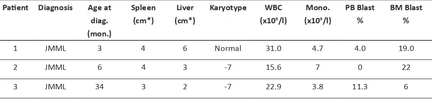
The MNC cells from three patients with JMML were cultured in RPMI/10% FCS for 7 days. At day 7 the cells were analyzed using immunocytochemistry. First to determine the proliferation status of the cells, antibody against proliferation associated nuclear protein Ki 67 was used. In contrast to the quiescent cells, the proliferating cells, express Ki 67 protein in their nucleous. The figure 1B showes the nucleous of proliferating cells in brown color. A cell in mitosis is also recognizable in this figure, which is stained brown. Figure 1A shows a negative control. The immunostaining of the cells showed that 38% of the cells were in proliferating status in cells cultured from the JMML patient with normal karyotype (patient #1), whereas the percentage of the Ki 67 positive cells was 26% and 24% in other two patients with monosomy 7 (patients #2 and #3) respectively. The MNC at initial time of cell culture
Patient Diagnosis Age at
diag. (mon.)
Spleen (cm*)
Liver
(cm*)
Karyotype WBC
(x109/l)
Mono.
(x109/l)
PB Blast
%
BM Blast %
1 JMML 3 4 6 Normal 31.0 4.7 4.0 19.0
2 JMML 6 4 3 -7 15.6 7 0 22
3 JMML 34 3 2 -7 22.9 3.8 11.3 6
were CD1a negative. We have previously shown that the CD1a positive cells start to differentiate after 5 in vitro culture days and the pick of the differentiation is reached at day 7 of culture 5. At
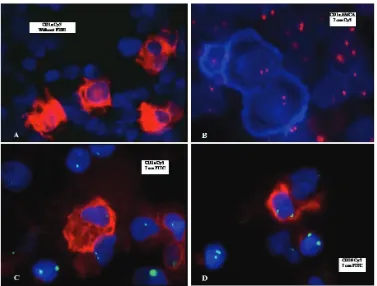
day 7 of culture, the cultured cells were analyzed using monoclonal antibody against CD1a, which is normally expressed in dendritic cells. The analysis showed that 31% of the cultured cells expressed CD1a molecule in the patient with normal karotype (figure 1 D), whereas the percentage of CD1a positive cells was 9% and 8% in two other patients with monosomy 7 (patients 2# and 3#) respectively. The analysis of the cultured cells with monoclonal antibody against CD20, a B cell marker, showed that 12% of the cells expressed CD20 moleculs on the cells cultured from patients 1# and 2#, whereas 5% of the cells were CD20 positive in cultured cells from patient 3# (figure 1C). Nearly the same data was observed by immunofluorescence staing of the cultured cells using corresponding antibodies (table 2). After checking the presence of CD1a and CD20 positive cells within the cells, the cells were analyzed using combined fluorescence immunophenotyping and interphase cytogenetics technique. To do this, the cells were first immunofluorescence stained with monoclonal antibody against CD1a or CD20. In second step, the chromosome from the same cells was hybridized with the Cy3 or FITC conjugated DNA prob specific for chromosome 7. The interphase cytogenetic analysis showed as expected that two chromosomes 7 were detected in the cells cultured from MNC of patient with normal karyotype. Thirty two and 12 percent of the cultured cells had monosomy 7 in patients 2# and 3# respectively (table 2). Figure 2A shows the immunofluoresence staining of the cells with antibody against CD1a. The membrane of the positive cells was stained
red. The Figure 2B shows the simultaneous staining of the cells from patient with normal karyotype with AMCA-conjugated CD1a and Cy3-conjugated chromosome 7 specific DNA probe. The CD1a positive cells stained blue and the chromosome 7 stained red. The analysis showed that all CD1a positive cells had normal karyotype in JMML patient with normal karyotype (figure 2B). Figure 2C shows simultaneous staining of the cultured cells from patient 2 with Cy3-conjugated CD1a antibody and FITC-cojugated chromosome 7 specific DNA probe. The CD1a positive cells were stained red and chromosome 7 was stained green. The analysis showed that nearly all CD1a positive cells generated in the culture from patient 2# had monosomy 7, whereas approximately 11% of the CD1a positive cells generated by patient3# had monosomy 7 (table 2). The cultured cells from all three patients were also simultaneously analyzed using Cy3-conjugated CD20 antibody and FITC-conjugated chromosome 7 specific DNA probe (figure 2D). No CD20 positive cells with monosomy 7 could be recognized in any of three patients.
Discussion
JMML is an aggressive myeloproliferative and myelodysplastic syndrome in childhood. The molecular mechanisms responsible for the pathogenesis of JMML are unknown. Some characteristics previously described for JMML are leukocytosis with monocytosis, presence of hematopoietic progenitor cells in peripheral blood, massive infiltration of various organs with the leukemic cells and consequently hepatosplenomegaly, lymphadenopathy, skin rash and damage of lung tissue 1, 2, 4, 13, 14. The myeloid progenitor cells of the patients with JMML show
Table 2: Immunoycytochemical analysis of MNC from spleen with antibodies against CD 1a, CD20 and Ki-67 and simultaneous staining of cells with antibody against CD1a or CD20 and chromosome 7 specific DNA probe.
JMML
Patients no.
Monosomy 7 Monosomy 7
(%)
CD1a
(%)
CD20 (%)
Ki 67 (%)
CD1a/-7 (%)
CD20/-7 (%)
1 No 0 31 12 38 0 0
2 Yes 32 9 12 26 99 0
3 Yes 12 8 5 24 11 0
an excessive in vitro proliferation and a selective hypersensitivity to the GM-CSF 4. It is known that the proliferation capability of the JMML cells is GM-CSF dependent, since the antibody against GM-GM-CSF has an inhibitory effect in colony growth in semi solid medium as well as an inhibitory effect on the proliferation of the JMML cells in liquid medium6,7.
Previously, we showed that a population of the cells withinh the MNC isolated from peripheral blood, bone marrow or spleen of JMML patients differentiate within 7 days to the CD1a positive cells harboring multilamellar structures resembling major histocompatibility complex (MHC) class II compartiments (MCIIC) 5. Interestingly, it was also
shown that the neutralization of GM-CSF using monoclonal antibody against human GM-CSF inhibites dose dependently the differentiation of the cells into the CD1a positive cells 7. Since the analysis of GM-CSF receptor fhas shown that the receptor is not altered in patients with JMML 15,
it is to speculate that downstream of the GM-CSF receptor could be involved in the pathogenesis
of JMML. Activating RAS point mutaions have been demonstrated in codon 12, 13 and 61 of N-RAS and K-RAS in 25% of patients with JMML [16]. Furtheremore, mutation in PTPN11, a gene encoding a non-receptor protein phosphatase, has been observed in 35% of JMML patients 17. Kratz et al. (2005) [18] found PTPN11 mutations in 74 out of 107 patients with JMML. Mutation in this gene leads to the activation of the GNEFs which can then activate RAS. Abnormality in c-CBL gene has also been studied. C-CBL is an E3 ubiquitin ligase, which is involved in intracellulare transport and degradation of some tyrosine kinase receptors. Mutation in this gene can lead to the continuous activation of RAS. Loh et al. (2009) 19 showed that 17% of the examined JMML patients had mutations in c-CBL gene. These results and the results from a study with the tumor suppressor gene PTEN together let to estimate that 80 percent of the patients with JMML have abnormality in the RAS pathway.
Another interesting aspect of JMML patients
Figure1: The MNC from patients with JMML was cultured in RPMI/10%FCS for 7 days. At day 7, the cells were ana
-lyzed using antibodies against Ki 67, CD1a and CD20 by immunoperoxidse method. A: negative control, B: immu
-nostaining with antibody against Ki 67, C: immu-nostaining with antibody against CD20 and D: immu-nostaining with antibody against CD1a.
is that not only mutations at gene level but also chromosomal aberrations occur amongsome patients. It has been demonstrated that about 25% of the JMML patients have monosomy 7 13.
Since the GM-CSF playes a prominent role in the in vitro generation of CD1a positive cells, it is interesting to analyze the karyotype of these cells generated in patient with monosomy 7. To achieve this we used MNC from three patients with JMML. One of these patients had normal karyotype and two other patients had monosomy 7. First we analyzed the proliferation status of the culture cells at day 7 using antibody against Ki 67. We could observe that a high nimber of the cells were in proliferating status in all three patients. The percentage of Ki67 positive cells was 38%, 26% and 24% in patient1#, 2# and 3# respectively. The study of the cultured cells with antibody against CD1a showed that
31%, 9% and 8% of the cultured cells were CD1a positive in patients 1#, 2# and 3# respectively. The interphase cytogenetic analysis showed that 32 and 12 percent of the cells had monosomy 7 in patients 2# and 3# respectively. As expected, no monosomy 7 could be detected in patient 1#. The simultaneous staining of the cultured cells with antibody against CD1a and chromosome 7 specific DNA probe demonstrated that, as expected, in patient 1# no monosomy 7 in generated CD1a positive cells is present. Interestingly, in patient 2# nearly all CD1a positive cells had only one chromosome 7, whereas in patient 3# only approximately 11 percent of the generated CD1a positive cells had monosomy 7. Since, CD1a positive cells were generated in all previously examined MNC from patients with JMML in RPMI/10%FCS without supplementation of additional growth factors and since it is believed
Figure 2: The MNC from patients with JMML was cultured in RPMI/10%FCS for 7 days. At day 7, the cells were analyzed simoultaneously using antibodies against CD1a or CD20 and fluoresence conjugated chromosome 7 specific DNA probe. A: The cells were stained with FITC conjugated antibody against CD1a. B: the cells were simultaneously immunostained using AMCA-conjugated antibody against CD1a (blue color), and hybridized using Cy3-conjugated chromosome 7 specific DNA probe (red color). C: the cells were simultaneously immunostained with Cy3-conjugated antibody against CD1a (red color), and hybridized using FITC-conjugated chromosome 7 specific DNA probe (green color). D: the cells were simultaneously immunostained with Cy3-conjugated antibody against CD20 (red color), and hybridized using FITC-conjugated chromosome 7 specific DNA probe (green color).
that JMML is a clonal malignancy, it is to assume that monosomy 7 is a secondary event in the pathophysiology of JMML. But the presence of monosomy 7 in the CD1a positive cells point to the CD1a positive cells as malignant cells in these patients. In the present study, the karyotype of the B cells was also analyzed. The analysis showed that the cultured B cells had two chromosomes 7. The molecular mechanisms leading to the removal of one chromosome 7 in cells are not understood. But there is some evidences for the involvement of viruses in the generation of chromosomal abnormalties. Human immunodeficiency virus (HIV), Epstein-Barr virus (EBV), herpes simplex virus (HSV-1), adenovirus, Simian virus 40 and human cytomegalovirus (CMV) have been reported to be able to induce DNA damage 20, 21, 22, 23, 24, 25, 26. In the
case of patients with JMML, it is believed that they suffer under clonal disorder with abnormality in RAS-RAF-MEK-ERK signal transduction pathway 9. On the other hand, the patients with JMML are very susceptible for viral infection, which can contribute to further complications such as chromosomal aberrations. The molecular mechnisms involved in the chromosomal alteration by patients with JMML are to be resolved in future studeis. Our study showed that the malignant myeloid cells retain the capability to differentiate to the dendritic cells in vitro and these cells may contribute to the clinical charactristis in patients with JMML.
Acknowledgments
We thank University of Theran, Pathobiology Group, the Zentrum für klinische Forschung I, University of Freiburg and investgating group “Molecular Biological System Transfer” (Iran, Tehran) for their support. We would like to specially thank Prof. Dr. Niemeyer at division of pediatric hematology and oncology, department of pediatrics and adolescent medicine, University of Freiburg for her scientific support. We would like to also thank Prof. Dr. Reiner Siebert and Dr. Stefan Gesk from Institute of Human Genetics, University Hospital Schleswig-Holstein, Campus Kiel for scientific and technical support dealing with the FICTION analysis.
References
1. Aricò M, Biondi A, Pui,CH. Juvenile myelomonocytic leukemia. Blood. 1997; 90: 479-488. 2. Niemeyer CM, Aricò M, Basso G, Biondi A, Cantù
Rajnoldi A, Creutzig U, Haas O, Harbott J, Hasle H, Kerndrup G, Locatelli F, Mann g, Stollmann Gibbels B, van t Verr Korthof ET, van Wering E, Zimmermann M. Chronic myelomonocytic leukemia in childhood: a report of 110 cases. Blood. 1997; 89: 3534 43. 3. Niemeyer CM, Fenu S, Hasle H, Mann G, Stary J, van
Wering E. Differentiating juvenile myelomonocytic leukemia from infectious disease (Reponse to letter to the editor). Blood. 1998; 91:365 7. 4. Emanuel PD, Bates LJ, Castleberry RP, Gualtieri RJ,
Zuckerman KS (1991) Selective hypersensitivity to granulocyte macrophage colony stimulating factor by juvenile chronic myeloid leukemia hematopoietic progenitors. Blood. 1991; 77: 925 9. 5. Shayan P. JMML cells spontaneously differentiate
into dendritic-cell like populations in vitro. Iranian Journal of pediatrics. 2008; 18:3: 213-21. 6. Emanuel PD, Bates LJ, Zhu SW, Castleberry RP, Gualtieri
RJ, Zuckerman KS. The role of monocyte-derived hemopoietic growth factors in the regulation of myeloproliferation in juvenile chronic myelogenous leukemia. Exp Hematol. 1991; 19: 1017-24. 7. Shayan P (2009) The role of GM-CSF in the generation
of CD1a positive cells in vitro by JMML patients. Iranian Journal of pediatrics. 2009; 19: 3: 213-23. 8. Iversen PO, Sioud M. Modulation of granulocyte-macrophage colony-stimulating factor gene expression by a tumor necrosis factor α specific ribozyme in juvenile myelomonocytic leukemic cells. Blood 1998; 92: 4263-8. 9. Iversen PO, Emanuel PD, Sioud M. Targeting
Raf-1 gene expression by a DNA enzyme inhibits juvenile myelomonocytic leukemia cell growth. Blood. 2002; 1: 99: 11: 4147-53 10. Emanuel PD. RAS pathway mutations
in juvenile myelomonocytic leukemia. ActaHaematol. 2008; 119: 4: 207-11. 11. Hasle H, Aricò M, Basso G, Biondi A, Cantù Rajnoldi
A, Creutzig U, Fenu S, Fonatsch C, Haas OA, Harbott J, Kardos G, Kerndrup G, Mann G, Niemeyer CM, Ptoszkova H, Ritter J, Slater R, Starý J, Stollmann-Gibbels B, Testi AM, van Wering ER, Zimmermann M. Myelodysplastic syndrome, juvenile myelomonocytic leukemia, and acute myeloid leukemia associated with complete or partial monosomy 7. European Working Group on MDS in Childhood (EWOG-MDS). Leukemia. 1999; 13: 3: 376-85. 12. Weber Matthiesen K, Winkemann M, Muller
Hermelink A, Schlegelberger B, Grote W. Simultaneous fluorescence immunophenotyping
and interphase cytogenetics: a contribution to the characterization of tumor cells. Journal of Histochemistry & Cytochemistry. 1992; 40:171 5. 13. Freedman MH, Estrov Z, Chan HS. Juvenile chronic myelogenous leukemia. American Journal of Pediatric Hematology Oncology. 1988; 10:261 7. 14. Estrov Z, Grunberger T, Chan HS, Freedman MH. Juvenile chronic myelogenous leukemia: characterization of the disease using cell cultures. Blood. 1986; 67: 5:1382 7. 15. Freeburn, R.W., Gale, R.E., Wagner, H.M., Linch,
D.C Analysis of the coding sequence for the GM-CSF receptor α and ß chains in patients with juvenile chronic myeloid leukemia (JCML). Experimental Hematology. 1997; 25:306-11. 16. Flotho C, Valcamonica S, Mach-Pascual S, Schmahl
G, Corral L, Ritterbach J, HasleH, Aricò M, Biondi A and Niemeyer CM. RAS mutations and clonality analysis in children with juvenile myelomonocytic leukemia (JMML). Leukemia. 1999; 13:1: 32-7. 17. Tartaglia M, Niemeyer CM, Fragale A, Song X, Buechner
J, Jung A, Hählen K, Hasle H, Licht JD, Gelb BD. Somatic mutations in PTPN11 in juvenile myelomonocytic leukemia, myelodysplastic syndromes and acute myeloid leukemia. Nat Genet. 2003; 34: 2:148-50. 18. Kratz CP, Niemeyer CM, Castleberry RP, Cetin
M, Bergsträsser E, Emanuel PD, Hasle H, Kardos G, Klein C, Kojima S, Stary J, Trebo M, Zecca M, Gelb BD, Tartaglia M, Loh ML. The mutational spectrum of PTPN11 in juvenile myelomonocytic leukemia and Noonan syndrome/myeloproliferative disease. Blood. 2005; 106: 6: 2183-5. 19. Loh ML, Sakai DS, Flotho C, Kang M, Fliegauf
M, Archambeault S, Mullighan CG, Chen L,
Bergstraesser E, Bueso-Ramos CE, Emanuel PD, Hasle H, Issa JP, van den Heuvel-Eibrink MM, Locatelli F, Stary J, Trebo M, Wlodarski M, Zecca M, Shannon KM, Niemeyer CM. Mutations in CBL occur frequently in juvenile myelomonocytic leukemia. Blood. 2009; 114: 9: 1859-63. 20. Kamranvar SA, Gruhne B, Szeles A, Masucci MG.
Epstein-Barr virus promotes genomic instability in Burkitt’s lymphoma. Oncogene. 2007; 26: 5115-23. 21. Everett RD. Interactions between DNA
viruses, ND10 and the DNA damage response. Cell Microbiol. 2006; 8: 365-74. 22. Lilley CE, Schwartz RA, Weitzman MD. Using or
abusing: viruses and the cellular DNA damage response. Trends Microbiol. 2007; 15:119-26. 23. Sinclair A, Yarranton S, Schelcher C.
DNA-damage response pathways triggered by viral
replication. Expert Rev Mol Med. 2006; 8: 1-11. 24. Weitzman MD, Carson CT, Schwartz RA, Lilley CE. Interactions of viruses with the cellular DNA repair machinery. DNA repair 2004; 3: 1165-73. 25. Voon-Kwan Siew, Chang-Yih Duh and Shang-Kwei Wang. Human cytomegalovirus UL76 induces chromosome aberrations. Journal of Biomedical Science 2009, 16: 107. 26. Gruhne B, Sompallae R, Marescotti D, Kamranvar
SA, Gastaldello S and Masucci MG. The Epstein-barr virus nuclear antigen-1 promotes genomic instability via induction of reactive oxygen species. Proc Natl Acad Sci USA. 2009; 106: 7: 2313-8.