Ali Dastjerdi1 , Omid Shahpari1 * , Mahdi Mazloumi1 , Nafise Elahpour1
1. Orthopedic Research Center, Ghaem Hospital, Mashhad University of Medical Science, Mashhad, Iran.
* Corresponding Author: Omid Shahpari, PhD.
Address: Orthopedic Research Center, Ghaem Hospital, Mashhad University of Medical Science, Mashhad, Iran. Phone: +98 (51) 38417453
E-mail: [email protected]
Case Report:
Acetabular Aneurysmal Bone Cyst
Background:Aneurysmal Bone Cysts (ABCs) are benign destructive tumors. Their diagnosis is sometimes challenging, and their treatment in unusual sites, especially in the pediatric age group is difficult, too. This study aims to support the paraclinical measures along proper consultation to approach the pelvic ABC.
Methods:We present a 9-year-old female who suffered from pain in the left hip 3 months before the referral. A pelvic mass was diagnosed and unfortunately misdiagnosed with an ovarian mass. It was led to a wrong operation with a wrong approach by a general surgeon and result in intra-operative bleeding and delay in diagnosis and treatment. the final diagnosis was massive ABC in the supra-acetabular area with extension to the ilium and pubic.
Conclusion:Pelvis is one of the most challenging sites for the diagnosis and treatment of bone tumors. Large ABC tumors in the hip are unusual and may be presented by non-specific signs and symptoms. Therefore complete clinical and radiological evaluations should be done before any attempt to surgically remove the pelvic mass. Prognosis is excellent if the pelvic mass remove wisely by an experienced hip surgeon.
A B S T R A C T
Keywords:
Aneurysmal bone cysts, Bone tumor, Surgical treatment
Citation ِDastjerdi A, Shahpari O, Mazloumi M, Elahpour N. Acetabular Aneurysmal Bone Cyst. Journal of Research in Orthopedic Science. 2019; 6(4):35-40. http://dx.doi.org/10.32598/JROS.6.4.77
:
: http://dx.doi.org/10.32598/JROS.6.4.77
Use your device to scan
and read the article online
Article info:
Received: 07 Mar 2019
Revised: 27 Mar 2019
Accepted: 23 Aug 2019
Available Online: 01 Nov 2019
1. Introduction
he Aneurysmal Bone Cyst (ABC) rep-resent around 1.5% of primary bone tu-mors, which were first described by Jaffe and Lichtenstein in 1942. Today they are not considered as true neoplasms. Most ABCs are found in the long bones, especially lower extremities [1-3]. About 8% to 12% of all ABCs oc-cur in the pelvis. Even though every bone could be in-volved with this lesion, the most common sites of the involvement include proximal humerus metaphysis, distal femur, proximal tibia, and vertebrae. However,
the reports on the pelvic bone in child age are limited, especially the acetabulum [1-3].
It has a higher prevalence in females younger than 20 years old. ABCs in patients younger than 6 years
are rare [4]. Bone expansion and deformation, in many cases, may grow in an aggressive shape that can seem malignant [5].
Magnetic Resonance Imaging (MRI) is the best mo-dality to the diagnosis of ABCs. On plain radiography, ABCs are usually eccentric lesions and appear osteolyt-ic. The periosteum is elevated, and the cortex is eroded to
a thin margin. The expansive nature of the lesion is often reflected by a “blow-out” or “honeycomb” appearance on plain radiography [3]. Computerized tomography and MRI scans are helpful modalities when plain film imaging is inadequate. Characteristic multiple air-fluid levels in MRI within a multiloculated nonhomogeneous lesion are the best landmarks. However, this finding is not pathognomonic for ABC. The thin cortical osseous shell is represented by a low signal rim, encircling the cystic lesion on T1- and T2-weighted images [3].
It is challenging to remove pelvic ABCs by surgical means, as they are intractable tumors with the possibil-ity of affecting either acetabular or sacral stabilpossibil-ity [6]. However the preferred treatment for ABC is extended. The selected treatment of ABC is surgical, including ex-tended curettage and bone grafting. Embolization and low-dose radiotherapy are only indicated in inoperable ABC cases.
This study aims to show how pelvic CT scan and/or MRI could be helpful in ABC diagnosis.This case report illustrates some important features of the lesion.
2. Case Presentation
We present a 9-year-old female, who suffered from a dull pain in the left hip and abdomen started 3 months before the referral and increased gradually. This patient had developed lameness 1 month before the visit. She complained of increasing pain at night and during activ-ity, and she recently reported some vague symptoms of urgency and fullness. The patient did not have general symptoms, including fever, weight loss, etc.
Before referring to an orthopedic surgeon, the pelvic tumor was misdiagnosed with ovarian pathology by a general surgeon based on unreliable sonography, and she went under the wrong operation by midline abdominal incision. After making an incision, the surgeon noticed no visceral origin, and the bone was involved. Because of the intraoperative bleeding, emergency vascular em-bolization was done and the incision was closed. In other words, the surgeon did not access the cyst’s pathology and referred the patient to our clinic.
a b
c d
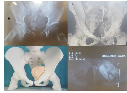
Figure 1. Radiological evaluations
a. Simple pre-operative radiographic demonstration of the tumor; b. 3-D CT scan; c. 3-D printed anatomical shape of the tumor;
In our department, simple radiography was done that demonstrated huge radiolucency in the acetabular dome extending to the ilium and pubic with pressure on the bladder. The laboratory data in blood tests were normal, but anemia and leukocytosis were reported in cell count.
MRI showed that the lesion of the left hip was con-sistent with multiple cysts and fluid-fluid levels arising bone that extended to the pelvic space. Also, we did 3-dimensional reconstruction, using a fine-cut CT scan before the operation to have a better approach (Figure 1).
Diagnostic angiography was done to examine all parts of the mass and its vascular relation with pelvic vessels. The lesion was fed from all branches of iliac (including posterior, anterior, and internal iliac artery) and external iliac arteries (branches of the pudendal and inguinal ar-eas). Embolization in the main vessels was done to re-duce bleeding during the operation.
With the high suspicion of ABC tumor, the operation was performed by the ilioinguinal approach, and the tu-mor was removed with extensive curettage. Medial wall perforation of acetabulum had not disturbed the ana-tomic stability covered by local soft tissue and muscles. After the operation, a sample of the tumor was sent for culture and pathological examination. The culture re-sult of secretions was negative and the pathology report confirmed the ABC tumor. The hip range of motion and strengthening exercises were started after pain relief.
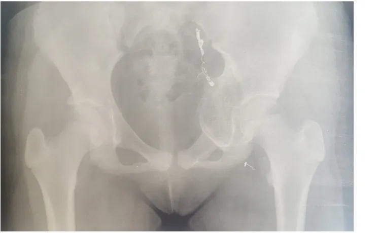
After 2-year follow up, the patient’s general condition was good, her hip range of motion was full, and the lytic mass was completely exchanged by bone (Figure 2).
3. Discussion
Despite the long experience with ABCs, the cause of lesion is still unclear [7]. However, ABC may be associ-ated with some other bone lesions. The pathologic find-ings cannot support the degenerative vascular procedure in the lesion for some benign bone lesions. Few patho-logic specimens contain tissues that are highly character-istic or diagnostic for other benign tumors [8].
Challenging therapeutic problems are common in ABCs of the pelvis and sacrum. Acetabular lesions could be associated with some complications such as pathological fracture, protrusio acetabuli with medial migration of the femoral head into the pelvis, and hip subluxation and dislocation. Transcatheter embolization techniques are essential to the primary treatment for a selected group of benign tumors of bone [9].
Giuseppe Rossi et al. reported a case of pathologic frac-ture of the left femoral neck in a 5-year-old girl occurring through an eccentric expansile osteolytic lesion involv-ing the neck part of the greater trochanter and the head of the left femur. Because of the unfavorable anatomic lo-cation, selective embolization prior to surgical treatment was done. The reduced vascularized zone of inhibition besides the treatment of osseous parts made surgeons choose a different modality. A long-term follow-up of 5
years for the patient had shown the abnormality of the growth plate of the great trochanter. It was concluded that leg length discrepancy may be the result of the ex-pansive nature of the lesion itself [4].
In Syvanen et al. study, 1 out of 18 children had this complication in the pelvis, and the histologically-proven primary ABC was operated with curettage and bioactive glass filling. The mean age of the patients in this study was 11.3 years and the mean follow-up was 2 years. At last, 2 of the patients (11%) had shown recurrence and were re-operated. Bone remodeling with remaining growth and no intra- or post-operative complications were recorded. It was concluded that bioactive glass is a suitable material for primary ABCs, which will not affect bone growth [10].
Al Sobeai et al. had presented a case of ABC with the involvement of the right ilium and supra-acetabular area in a 24-year-old female patient with a growing mass in her right hip. As the mass was too large to be removed all at once, it was released in pieces. The defect in the dome of the acetabulum owing to tumor extension was recon-structed with calcium carbonate-based bone graft. The patient’s full weight-bearing mobilization was started a day after the operation [11]. The treatment method of ABC of the pelvis must be individualized depending on the location, aggressiveness, and extent of the lesion, but as a rule, a curettage is necessary to reduce the chance of recurrence.
We did 3-D reconstruction before the operation to have a better approach. The diagnosis of hip lesions is usually tricky. CTs and MRIs sometimes do not provide clear criteria for the diagnosis of the ABC, which is sometimes mistaken with other lesions like eosinophilic granuloma, giant cell tumor, nonossifying fibroma, unicameral bone cyst, fibrous dysplasia, chondroblastoma, chondrosar-coma, chondromyxoid fibroma, and so on. [12-16].
Most of the reported recurrences of the ABCs occur within 18 months after the primary intervention [14]. In this case, we concerned about hip joint destruction because of the medial wall perforation and risk of re-currence owing to the large extension of the tumor and difficult accessibility. After a 30-month follow-up, the patient had a normal hip joint without any complaint.
The correct diagnosis and suitable treatment of ABC in the pelvic area are difficult because of its location, and exact paraclinical studies are essential to examine the best approach. This challenging lesion is difficult to manage surgically since it depends on the lesion’s size
and stability. It is concluded that unlike the large lesions, small cysts could be managed more conservatively.
Ethical Considerations
Compliance with ethical guidelines
Written informed consent was obtained from the patient for the publication of this case report and any accompa-nying image. A copy of the written consent is available for review by the Editor-in-Chief of this journal.
Funding
This article is funded by Mashhad University of Medical Sciences
Authors' contributions
Conceptualization: Omid Shahpari; Writing–original draft: Ali Dastjerdi, Omid Shahpari; Writing–review & editing: Mahdi Mazloumi, Nafise Elahpour; Supervision: Omid Shahpari, Mahdi Mazloumi.
Conflict of interest
The authors declared no conflict of interests.
References
[1] JAffe HL, Lichtenstein L. Solitary unicameral bone cyst: With
emphasis on the roentgen picture, the pathologic appear-ance and the pathogenesis. Arch Sur. 1942; 44(6):1004-25.
[DOI:10.1001/archsurg.1942.01210240043003]
[2] Agarwal A, Goel P, Khan SA, Kumar P, Qureshi NA. Large
aneurysmal bone cyst of iliac bone in a female child: A case
report. J Orth Surg Res. 2010; 5(1):24-36.
[DOI:10.1186/1749-799X-5-24] [PMID] [PMCID]
[3] Elkattah R, Foulk B. A suspected pelvic aneurysmal bone cyst
in pregnancy. Case Rep Obstet Gynecol. 2013; 2013(676087):1-4. [DOI:10.1155/2013/676087] [PMID] [PMCID]
[4] Rossi G, Angelini A, Mavrogenis AF, Rimondi E, Ruggieri
P. Successful treatment of aneurysmal bone cyst of the hip in a child by selective transcatheter arterial embolization.
J Vasc Interv Radiol. 2010; 21(10):1591-5. [DOI:10.1016/j.
jvir.2010.06.016] [PMID]
[5] Capanna R, Bertoni F, Bettelli G, Present D, Biagini R,
Rug-gieri P, et al. Aneurysmal bone cysts of the pelvis. Arch
Orthop Trauma Surg. 1986; 105(5):279-84. [DOI:10.1007/
BF00449926] [PMID]
[6] Sobeai MA, Juhani WA, editors. Pelvic aneurysmal bone cyst.
[7] Dorfman HD and Czerniak B. Giant-cell lesions. In: Dorfman
HD, Czerniak B (editors). Bone Tumors. St Louis: Mosby; 1998. [8] Hadders HN, Oterdoom HJ. The identification of aneurysmal
bone cyst with haemangioma of the skeleton. J Pathol
Bac-teriol . 1956; 71(1):193-200. [DOI:10.1002/path.1700710125]
[PMID]
[9] Murphy WA, Strecker EB, Schoenecker PL. Transcatheter
embolisation therapy of an ischial aneurysmal bone cyst.
J Bone Joint Surg. 1982; 64(2):166-8.
[DOI:10.1302/0301-620X.64B2.7068734]
[10] Syvanen J, Nietosvaara Y, Kohonen I, Koskimies E, Haara
M, Korhonen J, et al. Treatment of aneurysmal bone cysts
with bioactive glass in children. Scandinavian J Surg. 2018;
107(1):76-81. [DOI:10.1177/1457496917731185] [PMID]
[11] Al Sobeai M, Al Juhani W. Pelvic aneurysmal bone cyst:
Case report and literature review. Int J Surg Case. 2015;
1(2):20-5. [DOI:10.15713/ins.jmrps.16]
[12] Papagelopoulos PJ, Choudhury SN, Frassica FJ, Bond JR,
Unni KK, Sim FH. Treatment of aneurysmal bone cysts of the pelvis and sacrum. J Bone Joint Surg Am. 2001; 83(11):1674-81.
[DOI:10.2106/00004623-200111000-00009] [PMID]
[13] Cottalorda J, Kohler R. Sales de Gauzy J, Chotel F, Mazda
K, Lefort G, et al. Epidemiology of aneurysmal bone cyst in children: A multicenter study and literature review. J
Pedi-atr Orthop B. 2004; 13(6):389-94.
[DOI:10.1097/01202412-200411000-00008] [PMID]
[14] Campanacci M, Capanna R, Picci P. Unicameral and aneu
-rysmal bone cysts. Clin Orthop Relat Res. 1986; 3(204):25-36.
[DOI:10.1097/00003086-198603000-00004]
[15] Mahnken AH, Nolte-Ernsting CC, Wildberger JE, Heussen
N, Adam G, Wirtz DC, et al. Aneurysmal bone cyst: Value
of MR imaging and conventional radiography. Eur Radiol.
2003; 13(5):1118-24. [PMID]
[16] Vergel De Dios AM, Bond JR, Shives TC, McLeod RA, Unni
KK. Aneurysmal bone cyst, a clinicopathologic study of
238 cases. Cancer. 1992; 69(12):2921-31.