R E S E A R C H
Open Access
Optimizing mesoderm progenitor selection
and three-dimensional microniche culture
allows highly efficient endothelial
differentiation and ischemic tissue repair
from human pluripotent stem cells
Fengzhi Zhang
1, Lin Wang
1, Yaqian Li
2, Wei Liu
2, Fuyu Duan
1, Rujin Huang
1, Xi Chen
1, Sophia Chia-Ning Chang
3,4,
Yanan Du
2and Jie Na
1*Abstract
Background:Generation of large quantities of endothelial cells is highly desirable for vascular research, for the treatment of ischemia diseases, and for tissue regeneration. To achieve this goal, we developed a simple, chemically defined culture system to efficiently and rapidly differentiate endothelial cells from human pluripotent stem cells by going through an MESP1 mesoderm progenitor stage.
Methods:Mesp1 is a key transcription factor that regulates the development of early cardiovascular tissue. Using an MESP1-mTomato knock-in reporter human embryonic stem cell line, we compared the gene expression profiles of MESP1+and MESP1−cells and identified new signaling pathways that may promote endothelial differentiation. We also used a 3D scaffold to mimic the in vivo microenvironment to further improve the efficiency of endothelial cell generation. Finally, we performed cell transplantation into a critical limb ischemia mouse model to test the repairing potential of endothelial-primed MESP1+cells.
Results:MESP1+mesoderm progenitors, but not MESP1−cells, have strong endothelial differentiation potential. Global gene expression analysis revealed that transcription factors essential for early endothelial differentiation were enriched in MESP1+cells. Interestingly, MESP1 cells highly expressed Sphingosine-1-phosphate (S1P) receptor and the addition of S1P significantly increased the endothelial differentiation efficiency. Upon seeding in a novel 3D microniche and priming with VEGF and bFGF, MESP1+cells markedly upregulated genes related to vessel development and regeneration. 3D microniches also enabled long-term endothelial differentiation and proliferation from MESP1+ cells with minimal medium supplements. Finally, we showed that transplanting a small number of endothelial-primed MESP1+cells in 3D microniches was sufficient to mediate rapid repair of a mouse model of critical limb ischemia.
Conclusions:Our study demonstrates that combining MESP1+mesoderm progenitor cells with tissue-engineered
3D microniche and a chemically defined endothelial induction medium is a promising route to maximizing the production of endothelial cells in vitro and augment their regenerative power in vivo.
Keywords:Human pluripotent stem cells, Endothelial cells, MESP1, 3D culture, Vascularization
* Correspondence:[email protected]
1Center for Stem Cell Biology and Regenerative Medicine, School of
Medicine, Tsinghua University, Beijing 100084, China
Full list of author information is available at the end of the article
Background
Human pluripotent stem cells (hPSCs) can self-renew indefinitely and differentiate into all types of tissues in the body. Therefore, it can be an unlimited source to provide specialized cells for biomedical research and cell-based therapy. hPSC-derived endothelial cells (ECs) may be used to study angiogenesis and regenerate blood vessels in ischemic diseases [1, 2]. Two methods of endothelial differentiation have been developed. One protocol uses embryoid bodies (EBs) as a differentiation intermediate, followed by EC purification using specific surface antibodies [1, 2]. As the cell types in EBs are highly heterogeneous, the differentiation efficiency of this protocol is relatively low. The second protocol was developed more recently, where, a monolayer culture and sometimes chemically defined conditions are used to achieve directed and more efficient EC differenti-ation. hPSCs are first induced to form mesoderm, then vascular endothelial growth factor (VEGF) and basic fibroblast growth factor (bFGF) are added to specify the
endothelial fate [3–7]. Such monolayer and serum-free
protocols can be modified to screen for small molecules that may enhance differentiation and are more compat-ible with requirements for clinical applications. The re-ported yields of ECs range from 20 to 80%, depending on the time period of differentiation and whether selec-tion and purificaselec-tion steps were included in the proto-col [3–7].
To achieve efficient in vitro differentiation and obtain cells more closely resembling in vivo counterparts, it is important to follow the developmental principle of the desired lineage. During embryo development, a sub-group of extraembryonic mesoderm cells forms primitive endothelial cells in the yolk sac [8]. Mesoderm posterior 1 (Mesp1) is a basic helix–loop–helix (bHLH) transcrip-tion factor first expressed in the nascent mesoderm dur-ing gastrulation [9, 10]. These cells then migrate laterally and anteriorly and give rise to the heart and great vessels [9, 10]. In addition, Mesp1 progenies also contribute to extraembryonic mesoderm [9]. The fate of this subgroup of cells is less well studied. Studies in mice and zebrafish convincingly demonstrated that Mesp1 represents the earliest marker of cardiovascular progenitors [9, 11, 12]. However, whether MESP1 progenitor cells can be a good source to generate ECs during hPSC in vitro differenti-ation is unknown.
Previously, we generated an MESP1 reporter human embryonic stem cell (hESC) line with mTomato knocked in just before the stop code of the MESP1 cod-ing sequence (manuscript submitted). In the latter study, we showed that in mouse embryos, Mesp1 pro-geny cells gave rise to vasculature in the yolk sac of the
mouse embryo. In hPSCs, MESP1-mTomato+ cells
could differentiate to ECs at high efficiency. Moreover,
plating MESP1+ cells in three-dimensional (3D)
micro-niches significantly improved the EC differentiation efficiency and the long-term survival ability, and it fa-cilitated cell transplantation repair of a critical limb is-chemia mouse model. A global gene expression study revealed novel genes and pathways that might regulate EC differentiation and proliferation during the meso-derm induction and endothelial fate specification stages. Our work suggests that following developmental cues and using 3D microniches that resemble the in vivo cellular microenvironment may greatly facilitate directed differentiation from hPSCs and augment the repairing power of transplanted cells.
Methods hPSC culture
H9 hESCs (WiCell Institute Inc., Madison, WI, USA), H9-MESP1-mTomato hESC reporter cell line (gener-ated by LW), and human induced pluripotent stem cells (hiPSCs) (CD34-iPSCs, generated by FD) were routinely maintained on MEF feeders in the hESC
medium: KnockOut Dulbecco’s modified Eagle’s
medium (DMEM) culture medium supplemented with 20% (vol/vol) KnockOut serum replacement, 1% non-essential amino acids, 1 mM L-GlutaMAX-I, 0.1 mM
β-mercaptoethanol, and 8 ng/ml bFGF. They were
passaged with 1 mg/ml collagenase IV (Invitrogen,
Carlsbad, CA, USA) and seeded onto a 25 cm2 flask
that had been previously coated with 0.1% gelatin so-lution (Sigma-Aldrich, St. Louis, MO, USA). To achieve feeder-free culture, hESCs were maintained on Matrigel (BD Biosciences, San Jose, CA, USA)-coated plates (Corning, Corning, NY, USA) in E8 medium (Stemcell Technologies, Vancouver, Canada).
Generation of MESP1-mTomato knocking-in reporter cell line
The homologous recombination donor vector was com-posed of the MESP1 left arm, T2A fused with a membrane-bound tdTomato (mTomato), PGK promoter-driving puromycin resistance gene (PGK-Puro), MESP1 right arm, and MC-1 promoter-driving TK gene. H9 cells were electroporated with TALEN and donor vectors by Neon microporator (Invitrogen). After puromycin selec-tion, targeted clones were picked and expanded for characterization. Detailed protocols (design of TALEN pair and synthesis of tandem arrays of TALE repeats) have been described elsewhere (manuscript submitted).
RNA isolation, Q-PCR and high-throughput RNA sequencing
Total RNA was isolated from undifferentiated hESCs, differentiation day 3 cells, and differentiation day 8 cells on two-dimensional (2D) plates and 3D microniches using RNeasy Plus Mini Kit (Qiagen, Hilden, Germany)
sample was reverse-transcribed with Superscript III (TransGen Biotech, Beijing, China). Quantitative real-time polymerase chain reaction (Q-PCR) reactions were performed using GoTaq qPCR Master Mix (Promega, Madison, WI, USA) in a CFX96 Real-Time System (Bio-Rad, Hercules, CA, USA) and results were analyzed with the Bio-Rad CFX Manager program. The sample input was normalized against the Ct (critical threshold) value of
GAPDH. Primer sequences are listed in Additional file 1: Table S1. For high-throughput RNA sequencing, comple-mentary DNA (cDNA) libraries were prepared using the
TruSeq™ RNA Sample Preparation kit (Illumina, San
Diego, CA, USA) and sequencing was performed at the Biopic sequencing facility of Peking University (http://
biopic.pku.edu.cn/english/). The clean reads were
mapped to human reference genome (hg19) using BWA software. Cluster analysis of gene expression pat-terns was performed using Cluster 3.0 and JavaTreeview software. Gene ontology (GO) term enrichment was ana-lyzed using the database for annotation, visualization, and integrated discovery (DAVID) (https://david.ncifcrf.gov). Data are publicly available at the National Center for Bio-technology Information with Gene Expression Omnibus (GEO), accession number GSE79470.
MESP1+mesoderm progenitor induction
MESP1+ mesoderm progenitor induction was performed
based on the method described by Cao et al. [13] with some modifications. Briefly, undifferentiated hESCs cul-tured in E8 medium were digested into single cells by Accutase (EMD Millipore, Billerica, MA, USA) and plated onto Matrigel-coated culture dishes at a density
of 6 × 10⁴ cells/cm2 in a medium containing DMEM/
F12, 1 × B27 supplement (without vitamin A), 1%
L-GlutaMAX-I, 50 μg/ml ascorbic acid (Sigma-Aldrich),
induced by 25 ng/ml bone morphogenetic protein
(BMP)4 and 10μM GSK3 inhibitor CHIR99021 (Tocris
Bioscience, Bristol, UK) for H9 and induced by 2 μM
CHIR99021 for CD34-iPSC. Rho-associated coiled-coil kinase (ROCK) inhibitor Y27632 (Tocris Bioscience)
(5μM) was added during cell dissociation and replating
for 24 h and it was removed during the medium change. The monolayer-cultivated cells were harvested at differentiation day 3 for further analysis.
Differentiation of MESP1+cells to endothelial cells in 2D culture system and 3D microniches
Day 3 MESP1+ cells were dissociated with Accutase for
10 min and plated onto Matrigel-coated culture dishes at a density of 2 × 10⁴cells/cm2in a basal medium and stimulated with 25 ng/ml VEGF plus 10 ng/ml bFGF (both Sino Biological, Inc., Beijing, China) for 5 d. For the 3D microniches culture, biodegradable gelatin microcryogels (GMs) were prepared using a microstencil
array chip as previously described [14]. Briefly, 6% (wt/vol) gelatin precursor solution was mixed with
0.3% glutaraldehyde, and then 200 μl of precursor
so-lution was pipetted and scraped onto the microstencil array chip. The microstencil array chip underwent cryogelation for 16 h and was then lyophilized for 30 min (Beijing Boyikang Laboratory Instruments, Beijing, China). GMs were then harvested by a PMMA Ejector Pin array fabricated with a desktop
3D milling machine (MDX-40A; Roland DG,
Shizuoka-ken, Japan). Before loading the cells, the harvested and dried GMs in the dish were sterilized by an ethylene oxide sterilization system that per-formed a 12 h degassing step under vacuum after 12 h of gas exposure (AN74j/Anprolene; Anderson
Products, Haw River, NC, USA) [15]. A 50 μl MESP1
+
cell suspension (1 × 10⁷ cells/ml) was subsequently
pipetted onto 500 tightly packed GMs and automatic-ally absorbed to hydrate the porous structures and then maintained in a humidified chamber and incu-bated at 37 °C for 2 h to allow cell attachment.
Sub-sequently, the basal medium supplemented with
25 ng/ml VEGF plus 10 ng/ml bFGF was added for long-term culture.
FACS analysis
Cells were dissociated into single cells as described be-fore, and resuspended in a FACS washing buffer (PBS with 5% fetal calf serum (FCS) and 2.5 mM EDTA). To perform direct flow cytometry, the samples were then stained for FITC-conjugated CD31 (1:20; Miltenyi Bio-tec, Bergisch Gladbach, Germany), and FITC-conjugated mouse IgG2a (for CD31, 1:20; Miltenyi Biotec) was used as isotype-matched negative control. Data were collected with a FACSCaliber flow cytometer (BD Biosciences) and analyzed using FlowJo software (Tree Star, Inc., Ashland, OR, USA).
Immunostaining analysis
Cells were fixed with 4% paraformaldehyde, perme-abilized in 0.5% Triton X-100 (Sigma-Aldrich), blocked in 10% normal goat serum (Origene, Rockville, MD, USA), and then incubated with primary antibodies against MESP1 (1:200; Abcam, Cambridge, MA, USA), CD31 (1:100; Santa Cruz Biotechnology, Dallas, TX,
USA), VE-cadherin (1:200; Abcam), αSMA (1:200;
Abcam), human nuclei antibody (1:100; Abcam) in 4 °C overnight and detected by DyLight 488- or 549-conjugated secondary antibodies (Thermo Fisher Scientific, Waltham,
MA, USA). Nuclei were stained with 4′,
hPSC-EC functional studies
The biological function of hPSC-ECs was assessed by
DiI-conjugated acetylated low-density lipoprotein
(DiI-ac-LDL) uptake and vascular tube formation. For the DiI-ac-LDL uptake assay, hPSC-ECs were incu-bated with 20 mg/ml of DiI-Ac-LDL (Yeasen Biotech, Hong Kong) at 37 °C for 6 h, washed with PBS, and stained with Hoechst 33258 (Dojindo Kumamoto,
Japan). For the vascular tube formation assay, 1 × 10⁵
cells were plated into one well of 24-well plates pre-coated with Matrigel (BD Biosciences) and incubated at 37 °C for 12 h.
Mouse model of hind limb ischemia fluorescence imaging
Procedures were performed on female BALB/c nude mice (20–25 g; 8–10 weeks) as previously described [16] with the approval from the Internal Review Board, La-boratory Animal Research Center, Tsinghua University. Unilateral left hind limb ischemia in mice was induced by ligating the proximal end of the femoral artery and its branches using 5-0 silk surgical suture as described in [14]. The overlying skin was then closed by 5-0 silk sur-gical suture. Cell transplantation was performed after skin closure. 300 medium-soaked GMs (acellular
con-trols) or 300 cell-load GMs (3 × 10⁵cells) were washed
with PBS, then resuspended in 300 μl gelatin solution
and intramuscularly injected into three sites of the graci-lis muscle around the artery incision using 1 ml syringes with a 23-gauge needle. For free cell injection, 1 × 10⁷ cells were mixed with Matrigel and injected as above.
Blood perfusion analysis was performed after surgery at the indicated days post operation on a home-made fluorescence imaging system [17] in reflectance geom-etry. Animals were injected with indocyanine green
(ICG, 0.1 ml of 100 μg/ml) through the tail vein and
time-series fluorescence images were collected for 100 min at intervals of 0.2 s.
Statistical analysis
Quantitative data are expressed as mean ± standard devi-ation. The statistical significance was determined using a Student’s t test (two-tailed) for two groups or one-way
ANOVA for multiple groups. A value of p< 0.05 was
considered statistically significant.
Results
MESP1 reporter marked cardiovascular progenitor cells
Mesp1 is a master regulator of early heart development
[9, 11, 18]. Using mouse embryos from Mesp1cre/+
crossed with ROSA26Sortm4(ACTB-tdTomato,-EGFP)Luo, we
found that in addition to the heart, Mesp1 progenies (marked by GFP) also formed yolk sac blood vessels, suggesting that a subgroup of Mesp1 cells may have strong endothelial differentiation potential given the
appropriate induction cues (Fig. 1a). To study MESP1 cells during endothelial differentiation in the human sys-tem, we generated an MESP1-mTomato reporter hESC line through TALEN-mediated homologous recombin-ation, which enabled us to monitor and purify MESP1-expressing mesoderm progenitor cells (Fig. 1b; manuscript submitted).
MESP1-mTomato reporter cells do not show any fluorescence in undifferentiated hESCs cultured in E8 medium, but strongly turned on red fluorescence in re-sponse to BMP4 and increasing concentrations of CHIR99021 (a small molecule inhibitor of GSK3 and activator of canonical Wnt signaling) treatment (Fig. 1c and d). We could obtain nearly 100% MESP1-mTomato-positive cells by treating cells with a combination of BMP4 (25 ng/ml) and glycogen synthase kinase 3
(GSK3) inhibitor CHIR99021 (8–10 μM) in a
chem-ically defined medium consisting of DMEM/F12 and B27(-VitA) supplement (Fig. 1c and d). To obtain a
homogeneous population of MESP1+cells,
undifferenti-ated cells cultured in E8 without feeders were dissoci-ated into single cells in the presence of Rho-associdissoci-ated coiled-coil kinase (ROCK) inhibitor Y27632, replated on Matrigel and then treated with BMP4 (25 ng/ml)
and CHIR99021 (10 μM). By day 3, FACS analysis
showed that most cells were positive for mTomato (Fig. 1e). We also examined the expression of mesoder-mal and cardiac lineage marker genes by Q-PCR.
MESP1+ cells significantly upregulated mesoderm
tran-scription factors T/BRACHYURY (T), MESP1, and
GATA4, as well as early cardiac markers TBX5 and
NKX2.5. In contrast, the expression of pluripotency,
endoderm and neuroectoderm marker genes, OCT4,
SOX2, NANOG, SOX17, and PAX6 were significantly
downregulated in MESP1+cells (Fig. 1f ).
Immunostain-ing confirmed that mTomato-positive cells co-localized with endogenous MESP1 protein detected by an anti-MESP1 antibody (Fig. 1g). Taken together, anti- MESP1-mTomato reporter cells reflected the expression of endogenous MESP1 and exhibited gene expression typ-ical of early cardiovascular progenitor cells.
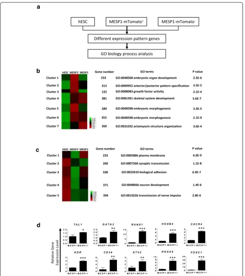
Next, we performed high-throughput RNA sequencing
of MESP1-mTomato positive cells (MESP1+
) at day 3 of differentiation and compared their gene expression
pro-file with MESP1-mTomato negative cells (MESP1–) and
undifferentiated hESCs (Fig. 2a). A total of 1951 genes showed a greater than 1.5-fold increase in
MESP1-mTomato+ versus undifferentiated hESCs, which were
grouped into seven clusters based on different dynamic
patterns in undifferentiated hESCs, MESP1+, and
MESP1– cells (Fig. 2b). Gene ontology (GO) analysis
showed that clusters 1, 2, 3, and 5 (upregulated in
MESP1+ compared with undifferentiated hESCs or
organ development, anterior/posterior pattern specifica-tion, growth factor activity, and embryonic morphogen-esis, respectively, which is in accordance with MESP1 functions during embryo development in vivo (Fig. 2b
and Additional file 2: Table S2 and Additional file 3:
Table S3). A total of 1596 genes in MESP1+cells showed
more than 1.5-fold decrease compared to undifferenti-ated hESCs and they were divided into five clusters
Fig. 1Generation of MESP1+cells from human embryonic stem cells.aMesp1 progeny cells formed yolk sac blood vessels. Images of Mesp1
lineage cells (green) on the yolk sac of E9.5 embryos of Mesp1cre/+/ROSA26Sortm4(ACTB-tdTomato,-EGFP)Luogenetic background.bImage showing
MESP1-mTomato knock-in reporter cells.cProtocol to derive MESP1+cells from hESCs by treatment with BMP4 and CHIR99021.dPercentage of
MESP1+cells on day 3 under different induction conditions (n= 3,****p< 0.0001.).eFACS analysis showing, on day 3, that 97.93% of cells turned
according to their different dynamic patterns (Fig. 2c and Additional file 2: Table S2 and Additional file 3: Table S3). GO analysis showed that clusters 4 and 5 were closely related to neural differentiation, which reflects that the one important aspect of mesoderm induction is to in-hibit neural fate [19]. Interestingly, the expression of genes involved in the plasma membrane and biological adhesion obviously decreased. This is in agreement with the meso-derm differentiation process that involves an epithelial-to-mesenchymal transition and dramatic downregulation of cell–cell adhesion and selected extracellular matrix (ECM) genes [18]. Genes important for EC differentiation such as
TAL1, GATA2, RUNX1, HOXB4, CXCR4, KDR, CD34,
ETV2,HOXA9, andFOXC1were among the most
signifi-cantly upregulated genes in MESP1-mTomato+ cells, as
confirmed by Q-PCR analysis (Fig. 2d).
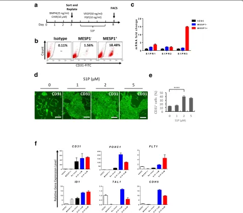
Sphingosine-1-phosphate significantly enhanced CD31 endothelial differentiation
To test whether MESP1-mTomato+ cells have stronger
endothelial differentiation potential, we used a mono-layer, serum-free, and chemically defined differentiation
system as shown in Fig. 3a. Sorted MESP1+and MESP1–
were replated on Matrigel, and VEGF (50 ng/ml) and
Fig. 3S1P greatly enhanced CD31 expression and upregulated endothelial marker genes expression.aProtocol for endothelial differentiation.
bMESP1+cells have significantly greater ability to differentiate to endothelial cells compared with MESP1–cells.cQ-PCR analysis of S1P receptor
expression in undifferentiated hESC, differentiation day 3 MESP1-mTomato–(MESP1–) and MESP1-mTomato+cells (MESP1+).dImmunostaining analysis
showing that S1P treatment greatly enhanced the generation of CD31+cells on differentiation day 8. Scale bars: 100μm.eQuantification of
FACS analysis of CD31+cells percentage after S1P treatment on differentiation day 8 (n= 3,****p< 0.0001).fQ-PCR analysis showing S1P treatment
bFGF (10 ng/ml) were added. After 5 days, cells were harvested for FACS analysis of endothelial cell surface
marker CD31 expression. Some 18.5% of MESP1+sorted
cells expressed CD31 compared to only 1.5% from MESP1– cells (Fig. 3b). This result suggested that, as in
the embryo, MESP1+cells indeed had stronger
endothe-lial differentiation potential. From our RNA-seq data, we found that Sphingosine-1-phosphate receptor 3 (S1PR3), but not S1PR1 and S1PR2, was significantly upregulated
in MESP1+ cells. Q-PCR analysis confirmed the
expres-sion patterns of S1PR1, S1PR2, and S1PR3. S1PR3 was
nearly 15-fold higher in MESP1+ cells compared with
hESCs and MESP1–(Fig. 3c). We next wanted to
deter-mine whether Sphingosine-1-phosphate (S1P) treatment could also affect hPSC endothelial differentiation. S1P
was added during day 3–8 of differentiation, together
with VEGF and bFGF. Immunostaining and FACS ana-lysis showed that S1P greatly enhanced CD31 expres-sion. Without S1P, CD31 was detected on 13.5% of cells,
while after treatment with 2 μM of S1P, 41.6% of cells
expressed CD31 (Fig. 3d and e). In addition, Q-PCR ana-lysis showed that S1P treatment significantly upregulated
endothelial marker genes, including TAL1, CDH5, ID1,
FLT1, FOXC1, and CD31 (Fig. 3f ). A similar result was obtained in human iPSC (Additional file 4: Figure S1).
Thus, optimizing the MESP1+population percentage
dur-ing the mesoderm induction stage and co-stimulation with S1P during the EC generation stage could markedly boost the CD31+EC differentiation efficiency.
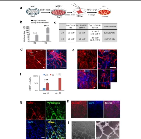
Microniches dramatically improved the endothelial differentiation efficiency and cell survival
In vivo vasculogenesis and angiogenesis occur in 3D mi-croenvironments. Next, we tested whether a 3D scaffold
could improve endothelial differentiation from MESP1+
progenitor cells. To this end, we employed a biodegrad-able 3D microscale scaffold, which was fabricated by cryogelation of gelatin, and hence was called gelatin microcryogels (GMs) [14]. These 3D GMs had an inter-connected macroporous structure with pore sizes in the
range of 30–80μm. They had been shown to be
particu-larly suitable for human mesenchymal stem cell (hMSC) growth [14]. We developed a simple differentiation as depicted in Fig. 4a. Undifferentiated hESCs were dissoci-ated into single cells and cultured in E8 medium to about 30% confluency. Then BMP4 and CHIR99021 were added for 3 days to induce a high percentage of
MESP1-mTomato+ cells. MESP1+ cells were then
digested to single cell suspension and loaded onto 500 tightly packed 3D GMs and incubated at 37 °C for 2 hours, during which time cells were absorbed into GMs spontaneously. Afterwards, differentiation medium supplemented with VEGF and bFGF was added for more than 5 days (Fig. 4a). Culturing in 3D GMs significantly
promoted cell proliferation, from identical plating hESC numbers (1 × 10⁶cells), after 15 days of differentiation,
the 3D GM group contained an average of 4.8 × 10⁸
cells, while the conventional 2D culture group had only 1.2 × 10⁸cells (Fig. 4b and c). Moreover, 3D GM differ-entiation required less frequent medium changes, only once every 3 days, compared to daily medium changes in 2D differentiation during a 12-day window. The quan-tities of VEGF and bFGF growth factors used were much smaller (Fig. 4c). Based on our calculation, about 320 million ECs could be obtained from one million input hESCs after 15 days of differentiation in 3D, while only 17 million ECs could be obtained in 2D culture during the same time window. Thus, the 3D GM culture ap-peared to be significantly more cost-effective. Immuno-staining revealed that cells formed a tubular network strongly positive for CD31 both in 2D and 3D (Fig. 4d and e). FACS analysis showed that, on differentiation
day 10, the 3D GMs contained 49.1 ± 5.9% CD31+ cells
while the 2D culture only had 14.1 ± 1.8% CD31+ cells.
By day 27, 86.7 ± 5.0% of cells in 3D GMs were CD31+,
while only 1.9 ± 0.5% of cells on the 2D surface were
CD31+ positive owing to overgrowth and cell death
(Fig. 4f ). Additional experiments using human iPSCs showed similar results (Additional file 5: Figure S2). We also performed tube formation and Ac-LDL absorption assays using cells derived from hESC and iPSC through
the MESP1+progenitor stage. ECs digested out from 3D
GMs after 12 days strongly expressed CD31 and VE-cadherin (Fig. 4g). They took up Ac-LDL and could organize into a tubular network typical of ECs (Fig. 4h and i). Thus, 3D GMs appeared to significantly boost long-term cell survival of ECs differentiated from MESP1+cells.
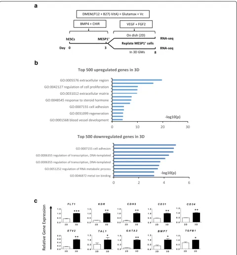
Global gene expression analysis of MESP1+cells endothelial differentiation in 2D and 3D
To investigate how the 3D microenvironment influences gene expression, we performed high-throughput RNA
sequencing of MESP1+cells differentiated in 2D and 3D,
respectively, on day 8, as shown in Fig. 5a. A total of 2102 genes were upregulated more than 1.5-fold in 3D. Among the top 500 genes upregulated in 3D, GO
ana-lysis showed a significant enrichment (p≤0.01) of the
genes upregulated in 3D, as shown Fig. 5c. Endothelial
lineage surface markers FLT1, KDR, CDH5, CD31, and
CD34, growth factors BMP7 and TGFB1, and
transcrip-tion factors ETV2, TAL1, and GATA2 all exhibited
en-hanced expression of a selection of the genes upregulated in the RNA-seq data (Fig. 5c). These results suggested
that a 3D microenvironment might enhance EC differ-entiation and survival from multiple aspects such as enrichment of secreted growth factors, remodeling of cell adhesion and extracellular matrix, and reorganization of the cytoskeleton, which directly or indirectly affect gene expression.
Fig. 43D microniche significantly improved endothelial differentiation efficiency and helped to maintain long-term cell survival.aProtocol for endothelial differentiation from hESC in 3D microniches.bBar graph quantification of CD31+ cell number obtained from 2D and 3D cultures on day 15.cCell numbers at different times and culture medium used in 2D and 3D are listed in the table (n= 3,***p< 0.001).dImmunostaining
showing CD31 expression in 2D condition on differentiation day 10. Scale bars: 100μm.eImmunostaining showing CD31 expression in 3D microniches on differentiation day 10 visualized by multiphoton microscopy. Scale bars: 100μm.fPercentage of CD31+cells during endothelial differentiation under
2D condition and 3D microniches on day 10 and day 27 (n= 3,***p< 0.001).gECs differentiated from hiPSCs were immunostained with VE-cadherin
MESP1 progenitor cell-derived ECs enhanced vascular repair of critical limb ischemia
To evaluate the therapeutic potential of ECs derived
from MESP1+ cells, we employed a critical limb
ische-mia (CLI) mouse model where the ischeische-mia was induced by ligation of the left femoral artery. After surgery, GMs
alone (control), MESP1+ cells induced with VEGF +
bFGF for 5 days (1 × 10⁷cells) or 3 × 10⁵ MESP1+ cells loaded in 3D GMs and induced with VEGF + bFGF for 5 days (Cell + GM) were injected into the ligation site (Fig. 6a). The therapeutic efficacy of cells was examined by evaluating the appearance and physiological status of ischemic limbs at 0, 7, 14, 21, and 28 day after the sur-gery. Both 1 × 10⁷ cells alone (n= 6) and 3 × 10⁵ cells
loaded in 3D GMs (Cell + GM) salvaged the ischemia limb within 7 days, while no limb salvage was observed in GM only control group (n= 6) after 28 days (Fig. 6b). The Cell + GM combination significantly reduced the
number of cells needed to salvage the CLI, since trans-planting 3 × 10⁵cells alone failed to salvage the ischemic hindlimb (data not shown). A total of 67% of mice com-pletely recovered their legs in the Cell + GM group and
none of them suffered from limb loss (Fig. 6c and d). In addition, the blood flow was significantly improved by Cell + GM or a high dose (107) of free cells (Fig. 6e). Vis-ual inspection revealed that more blood vessels formed across the ligation site in Cell + GM and high-dose (107) free cell-transplanted CLI mice by day 28 (Fig. 6f ). We
also confirmed that MESP1+ cells do not have
tumori-genicity. Undifferentiated MESP1-mTomato reporter cells formed teratoma after subcutaneous injection to the groin region of immune-deficient mice, while
differ-entiated MESP1+ cells did not form any teratoma after
8 weeks (Additional file 5: Figure S4). Thus, MESP1 pro-genies transplanted in 3D GMs can form functional blood vessels in vivo and salvage the CLI mice model.
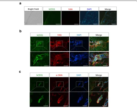
To check whether injected human cells formed blood vessels in CLI mice, we performed immunostaining of a frozen tissue section using anti-human CD31 antibody and anti-human nuclei antibody. As shown in Fig. 7, no
signal was detectable in tissues from GM (Control) transplanted mice (Fig. 7a), while numerous blood ves-sels were labeled with human CD31 and human-specific nuclei antibodies in tissue sections from cells or Cell +
GM transplanted mice, indicating that MESP1+
cell-derived ECs had formed human blood vessels in CLI mice, which are responsible for limb salvation (Fig. 7b
and c). Thus MESP1+cell-derived ECs could be a
poten-tially useful cell source for blood vessel regeneration and the treatment of ischemic diseases.
Discussion
In this study, we found that by optimizing the meso-derm induction stage using MESP1-mTomato reporter cells, we can significantly improve the efficiency of EC generation from hESCs and iPSCs. Previous reports mainly focused on vital functions of Mesp1 during heart development, since it is considered the earliest
marker of cardiovascular development in vertebrates
(Bondue and Blanpain, [11]). Our Mesp1cre/+
/ROSA26-Sortm4(ACTB-tdTomato,-EGFP)Luo lineage tracing experi-ments in mice clearly showed that Mesp1 progenies contributed extensively to yolk sac blood vessels, sug-gesting that Mesp1 cells are also very amenable to tak-ing an endothelial fate given the right induction signal. This is further supported by our RNA-seq results: almost all key regulators of endothelial differentiation
were upregulated in MESP1-mTomato+ cells, such as
ETV2, TAL1, GATA2, HOXA9, FOXC1, KDR, and
CXCR4. Thus, by monitoring MESP1-mTomato
re-porter, when MESP1+ cells were enriched to a high
per-centage by adjusting the concentration of BMP4 and GSK3 inhibitor CHIR99021, we can obtain the highest EC formation efficiency. In our hands, 1 hPSC can gen-erate 10 MESP1 positive cells and subsequently 325.34 ± 21.87 ECs after 15 days of induced differentiation. Similar results can be obtained from human iPSCs and other hESC lines, and thus the EC differentiation effi-ciency positively correlated with the MESP1 expression level during the mesoderm induction stage. Our proto-col is also very simple. We used dissociated single hPSCs cultured in E8 medium as the starting popula-tion, followed by mesoderm induction by only BMP4 and CHIR99021 for 3 days. Then cells can be replated on a 2D surface or in a 3D scaffold for EC induction and expansion. This protocol is cost-effective and can be modified for large-scale production.
Our RNA-seq data of MESP1+ cells provided a
valu-able resource to uncover signaling pathways that may regulate EC differentiation. We found that S1PR3 is specifically upregulated on MESP1+cells. S1P is a lyso-phospholipid mediator found in the blood which has been shown to participate in a wide range of biological responses, including stimulation of cell proliferation, inhibition of apoptosis and regulation of cell shapes and motility [20]. Biological activities of S1P are medi-ated through the S1P receptors, a family of G protein-coupled cell surface receptors. S1PR1, 2, and 3 triple knock-out mice displayed abnormal bleeding and died early during embryogenesis [21]. Mutant embryos had an immature vascular network and the authors con-cluded that although S1P receptors were not required for endothelial fate specification, they play important roles during angiogenesis [21]. Adding S1P combined with VEGF and bFGF during stage II of the differenti-ation significantly upregulated many key factors that
can promote EC formation, such asTAL1,CDH5,ID1,
FLT1, FOXC1, and ETV2, and, as expected, markedly
enhanced CD31+EC generation.
In this study, we also found that growing MESP1+cells in 3D GMs can further boost the EC differentiation effi-ciency. Moreover, 3D GMs can serve as an enrichment
platform, since during long-term culture ECs survived better and 85% of cells were strongly CD31 positive after about 4 weeks in 3D GMs. Another advantage of using a 3D GM culture is that the medium did not turn yellow as fast as the medium used for our 2D cultures, despite rapid cell proliferation. Hence, we only needed to change the medium every 3 days compared to daily changes for the 2D culture. Thus the amounts of VEGF and bFGF required for EC differentiation and maintenance were much smaller for our 3D culture. The RNA-seq results revealed that many genes related to ECM, cell adhesion, and blood vessel development were significantly upregu-lated in 3D. The 3D GMs used in our experiments were fabricated by cryogelation of gelatin. It had an intercon-nected macroporous structure with pore sizes in the
range of 30–80 μm and a high ratio of porosity [14].
These 3D GMs have been shown to attract deposition of ECM proteins and angiogenic growth factors from hu-man mesenchymal stem cells [14]. Endothelial matrix proteins have been shown to regulate EC sprouting, pro-liferation, and survival [22]. In our study, multiphoton microscopy demonstrated that 3D GM is ideal for the formation of a tubular endothelial network. Thus, the unique characteristics of 3D GMs may promote EC
dif-ferentiation, proliferation, and long-term survival
through both enrichment and stabilization of matrix proteins and angiogenic cytokines and remodeling of cell–matrix and cell–cell adhesion.
3D GM can also serve as a protective niche during cell transplantation. Previously, adult human mesen-chymal stem cells were loaded into the same type of 3D GMs and injected into the CLI mouse model. Limb sal-vation was achieved with as few as 1 × 10⁵
cells [14]. In our case, 67% of mice completely recovered their hind limbs with significantly improved blood flow when only
3 × 10⁵ MESP1+ cells were loaded into GMs. Clear
revascularization can be seen across the ligation site in either Cell + GM transplanted or 1 × 10⁷free cell
trans-planted, but not in 3 × 10⁵ free cell transplanted CLI
mice by day 28. It is likely that 3D GM prevented cell loss during injection and after transplantation to the is-chemic site in the animal. Interestingly, endothelial
primed MESP1+ cells formed human blood vessels at
the ligation site. Our results suggest that endothelial-primed MESP1 mesoderm progenitor cells combined with 3D GM could be an ideal approach to revasculate ischemic tissues.
Conclusions
In summary, using MESP1-mTomato reporter cells, we
showed that MESP1+ mesoderm progenitor cells could
tissue-engineering approach, we developed a simple, fast, cost-effective, and chemically defined protocol to obtain a high percentage of ECs in biodegradable 3D scaffolds in vitro and demonstrated their superior therapeutic effi-cacy in treating a CLI mouse model. Our study has opened a new route for cell-based therapy, where
patient-specific iPSCs can be converted into MESP1+
mesoderm progenitors, then primed toward endothelial fate to treat severe ischemia diseases.
Additional files
Additional file 1: Table S1.Primers used in this study for gene expression analysis. (DOCX 25 kb)
Additional file 2: Table S2.Excel tables for genes significantly upregulated in MESP1+cells compared with undifferentiated hESCs and
genes significantly downregulated in MESP1−cells compared with undifferentiated hESCs. (XLSX 53 kb)
Additional file 3: Table S3.Gene ontology classes overrepresented in day 3 positive cells versus day 3 MESP1-mTomato-negative cells,p< 0.05. (DOCX 19 kb)
Additional file 4: Figure S1.Differentiation of hiPSCs to endothelial cells through an MESP1+mesoderm progenitor stage. (a) FACS analysis
showing 95.42% of cells stained positive for MESP1+antibody after
treating with CHIR99021 (2μM) for 3 days. (b) Q-PCR analysis showing the downregulation of pluripotency, endoderm and ectoderm marker gene, and the upregulation of mesoderm and cardiac marker genes in MESP1+cells vs. MESP1-cells (n= 3). (c) Q-PCR analysis of S1P receptor
expression in undifferentiated iPSCs in differentiation day 3 MESP1+and
MESP1-cells (n= 3). (d) FACS analysis showing S1P treatment greatly enhanced the generation of CD31+cells on differentiation day 12. (PDF 175 kb)
Additional file 5: Figure S2.3D microniche dramatically improved endothelial differentiation efficiency from hiPSCs. (a and b)
Immunostaining showing CD31 expression in 2D (a) and 3D GM (b) on differentiated day 12 respectively. Scale bars: 100μm. (c) Percentage of CD31+cells on differentiated day 12 in 2D and 3D GM (n= 3,***p< 0.001). Figure S3. Signaling pathway analysis of enriched genes in 3D conditions. (a) Heatmaps of 3D enriched genes belonging to the following GO classes: extracellular matrix, vasculature development, regeneration, response to steroid hormone, response to wounding, and regulation to cell proliferation. (b) Q-PCR validation of representative endothelial marker genes enriched in 3D GM differentiation in iPSCs (n= 3,**p< 0.005,***p< 0.001, 3D versus 2D; ttest), related to Fig. 5.Figure S4. In vivo tumorigenicity test of MESP1+
cells. (a) Representative images showing teratoma formation in the right leg of the mouse injected with undifferentiated MESP1-mTomato cells (red cir-cle). No teratoma was found in immune-deficient mice injected with the same number of differentiated MESP1-mTomato+cells. (b) Table
summariz-ing the teratoma formation result from undifferentiated MESP1-mTomato and differentiated MESP1-MESP1-mTomato+cells. Similar results were obtained with human iPSCs and other ESC lines (H1 and H7) (data not shown). (ZIP 10253 kb)
Additional file 6: Table S4.Gene ontology classes overrepresented in MESP1+cells differentiated in 3D microniches compared with cells
differentiated in 2D culture condition for 5 days,p< 0.05. (DOCX 20 kb)
Abbreviations
2D:two-dimensional; 3D: three-dimensional; bFGF: basic fibroblast growth factor; bHLH: basic helix–loop–helix; BMP: bone morphogenetic protein; cDNA: complementary DNA; CLI: critical limb ischemia; DAPI: 4′, 6-diamidino-2-phenylindole; DAVID: database for annotation, visualization, and integrated discovery; DMEM: Dulbecco’s modified Eagle’s medium; EB: embryoid bodies; EC: endothelial cells; ECM: extracellular matrix; FACS: fluorescence-activated cell sorting; GM: gelatin microcryogels; GO: gene ontology; hESC: human embryonic stem cell; hiPSC: human induced pluripotent stem cell; hPSC: human pluripotent stem cell; MESP1: mesoderm posterior 1;
PCR: polymerase chain reaction; Q-PCR: real-time quantitative polymerase chain reaction; ROCK: Rho-associated coiled-coil kinase; S1P: Sphingosine-1-phosphate; S1PR: Sphingosine-1-phosphate receptor; VEGF: vascular endothelial growth factor
Acknowledgements
We thank professor Jin Bai and Dr Fei Liu from Department of Biomedical Engineering, School of Medicine, Tsinghua University for assistance with fluorescence imaging and Dr. Richard de Grijs for helpful comments and English language editing of the manuscript.
Funding
This work was supported by the National Basic Research Program of China Grant 2012CB966701, the National Natural Science Foundation of China (NSFC) grants 31171381 and 51273106 (to JN and YD), Tsinghua University Initiative Scientific Research Program grant 20141081264 to SCC and funding from the Tsinghua–Peking Center for Life Sciences.
Availability of data and materials
The supporting data are included within the article. RNA-seq data are publicly available at the Gene Expression Omnibus (GEO), accession number GSE79470.
Authors’contributions
FZ, SCC, YD, and JN conceived the study and designed the experiments. FZ performed MESP1-mTomato cell and other hPSC endothelial differentiation and all functional studies. LW generated and characterized the MESP1-mTomato H9 hESC line. YL and WL made the 3D microgel and performed cell transplantation to the CLI mouse model. SCC instructed the design and experiments of the CLI mouse model cell transplantation study. FD generated human iPSCs and performed the teratoma assay. FZ and RH performed the bioinformatics analysis. XC performed the Mesp1 cell lineage tracing experiment in mouse embryos. FZ, SCC, YD, and JN wrote and revised the manuscript. All authors read and approved the final version of the manuscript.
Authors’information
FZ, LW, FD, RH, XC, and JN are from Center for Stem Cell Biology and Regenerative Medicine, School of Medicine, Tsinghua University, Beijing, 100084, China. YL, WL and YD are from the Department of Biomedical Engineering, School of Medicine, Collaborative Innovation Center for Diagnosis and Treatment of Infectious Diseases, Tsinghua University, Beijing 100084, China. SCC is from the Department of Plastic Surgery, Beijing Tsinghua Changgung Hospital, Beijing 102218, and School of Medicine, Tsinghua University, Beijing 100084, China.
Competing interests
The authors declare that they have no competing interest.
Consent for publication
All authors have contributed to, read, and approved the manuscript for submission.
Ethics approval
This article does not contain any studies with human subjects performed by any of the authors. All animal experiments were conducted in accordance with the Guide for the Care and Use of Animals for Research Purposes. The protocol for mouse embryo isolation and teratoma formation in nude mice was approved by Institutional Animal Care and Use Committee and Internal Review Board of Tsinghua University.
Author details
1Center for Stem Cell Biology and Regenerative Medicine, School of
Received: 20 August 2016 Revised: 5 November 2016 Accepted: 10 December 2016
References
1. Levenberg S, Golub JS, Amit M, Itskovitz-Eldor J, Langer R. Endothelial cells derived from human embryonic stem cells. Proc Natl Acad Sci U S A. 2002; 99(7):4391–6.
2. Balconi G, Spagnuolo R, Dejana E. Development of endothelial cell lines from embryonic stem cells: a tool for studying genetically manipulated endothelial cells in vitro. Arterioscler Thromb Vasc Biol. 2000;20(6):1443–51. 3. Sahara M, Hansson EM, Wernet O, Lui KO, Spater D, Chien KR. Manipulation
of a VEGF-Notch signaling circuit drives formation of functional vascular endothelial progenitors from human pluripotent stem cells. Cell Res. 2014; 24(7):820–41.
4. Lian X, Bao X, Al-Ahmad A, Liu J, Wu Y, Dong W, Dunn KK, Shusta EV, Palecek SP. Efficient differentiation of human pluripotent stem cells to endothelial progenitors via small-molecule activation of WNT signaling. Stem Cell Rep. 2014;3(5):804–16.
5. Wu YT, Yu IS, Tsai KJ, Shih CY, Hwang SM, Su IJ, Chiang PM. Defining minimum essential factors to derive highly pure human endothelial cells from iPS/ES cells in an animal substance-free system. Scientific Rep. 2015;5:9718.
6. Wang ZZ, Au P, Chen T, Shao Y, Daheron LM, Bai H, Arzigian M, Fukumura D, Jain RK, Scadden DT. Endothelial cells derived from human embryonic stem cells form durable blood vessels in vivo. Nat Biotechnol. 2007;25(3): 317–8.
7. Orlova VV, van den Hil FE, Petrus-Reurer S, Drabsch Y, Ten Dijke P, Mummery CL. Generation, expansion and functional analysis of endothelial cells and pericytes derived from human pluripotent stem cells. Nat Protoc. 2014;9(6):1514–31.
8. Risau W, Flamme I. Vasculogenesis. Annu Rev Cell Dev Biol. 1995;11:73–91. 9. Saga Y, Miyagawa-Tomita S, Takagi A, Kitajima S, Miyazaki J, Inoue T. MesP1
is expressed in the heart precursor cells and required for the formation of a single heart tube. Development. 1999;126(15):3437–47.
10. Saga Y, Hata N, Kobayashi S, Magnuson T, Seldin MF, Taketo MM. MesP1: a novel basic helix-loop-helix protein expressed in the nascent mesodermal cells during mouse gastrulation. Development. 1996;122(9):2769–78. 11. Bondue A, Blanpain CD. Mesp1: A key regulator of cardiovascular lineage
commitment. Circ Res. 2010;107(12):1414–27.
12. Bondue A, Tannler S, Chiapparo G, Chabab S, Ramialison M, Paulissen C, Beck B, Harvey R, Blanpain C. Defining the earliest step of cardiovascular progenitor specification during embryonic stem cell differentiation. J Cell Biol. 2011;192(5):751–65.
13. Cao N, Liang H, Huang J, Wang J, Chen Y, Chen Z, Yang HT. Highly efficient induction and long-term maintenance of multipotent cardiovascular progenitors from human pluripotent stem cells under defined conditions. Cell Res. 2013;23(9):1119–32.
14. Li Y, Liu W, Liu F, Zeng Y, Zuo S, Feng S, Qi C, Wang B, Yan X, Khademhosseini A, et al. Primed 3D injectable microniches enabling low-dosage cell therapy for critical limb ischemia. Proc Natl Acad Sci U S A. 2014;111(37):13511–6.
15. Guo X, Park H, Young S, Kretlow JD, van den Beucken JJ, Baggett LS, Tabata Y, Kasper FK, Mikos AG, Jansen JA. Repair of osteochondral defects with biodegradable hydrogel composites encapsulating marrow mesenchymal stem cells in a rabbit model. Acta Biomater. 2010;6(1):39–47.
16. Yang F, Cho SW, Son SM, Bogatyrev SR, Singh D, Green JJ, Mei Y, Park S, Bhang SH, Kim BS, et al. Genetic engineering of human stem cells for enhanced angiogenesis using biodegradable polymeric nanoparticles. Proc Natl Acad Sci U S A. 2010;107(8):3317–22.
17. Liu F, Liu X, Wang D, Zhang B, Bai J. A parallel excitation based fluorescence molecular tomography system for whole-body simultaneous imaging of small animals. Ann Biomed Eng. 2010;38(11):3440–8.
18. Chan SS, Shi X, Toyama A, Arpke RW, Dandapat A, Iacovino M, Kang J, Le G, Hagen HR, Garry DJ, et al. Mesp1 patterns mesoderm into cardiac, hematopoietic, or skeletal myogenic progenitors in a context-dependent manner. Cell Stem Cell. 2013;12(5):587–601.
19. Freund C, Ward-van Oostwaard D, Monshouwer-Kloots J, van den Brink S, van Rooijen M, Xu X, Zweigerdt R, Mummery C, Passier R. Insulin redirects differentiation from cardiogenic mesoderm and endoderm to
neuroectoderm in differentiating human embryonic stem cells. Stem Cells. 2008;26(3):724–33.
20. Takuwa Y, Okamoto Y, Yoshioka K, Takuwa N. Sphingosine-1-phosphate signaling and biological activities in the cardiovascular system. Biochim Biophys Acta. 2008;1781(9):483–8.
21. Kono M, Mi Y, Liu Y, Sasaki T, Allende ML, Wu YP, Yamashita T, Proia RL. The sphingosine-1-phosphate receptors S1P1, S1P2, and S1P3 function coordinately during embryonic angiogenesis. J Biol Chem. 2004;279(28):29367–73. 22. Davis GE, Senger DR. Endothelial extracellular matrix: biosynthesis,
remodeling, and functions during vascular morphogenesis and neovessel stabilization. Circ Res. 2005;97(11):1093–107.
• We accept pre-submission inquiries
• Our selector tool helps you to find the most relevant journal
• We provide round the clock customer support
• Convenient online submission
• Thorough peer review
• Inclusion in PubMed and all major indexing services
• Maximum visibility for your research
Submit your manuscript at www.biomedcentral.com/submit