R E S E A R C H
Open Access
Bone morphogenetic protein 2 gene transduction
enhances the osteogenic potential of human
urine-derived stem cells
Junjie Guan
1†, Jieyuan Zhang
1†, Zhenzhong Zhu
1, Xin Niu
2, Shangchun Guo
2, Yang Wang
1,2*and Changqing Zhang
1,2*Abstract
Introduction:Urine-derived stem cells (USCs) have the ability to differentiate into osteogenic lineage. Previous studies have raised the possibility that USCs could be used for bone repair. To harness the power of USCs in promoting bone regeneration, methods must be developed to induce USCs to osteogenic lineage efficiently. The present study investigates the effect of lentivirus-encoded bone morphogenetic protein 2 (BMP2) gene transduction on the osteogenic potential of USCs.
Methods:USCs were isolated from voided urine and transduced with Lentiviral vector encoding BMP2. Anin vitro study was performed to detect Lentiviral-BMP2 transduced USCs differentiated towards osteogenic lineage. Furthermore, Lentiviral-BMP2 transduced USCs were transplantedin vivoto examine the ectopic bone formation ability. After six weeks, retrieval samples were obtained for immunostaining and histological analysis.
Results:The results showed that the transduction efficiencies were over 90%, and transduced USCs had high expression levels of the BMP2 gene and secreted BMP2 protein. Alkaline activity and mineral deposition staining demonstrated that transduced USCs differentiate into osteogenic lineages without the addition of osteogenic supplements. Transduced USCs also showed high expression of bone-related markers, including runt-related protein-2 (Runx2) and osteocalcin (OCN), confirming this lentiviral-BMP2 construct provides sufficient stimuli for osteogenic differentiation. Histological analysis indicated that the transduced USCs induced robust new bone formation in nude mice. Six weeks after transplantation, human derived cells were observed to participate in bone formation.
Conclusions:These results demonstrate that BMP2 gene transduction provides an effective method to enhance the osteogenic potential of USCs.
Introduction
Urine has recently been determined to be a source of adult stem cells. Zhanget al. reported that urine-derived stem cells (USCs) can be expandedin vitroand differen-tiated into multiple cell lineages [1]. Our previous stud-ies demonstrated that USCs express similar surface markers as adipose-derived stem cells (ASCs). Under certain induction conditions, USCs can differentiate
into osteoblasts, chondrocytes and adipocytes [2]. Com-pared with other cell sources, urine is vastly available and can be obtained by non-invasive methods. The poten-tial for USCs-based cytotherapy has gained the attention of researchers. USCs seeded onto bacterial cellulose could express urothelial markers [3]. USCs with a small intes-tinal submucosal scaffold could form tissue that is similar to the native urethral structure [4]. Those results demon-strated that USCs hold promise for use in urinary con-struction. We first combined USCs and a β-tricalcium phosphate (β-TCP) scaffold to heal critically sized bone defects in rats (data unpublished). The results showed that USCs hold tremendous promise for bone tissue engineer-ing; however, there is considerable room to enhance their * Correspondence:wang63cn@126.com;zhangcq@sjtu.edu.cn
†Equal contributors
1
Department of Orthopedics, Shanghai Jiao Tong University Affiliated Sixth People’s Hospital, 600 Yishan Road, Shanghai 200233, China
2
Institute of Microsurgery on Extremities, Shanghai Jiao Tong University Affiliated Sixth People’s Hospital, 600 Yishan Road, Shanghai 200233, China
repair ability. An alternative approach to promote the osteogenic potential of USCs may be to transduce them so that they express growth factors that direct USCs differenti-ation toward the osteoblasts cell fate. Bone morphogenetic protein 2 (BMP2), an essential growth factor for bone formation, is a possible candidate for this application.
BMP2 belongs to the transforming growth factor beta superfamily and has been used in spinal fusions, long bone defects and non-union bone fractures [5]. How-ever, human trials have been less successful than animal studies. Researchers have found that the low biological activity and short in vivo half-life of BMP2 may cause this reduced success [6]. In addition, recombinant BMP2 protein is quite expensive and would represent an in-creased health cost for treatment [7]. Therefore, re-searchers have turned to gene therapy as an approach to deliver therapeutic proteins in a more persistent and physiological manner. ASCs can be transduced with the BMP2 gene to form bone in vivo[8]. Wrightet al. transduced a retrovirus encoding BMP2 into muscle-derived stem cells (MDSCs), and their results demon-strated that MDSCs undergo osteogenic differentiation. Furthermore, the transduced MDSCs induce significant new bone formation [9]. Currently, it is still unknown whether BMP2 gene transduction can improve the osteo-genic ability of USCs.
The aims of present study were to: (1) determine if human USCs can be efficiently transduced with a lentivirus encoding BMP2; (2) determine if these transfected cells can undergo osteogenic differentiation; and (3) determine if transduced USCs can be used to form bone in vivo.
Methods
Isolation of human USCs
Primary USCs were harvested from five healthy adult donors (male, age range 23 to 30 years) using methods described previously [2]. The Ethics Committee of Shanghai Six People’s Hospital approved the use of human urine. All volunteers signed the informed consent. A total of 200 ml sterile urine samples were centrifuged and washed with 80 ml phosphate-buffered saline (PBS), and cell pel-lets were resuspended and plated in 24-well plates with mixed culture medium. The medium included (Dulbecco’s) modified Eagle’s medium ((D)MEM) culture medium sup-plemented with 2% (vol/vol) fetal bovine serum (FBS) (Gibco, Invitrogen, Grand Island, NY, USA), 10 ng/ml hu-man epidermal growth factor (hEGF, Peprotech, NJ, USA), 2 ng/ml platelet-derived growth factor (PDGF, Millipore, MA, USA), 1 ng/ml transforming growth factor-β(TGF-β, Peprotech), 2 ng/ml basic fibroblast growth factor (bFGF, Sigma-Aldrich, St. Louis, MO, USA), 0.5 μM cortisol (Sigma-Aldrich), 25 μg/ml insulin (Humulin, Eli Lilly, Indianapolis, USA), 20μg/ml transferrin, 549 ng/ml adren-aline, 50 ng/ml triiodothyronine, L-glutamine and antibiotics.
Five to seven days later, the non-adherent cells were washed out using PBS. Medium was changed every three days. After reaching subconfluence, the cells were passaged using trypsin.
Characterization of USCs
Surface marker expression of the USCs
To detect the surface antigens expressed on USCs, the cells were subjected to flow cytometry analysis. Briefly, USCs were harvested, washed and incubated with the following fluorochrome-conjugated antibodies: CD29, CD34, CD44, CD45, CD73, CD90, CD133 and HLA-DR for 30 minutes at 4°C (all from BD, Biosciences, CA, USA). The negative control received an equivalent amount of isotype-matched antibodies. Flow cytometry was performed using a Guava Technologies flow cytometer (Guava, Easy Cyte HT,MA, USA). The data were analyzed using Cytosofe (Version 5.2, Guava Technologies).
Multilineage differentiation potential
USCs were seeded onto six-well plates. The osteogenic medium (Invitrogen, Gibco) was added when cells reached 90% confluence and the medium was replaced twice a week. After 21 days of osteogenic differentiation, calcium depo-sition was detected by Alizarin Red S staining. To induce adipogenic differentiation, adipogenic medium was added when cells reached 90% confluence (Invitrogen, Gibco), and the medium was replaced twice a week. Intracellular lipid droplets were confirmed by Oil Red O staining.
Construction of the lentiviral vector
Lentiviral vectors carrying human BMP2 and green fluorescent protein (GFP) were constructed. One vector encoded BMP2 and GFP. However, the second vector only encoded GFP. Total RNA was harvested using the PrimeScript RT reagent kit according to the manufac-turer’s instructions (TAKARA-BIO, Shiga, Japan). The RNA was converted into complementary DNA (cDNA) using a Reverse Transcription System Kit (Invitrogen, Carlsbad, CA, USA). Based on the cDNA sequences of BMP2 from the Genebank database (Gen-Bank, Accession No.NM001200), the primers were synthesized using Primer Premier 5.0. software. The BMP2 gene was amplified by PCR and inserted into the LV5 vector (GenePharma Co., Ltd, Shanghai, China). The Cytomegalovirus (CMV) pro-moter was used to drive gene expression. The shuttle vec-tor and packaging plasmids pGag/Pol, pRev and pVSV-G were transfected into 293 T cells for lentiviral production. The viruses were collected on Day 3 after the transfection and were concentrated by ultracentrifugation.
Gene transduction
lentiviruses at a multiplicity of infection (MOI) of 10, 50 and 100. A total of 8μg/ml Polybrene (Sigma) was added to enhance the transduction efficiency. The transduction medium was replaced with basal medium after 48 hours.
Gene transduction efficiency and cell viability assay
Cells were observed under a fluorescence microscope three days after transduction (Nikon, ECLIPSE, Ti). The gene transduction efficiency was further analyzed using flow cytometry (Guava, easy Cyte HT). The cells were trypsinized and centrifuged before analysis. Then cells were subjected to flow cytometry (Guava easyCyte™). The percentage of GFP-positive cells was analyzed using Cytosofe (Version 5.2, Guava Technologies).
Cell viability was evaluated using a CCK-8 assay kit ac-cording to the manufacturer’s instructions. In brief, 5,000 cells were plated on a 96-well assay plate containing 100μL culture medium. At 1, 3 and 7 days after plating, 10 μL CCK-8 solution was added, and the plate was incubated for two hours at 37°C in a humidified, 5% carbon dioxide atmos-phere. The absorbance at 450 nm was then recorded using a microplate reader (iMark™; Bio-Rad, Hercules, CA, USA).
Quantitative RT-PCR analysis
Quantitative RT-PCR was performed to detect the related genes expression in Lentiviral-BMP2 transduced USCs at 3, 7 and 14 days after transduction. Total RNA was iso-lated from the cells using the Trizol method (Invitrogen). After the reverse transcription reaction, quantitative real-time PCR was carried out using TaqMan Universal PCR Master Mix (Roche, Branchburg, NJ, USA). The product was quantified using a standard curve, and GAPDH was used as control. The sequences for the primers used are listed in Table 1. Normal USCs and Lentiviral-GFP trans-duced USCs were analyzed as control. Each assay was per-formed in triplicate.
BMP2 ELISA
The secretion of BMP2 was measured in the culture medium of normal USCs, Lentiviral-GFP transduced
USCs and Lentiviral-BMP2 transduced USCs. Briefly, the cells were trypsinized and seeded at a density 1 × 10⁵ cells/well in six-well plates. Culture medium was collected and underwent analysis according to manufac-turer’s instruction (Quantikine, R&D Systems, Minneapolis, MN, USA). The protein content was determined with a BCA protein assay kit (Pierce Biotechnolog, Rockford, IL, USA). All experiments were performed in triplicate.
Osteogenic differentiation of Lentiviral-BMP2 transduced USCs
To investigate the effect of inductive osteogenesis by gene transfection only, lentiviral-BMP2 transduced USCs were cultured in basal medium as described in the Section
‘Isolation of human USCs’. The medium was replaced every three to four days. Osteogenic differentiation was assessed by measuring alkaline phosphatase (ALP) activity, mineral calcium deposition, and gene and protein expres-sion of bone-relate markers.
Alkaline Phosphatase activity
On days 7 and 14, cells were washed twice with PBS and then 0.1% Triton X-100 was added to dissolve the cells. The solution was transferred into a 1.5 mL tube, and the sam-ples were then centrifuged at 14,000 rpm at 4°C for 20 mi-nutes. The supernatants were transferred to fresh 1.5 mL tubes and ALP reaction buffer (100μl 1 M Tris–HCl, 20μl 5 mM MgCl2, and 20μl 5 mM p-nitrophenyl phosphate)
was added. After incubation for 30 minutes at 37°C the re-action was stopped by the addition of 50μL of 1 N NaOH. Using p-nitrophenol as a standard, the absorbance was measured at 410 nm with a spectrophotometer. The ALP activity was expressed as the value of the p-nitrophenol duction quantity divided by the reaction time and the pro-tein synthesis quantity, as measured by a BCA Propro-tein assay kit (Thermo Scientific, Rockford, IL, USA).
Mineral calcium deposition
After 14 days of incubation, calcium deposition was de-tected by Alizarin red S and von Kossa staining. Briefly,
Table 1 Primers used in RT-PCR and production sizes
Amplication Ann.temp
Gene Primer sequence (5′-3′) Size (°C)
BMP2 Forward GAGAAGGAGGAGGCAAAGAAA 161 56
Reverse AGCAGCAACGCTAGAAGACAG
OCN Forward CCCCCTCTAGCCTAGGACC 151 56.4
Reverse ACCAGGTAATGCCAGTTTGC
Runx2 Forward CCAACCCACGAATGCACTATC 71 58.9
Reverse TAGTGAGTGGTGGCGGACATAC
GAPDH Forward ATCCCATCACCATCTTCC 293 51
cells were washed twice with PBS and fixed with 4% paraformaldehyde (PFA) for 20 minutes. The fixed cells were then washed with PBS. For the Alizarin red S stain-ing, the cells were treated with 2% Alizarin Red S for 15 minutes at room temperature. After the removal of unincorporated dye with distilled water, the cells were observed under a microscope. For the von Kossa stain-ing, cells were fixed in cold methanol for 20 minutes. After washing with PBS, the cells were incubated with 5% silver nitrate solution under UV light for 30 minutes. The cells were washed with PBS and treated with a 5% solution of sodium thiosulfate (Sigma-Aldrich, St. Louis, MO, USA). The mineralized nodules were labeled as black spots.
Gene and protein expression of osteogenic markers OCN and Runx2
The effect of BMP2 gene transduction on the gene expres-sion of bone-related markers Runx2 and OCN was mea-sured by RT-PCR. RT-PCR was performed as described before in‘Quantitative RT-PCR analysis’.
After 7 and 21 days of culture, cells were washed in PBS and fixed with 4% PFA in PBS at room temperature for 15 minutes. After that, the samples were rinsed in PBS and permeabilized in 0.1% Triton X-100 for 10 minutes. Non-specific antigen binding was blocked with 3% BSA/ PBS at 37°C for 20 minutes. Subsequently, the cells were incubated with primary antibody against Runx2 (Abcam, Cambridge, UK, ab76956, 1:200) and OCN (Abcam, Cam-bridge, UK, ab13420, 1:200) overnight at 4°C. After washing with PBS, the cells were incubated with Alexa Fluor 594 goat anti-mouse secondary antibody (Invitrogen, 1:200) for two hours at 37°C. Finally, the nuclei were stained with 4',6-diamidino-2-phenylindole (DAPI), and the image was ana-lyzed with a fluorescence microscope.
USCs transplantation and in vivo bone formation
All procedures were approved by the Shanghai Sixth People’s Hospital Committee on the Use and Care of Animals. USCs and Lentirival-BMP2 transduced USCs were seeded onto aβ-TCP scaffold. Two muscle pockets were made and two different implants were placed on each mouse. Twelve implants were placed and six mice were needed for the whole in vivo experiments. Trans-plantation surgery was performed as described previously [7]. Briefly, nude mice were adequately anesthetized with 10% chloral hydrate (0.35 to 0.4 mL/100 g). A skin inci-sion was made on the right hindlimbs, and muscle pockets were formed by blunt dissection. The composite was im-planted into the musculature, and the incisions were closed with 4–0 silk suture. Animals were allowed activity
ad libitum.
Six weeks after implantation, the transplants were har-vested from muscles, fixed in 10% formalin neutral buffer
solution and decalcified in 10% ethylenediaminetetraacetic acid (EDTA). The specimens were then dehydrated through a series of graded ethanol, infiltrated and em-bedded in paraffin wax. The tissues were cut into 6μm sections and stained with hematoxylin and eosin (H & E). The sections were observed and photographed with a microscope (TY9648; Leica).
For immunohistochemical staining, paraffin-embedded sections were deparaffinized, hydrated and incubated with 3% hydrogen peroxide, and then boiled in sodium citrate buffer for 10 minutes. After the sections were cooled at room temperature, the slides were blocked for 30 minutes with 1.5% horse serum. Subsequently, the slides were incubated with the primary BMP2 antibody (Abcam, ab6285, 1:200 dilution) or collagen I (Col I) (Sigma, C2456, 1:1000 dilution) overnight at 4°C. Next, the slides were incubated with biotinylated secondary antibody for 30 minutes at room temperature. Counter-staining was performed with hematoxylin. The slides were observed by three individuals who were blinded to the treatments.
To confirm the implantation of the transfected USCs in the β-TCP scaffold and their survival, sections were analyzed with a fluorescence microscope (Nikon, ECLIPSE, Ti). Briefly, the sections were treated with 0.2% Triton X-100 for 20 minutes. After washing, the sections were blocked with 5% normal goat serum in PBS for 30 minutes at room temperature. The sections were washed three times with PBS, and then incubated with the primary anti-body OCN (Abcam, ab13420, 1:200 dilution) at 4°C over-night, followed by incubation with Alexa Fluor 594 goat anti-mouse secondary antibody (Invitrogen, 1:200) for two hours. The sections were then stained with DAPI, and the slides were observed under a fluorescence micro-scope (MZFL III; Leica).
Statistical analysis
All quantitative data were expressed as means ± standard deviations. Statistical analysis was performed via one-way analysis of variance (ANOVA) using SPSS software (SPSS Inc., Chicago, IL, USA). A value of P <0.05 was considered statistically significant.
Results
USCs characterization
lineages. Alizarin Red S staining confirmed the osteogenic differentiation (Figure 1b). Oil Red O staining showed the lipid vesicles in cytoplasm of USCs, which demonstrated the adipogenic differentiation (Figure 1c).
The immunophenotypes of the USCs are shown in Figure 1d. USCs positively expressed CD29, CD44, CD73 and CD90. However, they were negative for CD34, CD45, CD133 and HLA-DR.
Transduction conditions and efficiency
The Lentiviral-BMP2 transduction conditions were opti-mized by considering both transduction efficiency and cell viability. As expected, the percentage of GFP-positive cells increased with the increased MOI (Figure 2a, b, and c). Transduction efficiency was further evaluated by flow
cytometry three days after transduction. As shown in Table 2, the percentage of GFP-positive cells increased as the MOI increased.
To determine the viability of USCs following Lentiviral gene transduction, CCK-8 analysis was performed. Figure 2d shows that Lentiviral-GFP and Lentiviral-BMP2 transduc-tion had no significant effect on cell viability. We concluded that the optimized Lentiviral-BMP2 transduction MOI was 100 because it resulted in the highest GFP-positive rate and had no significant effect on cell viability. All of our follow-ing experiments were performed at an MOI of 100.
Expression of BMP2 gene and protein
To examine the expression of BMP2 in Lentiviral-BMP2 transduced USCs, cells were harvested and analyzed by
RT-PCR. Lentiviral-BMP2 transduction significantly im-pacted the amount of BMP2 gene expression (Figure 2e). The results demonstrated the stable expression of BMP2 in Lentiviral-BMP2 transduced USCs. The amount of BMP2 protein secreted by these transduced cells was quantified using an ELISA assay kit (Figure 2f ). The mean secretion of BMP2 over 4, 7, 10 and 14 days was 40.5 ± 21.5, 108.1 ± 31.2, 87.4 ± 21.5 and 99.7 ± 28.7 ng/ mg protein/day in Lentirival-BMP2 transduced USCs, respectively.
In vitroosteogenic induction
ALP activity assay and mineral calcium staining
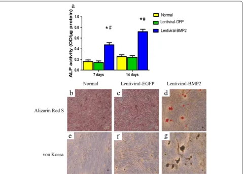
We assessed osteogenic differentiation by measuring ALP activity and mineralization. As shown in Figure 3a, the ALP activity of the Lentiviral-BMP2 transduced USCs increased gradually over 14 days of cell culture and was significantly higher than that of the normal USCs and Lentiviral-GFP transduced USCs (P<0.05).
Fourteen days after transduction, Alizarin Red S and von Kossa staining showed that the mineralized nodules were significantly greater in Lentiviral-BMP2 transduced USCs than in normal USCs and Lentiviral-GFP trans-duced USCs (Figure 3b,c,d,e, f and g). Taken together, these results demonstrate that Lentiviral-BMP2 trans-duction enhances the osteogenic activity of USCs.
Expression of osteogenic related marker OCN and Runx2 Expression of Runx2 and OCN genes in Lentiviral-BMP2 transduced USCs was detected by RT-PCR on days 3, 7 and 14. Transcription level of these genes was
Figure 2Lentiviral-BMP2 transduced USCs and the effect of transduction on USCs proliferation and BMP2 gene and protein expression.
USCs with Lentiviral-BMP2 vectors at different MOIs (a:10;b:50;c:100). Cells were assessed for the presence of positive GFP fluorescence microscopy three days after transduction. Scale bar = 250μm.(d)Cell proliferation assays were performed at 1, 3 and 7 days post-transduction. The means and standard deviations were calculated from three experiments.(e)RT-PCR analysis showed that the transduced USCs highly expressed the BMP2 gene at days 3, 7 and 14 after transduction.(f)ELISA results indicated that BMP2 production in Lentiviral-BMP2 transduced USCs was significantly increased as compared to that in Lentiviral-GFP transduced USCs or normal USCs. #, P <0.01 (compared with normal USCs) and *,P<0.01 (compared with Lentiviral-GFP transduced USCs). BMP2, bone morphogenic proteins 2; MOI, multiplicity of infection; USCs, urine-derived stem cells.
Table 2 The percentage of GFP-positive cells at different MOI
MOI Samples number Positive rate
10 3 30 ± 6%
50 3 53 ± 4%
100 3 90 ± 7%
significantly higher in Lentiviral-BMP2 transduced USCs than in normal USCs or Lentiviral-GFP transduced USCs (Figure 4a). To further detect the osteogenic differ-entiation of Lentiviral-BMP2 transduced USCs resulting from gene transduction, we used immunofluorescence to analyze the osteogenic-related proteins Runx2 and OCN. The results showed that Runx2 and OCN were expressed in Lentiviral-BMP2 transduced USCs at 7 and 21 days after transduction (Figure 4b), respectively. We conclude that the transduction of Lentiviral-BMP2 enhances the osteogenic differentiation of USCsin vitro.
In vivo bone formation
To further test the osteogenic potential of Lentiviral-BMP2 transduced USCs in vivo, Lentiviral-BMP2 transduced USCs were seeded onβ-TCP scaffolds and transplanted into the hind limbs of nude mice. Six weeks afterin vivo
implantation, mice were sacrificed and samples were
collected and examined. H & E staining revealed that Lentiviral-BMP2 transduced USCs generated bone tissues, whereas little ectopic bone formation was observed in control mice implanted with normal USCs (Figure 5a and b). The results showed Lentiviral-BMP2 transduction dramat-ically increased USCs osteogenic activity relative to USCs controlin vivo.
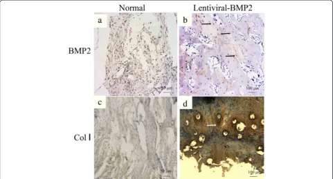
Immunohistochemistry targeting BMP2 and Collagen I (Col I) was performed. It revealed the negative staining of BMP2 and Col I in the USCs transplantation group (Figure 6a and c). However, positive immunohistochemi-cal staining for BMP2 and Col I was observed in the Lentiviral-BMP2 transduced USCs transplantation group (Figure 6b and d).
OCN immunofluorescence was performed to determine the osteogenic differentiation of USCs with or without Lentiviral-BMP2 transduction. Human derived OCN stained in red in the Lentiviral-BMP2 transduction
group (Figure 7a). However, no human derived OCN was detected in the normal USCs transplantation group (Figure 7a). This result indicated that Lentiviral-BMP2 transduced USCs were positive for the osteoblast marker OCN, which suggested that Lentiviral-BMP2 transduced USCs differentiate into osteoblasts in response to the secreted BMP2.
Discussion
In the present study, we demonstrated that the osteogenic activity of USCs can be enhanced by the gene transduction of Lentiviral-BMP2. Afterex vivotransduction, Lentiviral-BMP2 transduced USCs can secrete high levels of Lentiviral-BMP2 and inducede novobone formationin vivo. To the best of our knowledge, this study is the first to evaluate the
Figure 5H & E staining of histological sections formed by the implantation of Lentiviral-BMP2 transduced USCs/β-TCP and USCs/β-TCP into the hind limb of athymic mice for six weeks. (a)The USCs/β-TCP induced no bone formation, whereas the Lentiviral-BMP2 transduced USCs/β-TCP resulted in bone formation in the outer surface and inner pore of the implants(b). Scale bar = 50μm. TCP:β-TCP; FB: fibroblast tissue; NB: new bone; CA: cartilage tissue.Β-TCP,β-tricalcium phosphate; BMP2, bone morphogenic proteins 2; USCs, urine-derived stem cells.
transduction of the BMP2 gene into USCs and the effect of gene transduction on their osteogenic ability.
USCs are adult stem cells that have been characterized in recent years [1]. USCs have the capacity to differentiate into osteoblasts, chondrocytes, adipocytes, muscle and neurons. The large source for the cells and non-invasive method required to collect them has made USCs a suitable
choice for tissue engineering, including bone tissue en-gineering. The bone marrow is regarded as the natural reservoir of osteoprogenitor cells, and bone marrow-derived mesenchymal stem cells (BM-MSCs) are widely used to deliver growth factors for bone regeneration [10-12]. Nevertheless, bone marrow aspiration requires invasive surgery and causes additional pain to patients;
Figure 6Immunohistochemical staining of BMP2 and Collagen I of the transplanted scaffold at six weeks post-implantation. (a)BMP2 and(c)Collagen I staining in the normal USCs transplantation group.(b)BMP2 and(d)Collagen I staining in the Lentiviral-BMP2 transplantation group. The arrows indicate the positive staining of BMP2 or Collagen I in the Lentiviral-BMP2 transplantation group. Scale bar for a and c = 50μm; b and d = 100μm. BMP2, bone morphogenic proteins 2; USCs, urine-derived stem cells.
these factors have impeded the widespread use of the bone marrow for these applications. Therefore, cell sources that are easily accessible and relatively abundant have a pri-ority for cytotherapy. Due to their non-invasive procure-ment method and their high self-renewal capacity, USCs have gained increasing attention.
In the current study, we used culture media free of fetal bovine serum. Previous studies have demonstrated that various in the serum composition [13]. Additionally, the use of animal products has a high risk of virus trans-mission [14]. Currently, the serum-free media we used is mainly supplemented with approximately essential com-ponents including: insulin transferrin and selenium (ITS supplement), transferrin, glucocorticoids, triiodothyron-ine (T3), fibroblast growth factor-2 and glutamtriiodothyron-ine. We combined these supplement depend on the healthy ex-pansion of mesenchymal stem cells and the source envir-onment of USCs [15]. The fetal bovine serum free media pave the way for clinical application of USCs.
Stem cell-based gene therapy is a promising tool to enhance bone repair. Substantial studies have demon-strated that BMP2 gene transduction can be used to en-hance the bone formation ability of stem cells [7,16,17]. Our goal is to utilize BMP2 gene transduction to pro-mote the osteogenic potential of USCs. We integrated the BMP2 gene into lentiviral vectors and transduced them into USCs with an MOI of 10, 50 and 100. The re-sults showed that at an MOI of 100, over 90% of the USCs were transduced, which indicates a high transduc-tion efficiency. In additransduc-tion, the transductransduc-tion process was found to have no significant effect on cell viability.
Afterin vitrotransduction, USCs expressed high levels of the BMP2 gene and secreted a large amount of BMP2 protein. Furthermore, we present evidence that the se-creted BMP2 promoted USCs differentiation into osteo-blasts. ALP activity and mineral calcium deposition is a useful marker for osteogenic differentiation. Only osteo-blasts or cells with osteoblastic characteristics produce OCN [18]. ALP activity and mineral calcium deposition increased significantly after transduction with Lentiviral-BMP2. Lentiviral-BMP2 transduction also stimulated the gene and protein expression of Runx2 and OCN. These data showed that the BMP2 production was sufficient to lead to robust osteogenesis by USCs. Previous studies have demonstrated that BMP2 secreted by transduced cells can initiate a physiological effect on stem cells [12]. As expected, the transduction of the BMP2 significantly enhanced the osteogenic differentiation of USCsin vitro. We investigated whether Lentiviral-BMP2 transduced USCs combined with porous ceramic scaffolds made of
β-TCP could induce ectopic bone formation in vivo. When scaffolds were seeded with Lentiviral-BMP2 trans-duced USCs, bone was observed six weeks after implant-ation. The control scaffold with USCs failed to form
bone, indicating the crucial role of the Lentiviral-BMP2 transduced USCs during bone formation. In fact, previ-ous studies have demonstrated that BMP2 can enhance the ectopic bone formation of bone marrow-derived MSCs [19]. However, the fate of transplanted stem cells
in vivodiffered according to previous studies. Quintavalla
et al. reported the extensive loss of implanted MSCs on days 7 and 14 in osteochondral defects in goat [20]. Hasegawaet al. detected transplanted MSCs four weeks after implantation within the regenerated tissues [21]. In this study, we evaluated the survival of the trans-planted cells. The identification of human-derived OCN within the scaffold indicated that some of the planted cells survived. We demonstrated that the trans-planted cells had survived at six weeks and exerted their osteogenic stimulus on the implantation site to form bone.
Viral vectors currently represent the most efficient approach for gene delivery [22]. A number of viral con-structs have been investigated for gene delivery, with the most common including adenovirus, retrovirus, lentivirus and adeno-associated virus [23]. Lentiviral vectors facilitate long-term target gene expression and are attractive with regard to gene therapy [24]. In addition, the immunogenicity associated with Lenti-viral vectors is low. How to resolve the safety concerns about Lentiviral vectors is critical for their translation into clinical therapy. Several methods have been pro-posed to enhance the safety of Lentiviral vectors for gene transduction [25-27]. The results are encouraging and motivate further research to better apply Lentiviral vectors.
Conclusions
Our study shows that the osteogenic potential of USCs can be significantly enhanced by BMP2 gene transduction. These transfected cells can undergoin vitro osteogenic differentiation without osteogenic medium.In vivobone formation assays showed that the Lentiviral-BMP2 trans-duced USCs provide sufficient stimuli for bone formation. We conclude that the use of BMP2 gene therapy is a use-ful means of enhancing the osteogenic activity of USCs both in vitroand in vivo. These results provide a novel strategy for enhancing the osteoproductive ability of USCs.
Abbreviations
Competing interests
The authors declare that they have no competing interests.
Authors’contributions
JJG carried out most of the experiments and drafted the manuscript. JYZ and XN cultured the USCs and performed gene transduction. ZZZ and SCG carried out the animal experiments. YW and CQZ were responsible for conception and design of the experiments, drafting and final approval of the manuscript. All authors read and approved the final manuscript.
Acknowledgements
This work was supported by the Special Fund for Healthy-scientific Research in the Public Interest, China (201402016), Doctoral Innovation Fund of Shanghai Jiao Tong University School of Medicine (BXJ201341).‘Priority Among Priorities’ Clinical Medical Center Construction Project of the Shanghai Municipality.
Received: 9 July 2014 Revised: 16 December 2014 Accepted: 17 December 2014 Published: 7 January 2015
References
1. Zhang Y, McNeill E, Tian H, Soker S, Andersson KE, Yoo JJ, et al. Urine derived cells are a potential source for urological tissue reconstruction. J Urol. 2008;180:2226–33.
2. Guan J, Niu X, Gong F, Hu B, Guo S, Lou Y, et al. Biological characteristics of human urine derived stem cells: potential for cell-based therapy in neurology. Tissue Eng Part A. 2014;6:6.
3. Bodin A, Bharadwaj S, Wu S, Gatenholm P, Atala A, Zhang Y. Tissue-engineered conduit using urine-derived stem cells seeded bacterial cellulose polymer in urinary reconstruction and diversion. Biomaterials. 2010;31:8889–901. 4. Wu S, Liu Y, Bharadwaj S, Atala A, Zhang Y. Human urine-derived stem cells
seeded in a modified 3D porous small intestinal submucosa scaffold for urethral tissue engineering. Biomaterials. 2011;32:1317–26.
5. Bishop GB, Einhorn TA. Current and future clinical applications of bone morphogenetic proteins in orthopaedic trauma surgery. Int Orthop. 2007;31:721–7.
6. Haidar ZS, Hamdy RC, Tabrizian M. Delivery of recombinant bone morphogenetic proteins for bone regeneration and repair. Part A: current challenges in BMP delivery. Biotechnol Lett. 2009;31:1817–24.
7. Hsu WK, Sugiyama O, Park SH, Conduah A, Feeley BT, Liu NQ, et al. Lentiviral-mediated BMP-2 gene transfer enhances healing of segmental femoral defects in rats. Bone. 2007;40:931–8.
8. Dragoo JL, Lieberman JR, Lee RS, Deugarte DA, Lee Y, Zuk PA, et al. Tissue-engineered bone from BMP-2-transduced stem cells derived from human fat. Plast Reconstr Surg. 2005;115:1665–73.
9. Wright V, Peng H, Usas A, Young B, Gearhart B, Cummins J, et al. BMP4-expressing muscle-derived stem cells differentiate into osteogenic lineage and improve bone healing in immunocompetent mice. Mol Ther. 2002;6:169–78.
10. Tsuda H, Wada T, Ito Y, Uchida H, Dehari H, Nakamura K, et al. Efficient BMP2 gene transfer and bone formation of mesenchymal stem cells by a fiber-mutant adenoviral vector. Mol Ther. 2003;7:354–65.
11. Zou D, Zhang Z, Ye D, Tang A, Deng L, Han W, et al. Repair of critical-sized rat calvarial defects using genetically engineered bone marrow-derived mesenchymal stem cells overexpressing hypoxia-inducible factor-1alpha. Stem Cells. 2011;29:1380–90.
12. Sugiyama O, An DS, Kung SP, Feeley BT, Gamradt S, Liu NQ, et al. Lentivirus-mediated gene transfer induces long-term transgene expression of BMP-2 in vitro and new bone formation in vivo. Mol Ther. 2005;11:390–8. 13. van der Valk J, Mellor D, Brands R, Fischer R, Gruber F, Gstraunthaler G, et al.
The humane collection of fetal bovine serum and possibilities for serum-free cell and tissue culture. Toxicol in Vitro. 2004;18:1–12.
14. Wessman SJ, Levings RL. Benefits and risks due to animal serum used in cell culture production. Dev Biol Stand. 1999;99:3–8.
15. van der Valk J, Brunner D, De Smet K, Fex Svenningsen A, Honegger P, Knudsen LE, et al. Optimization of chemically defined cell culture media–replacing fetal bovine serum in mammalian in vitro methods. Toxicol In Vitro. 2010;24:1053–63.
16. Gao X, Usas A, Lu A, Tang Y, Wang B, Chen CW, et al. BMP2 is superior to BMP4 for promoting human muscle-derived stem cell-mediated bone regeneration in a critical-sized calvarial defect model. Cell Transplant. 2013;22:2393–408.
17. Musgrave DS, Pruchnic R, Wright V, Bosch P, Ghivizzani SC, Robbins PD, et al. The effect of bone morphogenetic protein-2 expression on the early fate of skeletal muscle-derived cells. Bone. 2001;28:499–506.
18. Hosseinkhani H, Yamamoto M, Inatsugu Y, Hiraoka Y, Inoue S, Shimokawa H, et al. Enhanced ectopic bone formation using a combination of plasmid DNA impregnation into 3-D scaffold and bioreactor perfusion culture. Biomaterials. 2006;27:1387–98.
19. Kim S, Jeon O, Lee J, Bae M, Chun HJ, Moon SH, et al. Enhancement of ectopic bone formation by bone morphogenetic protein-2 delivery using heparin-conjugated PLGA nanoparticles with transplantation of bone marrow-derived mesenchymal stem cells. J Biomed Sci. 2008;15:771–7. 20. Quintavalla J, Uziel-Fusi S, Yin J, Boehnlein E, Pastor G, Blancuzzi V, et al.
Fluorescently labeled mesenchymal stem cells (MSCs) maintain multilineage potential and can be detected following implantation into articular cartilage defects. Biomaterials. 2002;23:109–19.
21. Hasegawa N, Kawaguchi H, Hirachi A, Takeda K, Mizuno N, Nishimura M, et al. Behavior of transplanted bone marrow-derived mesenchymal stem cells in periodontal defects. J Periodontol. 2006;77:1003–7.
22. Betz VM, Betz OB, Harris MB, Vrahas MS, Evans CH. Bone tissue engineering and repair by gene therapy. Front Biosci. 2008;13:833–41.
23. Giacca M, Zacchigna S. Virus-mediated gene delivery for human gene therapy. J Control Release. 2012;161:377–88.
24. Apolonia L, Waddington SN, Fernandes C, Ward NJ, Bouma G, Blundell MP, et al. Stable gene transfer to muscle using non-integrating lentiviral vectors. Mol Ther. 2007;15:1947–54.
25. Palfi S, Gurruchaga JM, Ralph GS, Lepetit H, Lavisse S, Buttery PC, et al. Long-term safety and tolerability of ProSavin, a lentiviral vector-based gene therapy for Parkinson’s disease: a dose escalation, open-label, phase 1/2 trial. Lancet. 2014;383:1138–46.
26. Picanco-Castro V, de Sousa Russo-Carbolante EM, Tadeu CD. Advances in lentiviral vectors: a patent review. Recent Pat DNA Gene Seq. 2012;6:82–90. 27. Pluta K, Kacprzak MM. Use of HIV as a gene transfer vector. Acta Biochim
Pol. 2009;56:531–95.
doi:10.1186/scrt539
Cite this article as:Guanet al.:Bone morphogenetic protein 2 gene transduction enhances the osteogenic potential of human urine-derived
stem cells.Stem Cell Research & Therapy20156:5.
Submit your next manuscript to BioMed Central and take full advantage of:
• Convenient online submission
• Thorough peer review
• No space constraints or color figure charges
• Immediate publication on acceptance
• Inclusion in PubMed, CAS, Scopus and Google Scholar
• Research which is freely available for redistribution