PRIMARY RESEARCH
Regulatory effects of COL1A1
on apoptosis induced by radiation in cervical
cancer cells
Shurong Liu
1*, Gewang Liao
1and Guowen Li
2Abstract
Background: Cervical cancer is a common cancer of women in developing countries, and radiotherapy still remains its predominant therapeutic treatment. Collagen type I alpha 1 (COL1A1) has been shown to have a radioresistance effect in previous studies. However, such effect of COL1A1 has not yet been revealed in cervical cancer.
Methods: Expression of COL1A1 in cervical cancer tissues and normal tissues was assessed by qRT-PCR and immu-nohistochemistry. The effect of COL1A1 on radioresistance of human cervical cancer cell lines HeLa and CaSki was assessed using the colony formation assay. Apoptosis alterations were analyzed by flow cytometry. In addition, west-ern blotting was used assessed the alterations of several critical apoptosis and signaling pathway related proteins. Results: The expression of COL1A1 was significantly increased in cervical cancer tissues compared with normal tissues at the mRNA and protein level. Further, based on COL1A1 knock down and COL1A1 activation cell models, a negative correlation was observed between COL1A1 expression level and radiosensitivity. Moreover, the findings are further supported by apoptosis analysis that COL1A1 activation could inhibit the apoptosis of cervical cancer cells. Subsequently, a significantly decreased expression of p-AKT and Bcl-2, increased expression of Caspase-3 were observed in the LY294002 plus radiation group compared with radiation alone group, while these influences caused by LY294002 or X-ray radiation were reversed after COL1A1 activation.
Conclusions: To our knowledge, this is the only study to profile the mechanisms that COL1A1 plays a crucial role in cervical cells anti-apoptosis induced by radiation. Therefore, our identification of radioresistance-related COL1A1 in cervical cancer could be a starting point to explore the function of collagens, adding a new dimension to our under-standing of the cervical cancer, assisting cancer biologists and clinical oncologists in novel therapeutic strategies. Keywords: COL1A1, Cervical cancer, Radioresistance, Apoptosis
© The Author(s) 2017. This article is distributed under the terms of the Creative Commons Attribution 4.0 International License (http://creativecommons.org/licenses/by/4.0/), which permits unrestricted use, distribution, and reproduction in any medium, provided you give appropriate credit to the original author(s) and the source, provide a link to the Creative Commons license, and indicate if changes were made. The Creative Commons Public Domain Dedication waiver (http://creativecommons.org/ publicdomain/zero/1.0/) applies to the data made available in this article, unless otherwise stated.
Introduction
Cervical cancer is one of the most common cancers in women worldwide and the second most diagnosed can-cer of women in developing countries [1]. The treat-ment of cervical cancer is primarily based on the stage of disease, and conventional treatments include surgery, chemotherapy, or radiotherapy (RT) [2]. Previous studies have reported that RT is an excellent therapeutic method for cervical cancer patients, especially to the patients
with locally advanced disease. However, advanced or metastasized tumors with low radiosensitivity required an increasing radiation dose which may damage the sur-rounding healthy tissues and organs [3–5]. Therefore, the identification of novel methods to enhance tumor cell radiosensitivity has become a recent focus of medical radiation research [6].
Collagen type I alpha 1 (COL1A1) is a member of group I collagen which include COL1A1 and COL1A2 [7]. Collagen is the main protein of bones, tendons and teeth, and involved in tumor cell adhesion, gap junction and extracellular matrix (ECM) [8–10]. Previous stud-ies have reported that COL1A1 is upregulated in gastric
Open Access
*Correspondence: shurongliu2017@163.com
1 Department of Gynecologic Oncology, Hunan Cancer Hospital,
cancer, and plays important roles in cancer cell invasion and metastasis in this cancer [11, 12]. In addition, Kita-hara’s cDNA microarray analysis result has indicated that COL1A1 is involved in radioresistance of cervical cancer [13]. Moreover, a recent study confirmed that COL1A1 expression was altered after radiation [14].
In the present study, we hypothesize that COL1A1 is crucial for radioresistance in cervical cancer cells with RT. Here, based on the study of clinicopathological tis-sues and cell models, we found that COL1A1 played an important role in inhibiting apoptosis induced by radia-tion in cervical cancer cells. The findings provide a thera-peutic target as well as a diagnostic radioresistance in cervical cancer.
Materials and methods Clinical specimens
This study was permitted by the Ethical Committee of our hospital, and obtained consents from all the par-ticipants. A total of 20 patients with clinically diagnosed cervical cancer participated in, and their tissues were col-lected in our Hunan Cancer Hospital. Pathological sam-ples were contained two parts: cervical cancer tissues and tumor adjacent normal tissues.
Cell culture
The SiHa, CaSki and Hela cell lines were purchased from the Cell Center of the Xiangya School of Medicine, Cen-tral South University (Hunan, China), and maintained in high glucose Dulbecco’s modified Eagle’s Eagle (DMEM) (Gibco Life Technologies, USA) containing 10% fetal bovine serum (Gibco Life Technologies, USA) at 37 °C in a 5% CO2 incubator.
COL1A1-shRNA and activation plasmids were pur-chased from (Santa cruz, USA). Following transfection these two plasmids and their negative control plasmids into CaSki and Hela cells respectively.
Quantitative real time‑polymerase chain reaction (qRT‑PCR)
The expression of COL1A1 was confirmed by qRT-PCR. Briefly, Total RNA was extracted using total RNA isola-tion kit (Qiangen, Germany), and PCR was performed by Real Time Quantitative PCR SYBR Green detec-tion reagent (Takara, Japan). The relative expression of COL1A1 was calculated with 2−ΔΔCT method relative to GAPDH. COL1A1 primers: sense, CCC CTG GTG CTA CTG GTT TCC C; antisense, GAC CTT TGC CGC CTT CTT TGC. GAPDH primers: sense, ATT CCA CCC ATG GCA AAT TC; antisense, GAT GGG ATT TCC ATT GAT GAC A. All the procedures were repeated in triplicate.
Tissue microarray (TMA) and immunohistochemistry (IHC) Uterine cervical cancer tissue microarrays were pur-chased from Auragene (Changsha, China). This microar-ray contained cervical normal tissues (n = 5) and tumor
adjacent normal tissues (n = 5) and cancer tissues of
dif-ferent grades (n = 70). The IHC was performed using
ElivisionTM plus PolyerHRP (Mouse/Rabbit) IHC Kit (Fuzhou Maixin Biotech, China) according to the manu-facturer’s instructions. Briefly, the TMA was baked at 60 °C for 60 min, deparaffinized, and rehydrated through a series of ethanol with different concentrations. The slides were microwaved in Tris/EDTA pH 9.0 buffer solu-tion (10 mM Tris, 1 mM EDTA) for 10 min for antigen retrieval, and then quenched by immersing in 3% hydro-gen peroxide in distilled water for 20 min. After block-ing the nonspecific bindblock-ing with 10% normal goat serum in PBS buffer for 15 min, the slides were incubated with anti-Collagen I (1:100 dilution, Abcam, UK) and stored overnight at 4 °C. The slides were sequentially incubated with a secondary antibody (Maxim-Bio, Fuzhou, China). The slides were subsequently treated with 3′ 3-diamin-obenzidine tetra-hydrochloride, counterstained with haematoxylin, and finally mounted with neutral balata. Standardization of the incubation and development times allowed accurate comparisons in all cases. A nega-tive control was obtained by replacing the primary anti-body with a normal rabbit IgG.
Sections of tissue were observed under an Olympus microscope (Olympus Corporation, Japan) and images were taken at 400× magnification with the same light
intensity and exposure time. All images were then con-verted to 8-bit grayscale. The COL1A1 staining intensity of cervical tissues was semi-quantitatively compared by analyzing the integrated optical density value of each image, which was measured by the Image-Pro Plus image analysis software (Media Cybernetics, USA).
Western blotting
HRP-conjugated secondary antibody for 1 h at room temperature. The bound antibodies were visualized using chemiluminescence reagents following exposure to X-ray film. All experiments were performed in triplicate. The relative levels of target protein to control β-actin were analyzed by Quantity One 1-D Image Analysis Software (Bio-Rad).
Radiation
The CaSki and Hela cells were treated with the dose of 0, 2, 4, 6, and 8 Gy in a 6 MV X-rays at a dose rate of 200 cGY/min with a distance to the source irradiation about 100 cm. The cells were further cultured in com-plete medium for 12 h. The levels of COL1A1 expression were detected by RT-PCR to choose the minimal effec-tive radiation dose of X-ray.
Cell colony formation assay
Cell colony formation assays were performed using 30 mm cell culture plates coated with 0.5 ml bottom soft agar mixture (DMEM, 20% FBS, 0.6% soft agar). The cells treated with radiation at a dose of 4 Gy were mixed with top agar (DMEM, 20% FBS, 0.3% soft agar) and seeded into each plate, after the bottom layer had solidified. Two weeks later, the colonies were fixed with methanol and stained with 0.5% crystal violet. The number of colonies (>50 cells) was counted on an inverted microscope. The experiments were repeated in triplicate.
Apoptosis analysis
The CaSki and Hela cells were plated into 60 mm dishes and radiated with the dose of 4 Gy. Twenty-four hours later, the cells were harvested, and then the cellular apop-tosis was detected using the Annexin V-FITC Apopapop-tosis Kit (BD Biosciences, USA). The results were examined using the FACS flow cytometer. All these procedures were performed in triplicate.
Statistical analysis
Quantitative data were expressed as mean ± SD, and analyzed using SPSS 20.0 (IBM, USA). Significant differ-ences between groups were compared using ANOVAs and Student’s t test. P < 0.05 was considered to be statisti-cally significant.
Results
The expression level of COL1A1 is elevated in cervical cancer tissues
To examine the relationship between COL1A1 expres-sion level and cervical cancer, the difference of COL1A1 mRNA expression levels were measured in 20 patients with cervical cancer between their cervical cancer tis-sues and adjacent normal tistis-sues by qRT-PCR assay. Our
results demonstrated that COL1A1 expression was sig-nificant elevated in almost cancer tissues compared to adjacent normal tissues (Fig. 1a, P < 0.05). Meanwhile, we determined the COL1A1 protein levels in 70 cervical cancer tissues and 10 normal samples by high through-put IHC analyse. Representative sections of COL1A1 expression in all tissues are shown in Fig. 1b. Obviously, COL1A1 was expressed at substantially higher levels in the cancer samples by contrast to the weak but detectable expression in normal tissues with a statistical significance (P < 0.05) (Fig. 1c). Furthermore, a close relationship was observed between the increasing grade of lesion and the intensity of COL1A1 staining in cervical cancer tissues. For COL1A1 IHC staining in cervical cancer, immunore-activity was primarily observed in the ECM (extracellular matrix) of tumor cells. Therefore, our results suggest that COL1A1 expression was enhanced from normal cervical specimens to different grades cervical cancers gradually.
The expression of endogenous COL1A1 in cervical cells and constructed of COL1A1 cell models
The baseline expression of COL1A1 was explored in cer-vical cancer cell lines: Hela, CaSki and SiHa (Fig. 2a, b). QRT-PCR and western blotting analysis revealed that COL1A1 was increased expression apparent in Hela and CaSki cells.
Given the progressive up regulation of COL1A1 in human liver and gastric cancers [8], we investigated how COL1A1 influences cellular process by genetically manipulating the expression of COL1A1 in CaSki and Hela cells. COL1A1 knock down and COL1A1 activa-tion cell models were constructed. As shown in Fig. 3a, the expression of COL1A1 in CaSki and Hela cells were increased significantly when transfected with COL1A1 activation plasmids, while, COL1A1 expression lev-els were decreased significantly when transfected with COL1A1 shRNA plasmids. Moreover, these finding are further supported by western blotting analysis at pro-tein level (Fig. 3b). Thus, the results indicated that stably transfected lines were constructed successfully.
The effect of COL1A1 in cervical cells with radiation
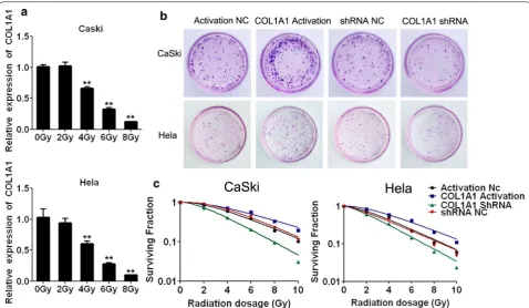
As indicated in Fig. 4a, the mRNA expression level of COL1A1 showed a dose dependent decreasing trend with the increasing of radiation in HeLa and CaSki cells. In particular, COL1A1 expression became significantly lower when irradiated with 4 Gy. Thus, 4 Gy was selected for further study.
colony formation (Fig. 4b). In addition, to further validate COL1A1 is related to radioresistance, Hela and CaSki with COL1A1 Activation and COL1A1 shRNA were sub-jected to X-ray radiation (0, 2, 4, 6, 8 and 10 Gy) (Fig. 4c). As shown in Fig. 4b, after 2 weeks the number of colonies formation in COL1A1-shRNA group was significantly decreased compared with that of negative control (NC) group. However, compared with NC cells, the number of colonies formation were observed significantly increased in Hela and CaSki cells with COL1A1 activation. These results suggested that COL1A1 shRNA cells were more sensitive to radiation than NC cells and COL1A1 Activa-tion cells were more tolerance to radiaActiva-tion than NC cells. Thus, these experiments indicated there is a close rela-tionship between radioresistance and COL1A1 in cervi-cal cells.
The regulated effect of COL1A1 on apoptosis induced by radiation
As shown in Fig. 5, compared with control cell lines, both COL1A1-shRNA and 4 Gy radiation induced sig-nificantly apoptosis in Hela and CaSki cells, meanwhile, COL1A1 activation suppressed apoptosis significantly in Hela and CaSki cells compared with NC cells. These data
suggested that COL1A1 is important for anti-apoptosis induced by radiation in cervical cancer cells.
Signaling transduction pathways involved in COL1A1 of anti‑apoptosis induced by radiation and the role of COL1A1 in signaling transduction pathways
Since several studies have shown cell apoptosis is asso-ciated with the activation of Caspase-3/PI3K/AKT path-way, in our study, western blotting was used to elucidate the mechanism involved in COL1A1 of anti-apoptosis induced by radiation. As shown in Fig. 6a, the COL1A1 activation led to the decreasing of Caspase-3 and Bax, and the increasing of Bcl-2, both in Hela and CaSki cells. However, an opposite tendency was observed in Hela and CaSki cells with COL1A1-shRNA. These results consist-ently indicated that COL1A1 may inhibit apoptosis of cervical cancer cells.
To further determine the role of COL1A1 in signal-ing transduction pathways, the expression of apoptosis-related molecules and COL1A1 were measured in Hela and CaSki cells with radiation treated by western blotting (Fig. 6b). As shown in Fig. 6b, X-ray radiation activated the expression of COL1A1, AKT and p-AKT in short time, but suppressed after treated 24 h. At the same time,
we used specific inhibitor (LY294002) specifically target-ing the pathway to elucidate the mechanism underlytarget-ing the COL1A1 antiapoptosis activity. As shown in Fig. 6c, LY294002 significantly suppressed the PI3K pathway and led to cell apoptosis, and suppressed the expression of COL1A1. While COL1A1 activation could reverse the cell apoptosis caused by LY294002, and COL1A1 shRNA could enhance the cell apoptosis as shown in Additional file 1: Figure S1 flow cytometry results. To further verify the important role of COL1A1 in signaling transduction pathways of apoptosis in cervical cancer cells with radia-tion. LY29400 and X-ray radiation were used to treat cer-vical cancer cells with COL1A1 activation. It was found that COL1A1 reversed the inhibiting effect of LY294002 on apoptosis induced by X-ray radiation (Fig. 6d).
Discussion
Recent studies have indicated overexpression of COL1A1 as an important event in a series of cancers. Aberrant COL1A1 expression has been found in some solid tumors including gastric and breast [11, 12, 15]. Furthermore, COL1A1 expression may represent an independent bio-marker for the prediction of prognosis of hepatocellular carcinoma [15].
Although the important roles of COL1A1 in tumor progression in multiple cancers are now being clarified,
it has not been studied in development of cervical can-cer. In the present study, we evaluated the possibility of COL1A1 as a therapeutic target of cervical cancer. First evidence from our group demonstrated, the expression of COL1A1 up regulation at mRNA and protein levels from grade I to III in cervical cancer tissues, in contrast to nor-mal cervical tissues. Similarly, previous study also found that the expression of COL1A1 is increased in ovarian serous carcinoma compared normal tissues [16].
It is well established that radiation sensitivity mecha-nisms in human cancers are determined by intracellular factors and interactions of cells with ECM or neighboring cells as well as the tumor microenvironment [3, 17, 18]. Meanwhile, COL1A1 is a member of collagen family and mainly expressed in the ECM [7–10], and collagens are overexpressed in the majority of human cancers. Thus, these finding indicated that COL1A1 is a radioresist-ance gene and play an important role in cellular defense irradiation [13]. But the mechanisms that how COL1A1 regulate radioresistance remain elusive. To address this question, we detected the levels of COL1A1 in Hela, CaSki and Siha cells. Interestingly, significant difference was observed among three cell lines, especially for Siha cells. This is likely because of the different radiosensitivity
Fig. 2 The expression of COL1A1 in Hela, CaSki, SiHa cells and cell models construction. a qRT-PCR analysis revealed that COL1A1 is differentially expressed in the cell lines. b Differentially expressed of COL1A1 was detected in three different cells by western blotting analysis. **P < 0.01 vs Hela, ##P < 0.01 vs Caski
Fig. 3 The construction of over expression and interference models.
in cells. Furthermore, COL1A1-shRNA and activation cell models were constructed, and we demonstrated that abundant COL1A1 could inhibit the cell death while low-abundant COL1A1 could promote cell death. The findings are further supported by apoptosis analy-sis. Our findings are consistent with results from other researchers, who also showed that cervical cells enhance radiosensitivity with lower colony survival and higher apoptosis in response to radiation [4, 5, 19].
Apoptosis as an ordered cellular process is of vital importance in regulating cell death [20] so that activation of apoptosis is widely considered as an anticancer strat-egy. Regarding to the molecular mechanism which lead-ing to the COL1A1 overexpression in cervical cancer, we are thinking that COL1A1 might be induced by activation of radiation dependent Caspase-3/PI3K/AKT signaling pathway. In our study, the expression of several critical apoptosis-related proteins, such as the Bcl-2 family pro-teins, AKT and Caspase-3, and COL1A1 were detected. Apoptosis regulator Bcl-2 is a family of regulator proteins that regulate cell death (apoptosis), by either inducing (pro-apoptotic) or inhibiting (anti-apoptotic) apoptosis. Bcl-2 is specifically considered an important anti-apop-totic protein [21, 22] and Bax has a pro-apoptotic effect
[23]. Caspase-3 plays a central role in the execution-phase of cell apoptosis and is considered as the most important performer of apoptosis in the caspase fam-ily [24]. Caspase-3 is activated in the apoptotic cell both by extrinsic (death ligand) and intrinsic (mitochondrial) pathways and caspase activity would kill cells indiscrimi-nately [25, 26]. Subsequently, a significantly decreased expression of p-AKT and Bcl-2 increased expression of Caspase-3 were shown in the LY294002 plus radiation group, compared with radiation alone. Meanwhile, a sig-nificantly increased expression of p-AKT and Bcl-2 and decreased expression of Caspase-3 were shown in the COL1A1 activation plus radiation group, compared with radiation alone. However, there was no significant differ-ence in the p-AKT, Bcl-2 and Caspase-3 expression level between the COL1A1 Activation plus LY294002 plus radiation group and radiation alone group. Therefore, we can get the conclusion that COL1A1 Activation may decrease apoptosis of cervical cancer cells by destroying the balance between anti-apoptotic and pro-apoptotic proteins and activating Caspase-3/PI3K/AKT pathway.
What’s more, COL1A1 is a fibril-forming collagen found in most connective tissues and is abundant in bone, cornea, dermis and tendon, and its expression level
is closely related to epithelial-to-mesenchymal transition (EMT) [27, 28]. While the EMT was known to be asso-ciated with radioresistance [29–31] and PI3K pathway [32–35]. Thus, these indicated that the radioresistance function of COL1A1 may be closely related to EMT.
As a conclusion, COL1A1 seems to modulate the radi-oresistance of cervical cells via complex mechanisms by affecting Caspase-3/PI3K/AKT pathways to regulate cell death. Afterwards we will try to detect the COL1A1-related radioresistance in animals and the protein that interacted with COL1A1 and the relationship among COL1A1, EMT and radioresistance.
Conclusions
This is the only study to profile that COL1A1 is a crucial radioresistance factor in cervical cancer cells and plays an important role in ant-apoptosis by Caspase-3/PI3K/ AKT pathways. Therefore, our identification of radiore-sistance-related COL1A1 in cervical cancer should be a starting point to explore the function of collagens, adding a new dimension to our understanding of the complex picture of cervical cancer and assisting cancer biologists and clinical oncologists in designing and testing novel therapeutic strategies.
Abbreviations
COL1A1: collagen type I alpha 1; ECM: extracellular matrix; EMT: epithelial-to-mesenchymal transition; IHC: immunohistochemistry; NC: negative control; qRT-PCR: quantitative real time-polymerase chain reaction; RT: radiotherapy; TMA: tissue microarray.
Authors’ contributions
SL performed in study design, vitro experiments, wrote this paper and paper modification. GL performed in vivo experiments. GL contributed to data analysis. All authors read and approved the final manuscript.
Author details
1 Department of Gynecologic Oncology, Hunan Cancer Hospital, Tongzipo
Road No. 283, Changsha 410011, Hunan, China. 2 Department of
Interven-tional Radiology, Hunan Cancer Hospital, Tongzipo Road No. 283, Chang-sha 410011, Hunan, China.
Acknowledgements
Not applicable. Additional file
Additional file 1: Figure S1. The regulated effect of COL1A1 on apopto-sis tested by flow cytometry. In CaSki and HeLa cells, both COL1A1 shRNA transfection and LY294002 could induce apoptosis. COL1A1 activation could inhibit the apoptosis caused by LY294002, and the combination of COL1A1 shRNA transfection and LY294002 could induce much more apoptosis.
Competing interests
Not available.
Availability of data and materials
Not applicable.
Consent for publication
Not applicable.
Ethics approval and consent to participate
Not applicable.
Funding
Not applicable.
Publisher’s Note
Springer Nature remains neutral with regard to jurisdictional claims in pub-lished maps and institutional affiliations.
Received: 13 March 2017 Accepted: 23 July 2017
References
1. Bermudez A, Bhatla N, Leung E. Cancer of the cervix uteri. Int J Gynaecol Obstet. 2015;131(Suppl 2):S88–95.
2. Fu JH, Gao Z, Ren CC, Shi YG. Comparison of clinical efficacy of three dif-ferent neoadjuvant approaches (chemotherapy combined vaginal intra-cavitary irradiation, neoadjuvant chemotherapy alone or radiotherapy)
• We accept pre-submission inquiries
• Our selector tool helps you to find the most relevant journal
• We provide round the clock customer support
• Convenient online submission
• Thorough peer review
• Inclusion in PubMed and all major indexing services
• Maximum visibility for your research
Submit your manuscript at www.biomedcentral.com/submit
Submit your next manuscript to BioMed Central
and we will help you at every step:
combined with surgery for patients with stage Ib2 and IIa2 cervical cancer. Asian Pac J Cancer Prev. 2013;14(4):2377–81.
3. Song L, Liu S, Zhang L, Yao H, Gao F, Xu D, Li Q. MiR-21 modulates radio-sensitivity of cervical cancer through inhibiting autophagy via the PTEN/ Akt/HIF-1alpha feedback loop and the Akt-mTOR signaling pathway. Tumour Biol. 2016;37(9):1–8.
4. Herd O, Francies F, Kotzen J, Smith T, Nxumalo Z, Muller X, Slabbert J, Vral A, Baeyens A. Chromosomal radiosensitivity of human immunodeficiency virus positive/negative cervical cancer patients in South Africa. Mol Med Rep. 2016;13(1):130–6.
5. Liu SS, Leung RC, Chan KY, Chiu PM, Cheung AN, Tam KF, Ng TY, Wong LC, Ngan HY. p73 expression is associated with the cellular radiosensitivity in cervical cancer after radiotherapy. Clin Cancer Res. 2004;10(10):3309–16. 6. Belfatto A, Riboldi M, Ciardo D, Cattani F, Cecconi A, Lazzari R,
Jereczek-Fossa BA, Orecchia R, Baroni G, Cerveri P. Kinetic models for predicting cervical cancer response to radiation therapy on individual basis using tumor regression measured in vivo with volumetric imaging. Technol Cancer Res Treat. 2016;15(1):146–58.
7. Chan TF, Poon A, Basu A, Addleman NR, Chen J, Phong A, Byers PH, Klein TE, Kwok PY. Natural variation in four human collagen genes across an ethnically diverse population. Genomics. 2008;91(4):307–14.
8. Zhao YP, Wang H, Fang M, Ji Q, Yang ZX, Gao CF. Study of the association between polymorphisms of the COL1A1 gene and HBV-related liver cir-rhosis in Chinese patients. Dig Dis Sci. 2009;54(2):369–76.
9. Marini JC, Forlino A, Cabral WA, Barnes AM, San Antonio JD, Milgrom S, Hyland JC, Korkko J, Prockop DJ, De Paepe A, et al. Consortium for osteo-genesis imperfecta mutations in the helical domain of type I collagen: regions rich in lethal mutations align with collagen binding sites for integrins and proteoglycans. Hum Mutat. 2007;28(3):209–21.
10. Carbone L, Harris RA, Gnerre S, Veeramah KR, Lorente-Galdos B, Huddles-ton J, Meyer TJ, Herrero J, Roos C, Aken B, et al. Gibbon genome and the fast karyotype evolution of small apes. Nature. 2014;513(7517):195–201. 11. Li AQ, Si JM, Shang Y, Gan LH, Guo L, Zhou TH. Construction of COL1A1
short hairpin RNA vector and its effect on cell proliferation and migration of gastric cancer cells. Zhejiang da xue xue bao Yi xue ban. 2010;39(3):257–63.
12. Sun H. Identification of key genes associated with gastric cancer based on DNA microarray data. Oncol Lett. 2016;11(1):525–30.
13. Kitahara O, Katagiri T, Tsunoda T, Harima Y, Nakamura Y. Classification of sensitivity or resistance of cervical cancers to ionizing radiation according to expression profiles of 62 genes selected by cDNA microarray analysis. Neoplasia. 2002;4(4):295–303.
14. Cruet-Hennequart S, Drougard C, Shaw G, Legendre F, Demoor M, Barry F, Lefaix JL, Galera P. Radiation-induced alterations of osteogenic and chondrogenic differentiation of human mesenchymal stem cells. PLoS ONE. 2015;10(3):e0119334.
15. Hayashi M, Nomoto S, Hishida M, Inokawa Y, Kanda M, Okamura Y, Nishi-kawa Y, Tanaka C, Kobayashi D, Yamada S, et al. Identification of the col-lagen type 1 alpha 1 gene (COL1A1) as a candidate survival-related factor associated with hepatocellular carcinoma. BMC Cancer. 2014;14:108. 16. Quinn MC, Wojnarowicz PM, Pickett A, Provencher DM, Mes-Masson AM,
Davis EC, Tonin PN. FKBP10/FKBP65 expression in high-grade ovarian serous carcinoma and its association with patient outcome. Int J Oncol. 2013;42(3):912–20.
17. Zhang Q, Gao M, Luo G, Han X, Bao W, Cheng Y, Tian W, Yan M, Yang G, An J. Enhancement of radiation sensitivity in lung cancer cells by a novel small molecule inhibitor that targets the beta-catenin/Tcf4 interaction. PLoS ONE. 2016;11(3):e0152407.
18. Bose MV, Rajkumar T. Assessment of the radiation sensitivity of cervical cancer cell lines. Methods Mol Biol. 2015;1249:351–62.
19. da Seong B, Hong S, Muthusami S, Kim WD, Yu JR, Park WY. Cordycepin increases radiosensitivity in cervical cancer cells by overriding or prolong-ing radiation-induced G2/M arrest. Eur J Pharmacol. 2016;771:77–83.
20. Lagadic-Gossmann D, Huc L, Lecureur V. Alterations of intracellular pH homeostasis in apoptosis: origins and roles. Cell Death Differ. 2004;11(9):953–61.
21. Tsujimoto Y, Finger LR, Yunis J, Nowell PC, Croce CM. Cloning of the chro-mosome breakpoint of neoplastic B cells with the t(14;18) chrochro-mosome translocation. Science. 1984;226(4678):1097–9.
22. Cleary ML, Smith SD, Sklar J. Cloning and structural analysis of cDNAs for bcl-2 and a hybrid bcl-2/immunoglobulin transcript resulting from the t(14;18) translocation. Cell. 1986;47(1):19–28.
23. Westphal D, Kluck RM, Dewson G. Building blocks of the apoptotic pore: how Bax and Bak are activated and oligomerize during apoptosis. Cell Death Differ. 2014;21(2):196–205.
24. Wong RS. Apoptosis in cancer: from pathogenesis to treatment. J Exp Clin Cancer Res. 2011;30:87.
25. Li X, Zhang Q, Cai L, Wang Y, Wang Q, Huang X, Fu S, Bai J, Liu J, Zhang G, et al. Inhibitor of growth 4 induces apoptosis in human lung adenocarci-noma cell line A549 via Bcl-2 family proteins and mitochondria apoptosis pathway. J Cancer Res Clin Oncol. 2009;135(6):829–35.
26. Mace PD, Riedl SJ, Salvesen GS. Caspase enzymology and activation mechanisms. Methods Enzymol. 2014;544:161–78.
27. Liu J, Eischeid AN, Chen XM. Col1A1 production and apoptotic resistance in TGF-β1-induced epithelial-to-mesenchymal transition-like phenotype of 603B cells. PLoS ONE. 2012;7(12):e51371.
28. Hosper NA, van den Berg PP, de Rond S, Popa ER, Wilmer MJ, Masereeuw R, Bank RA. Epithelial-to-mesenchymal transition in fibrosis: collagen type I expression is highly upregulated after EMT, but does not contribute to collagen deposition. Exp Cell Res. 2013;319(19):3000–9.
29. Halim TA, Farooqi AA, Zaman F. Nip the HPV encoded evil in the cancer bud: HPV reshapes TRAILs and signaling landscapes. Cancer Cell Int. 2013;13(1):61.
30. Theys J, Jutten B, Habets R, Paesmans K, Groot AJ, Lambin P, Wouters BG, Lammering G, Vooijs M. E-cadherin loss associated with EMT promotes radioresistance in human tumor cells. Radiother Oncol. 2011;99(3):392–7. 31. Marieegyptienne DT, Lohse I, Hill RP. Cancer stem cells, the epithelial
to mesenchymal transition (EMT) and radioresistance: potential role of hypoxia. Cancer Lett. 2013;341(1):63–72.
32. Liu X, Li Z, Song Y, Wang R, Han L, Wang Q, Jiang K, Kang C, Zhang Q. AURKA induces EMT by regulating histone modification through Wnt/β-catenin and PI3K/Akt signaling pathway in gastric cancer. Oncotarget. 2016;7(22):33152–64.
33. Lee YJ, Han HJ. Troglitazone ameliorates high glucose-induced EMT and dysfunction of SGLTs through PI3K/Akt, GSK-3β, Snail1, and β-catenin in renal proximal tubule cells. Am J Physiol Renal Physiol. 2010;298(5):F1263. 34. Ni J, Cozzi P, Hao J, Beretov J, Chang L, Duan W, Shigdar S, Delprado
W, Graham P, Bucci J. Epithelial cell adhesion molecule (EpCAM) is associated with prostate cancer metastasis and chemo/radioresist-ance via the PI3K/Akt/mTOR signaling pathway. Int J Biochem Cell Biol. 2013;45(12):2736–48.