R E S E A R C H A R T I C L E
Open Access
Influence of upper and temporal
transconjunctival sclerocorneal incision
on marginal reflex distance after cataract
surgery
Rikiya Tamaki
1, Masahiko Gosho
2, Kyoichi Mizumoto
1, Nahoko Kato
1and Masahiro Zako
1*Abstract
Background:Ptosis incidence following cataract surgery is reduced with a recently developed phacoemulsification technique using a small incision. However, it remains uncertain whether an upper transconjunctival sclerocorneal incision can cause minor blepharoptosis. In the present prospective study, patients underwent cataract surgery with either an upper or temporal 2.4-mm transconjunctival sclerocorneal incision. We measured the marginal reflex distance 1 (MRD1) preoperatively and postoperatively, and compared these measurements between the two different incision types. Further we explored the risk factors of the postoperative MRD1 reduction.
Methods:The study population included patients who underwent cataract surgery on both eyes at Aichi Medical University between October 2013 and September 2015. In each patient, one eye was operated using an upper 2. 4-mm transconjunctival sclerocorneal incision, and the other with a temporal incision. We prespecified that an MRD1 difference of≥0.5 mm between the pre- and post-surgical measurements indicated postoperative ptosis, which was a strict criterion. MRD1 was measured using digital photography, and we calculated the difference between the preoperative and postoperative MRD1 values. This change in MRD1 was compared between the groups with different incision locations. The change in MRD1 was analyzed by using the multivariate regression model including incision position (temporal or upper), preoperative MRD1, and preoperative distance between medial and lateral canthi.
Results:We assessed data from a total of 34 patients. The mean change in MRD1 from pre-operation to post-operation measurements was−0.26 ± 0.93 with the temporal incision and−0.24 ± 0.86 with the upper incision. The mean difference in the change in MRD1 between the different two incision types was−0.02, with a 95 % CI of−0.24 to 0.20, establishing equivalence between these incision types. The multivariate regression analysis showed that the preoperative MRD1 was significantly associated with the reduction of MRD1 after surgery (p= 0.034). Conclusions:Cataract surgery using upper and temporal 2.4-mm transconjunctival sclerocorneal incisions are clinically equivalent with regards to change in MRD1, and neither incision type caused critical postoperative ptosis. The longer preoperative MRD1 was significantly associated with the reduction of MRD1 after surgery. Trial registration:Current Controlled Trials UMIN000022310. Retrospectively registered 14 May 2016.
Keywords:Cataract, Marginal reflex distance, Ptosis, Transconjunctival sclerocorneal incision
* Correspondence:zako@aichi-med-u.ac.jp
1Department of Ophthalmology, Aichi Medical University, Nagakute,
480-1195 Aichi, Japan
Full list of author information is available at the end of the article
Background
Ptosis incidence following cataract surgery has been re-duced with the use of a recently developed phacoemulsifi-cation technique involving a small incision, with reported rates of 4–21 % [1–8]. Ptosis is generally defined as a de-crease in the marginal reflex distance 1 (MRD1) of 2 mm or more in the postoperative measurement compared to the preoperative measurement [9, 10]. The precise etiology of ptosis remains elusive, but is considered to be multifactorial. The most critical factor in postope-rative ptosis appeared trauma to the superior rectus/le-vator complex caused by local anesthesia, superior rectus bridle suture, and lid speculum [1–7]. Preoperative ptosis showed no effect on postoperative ptosis [1], but preoperative visible iris sign was shown as a clinical sign of severe involutional ptosis [10]. Puvanachandra et al. reported the incidence of postoperative ptosis has re-duced by changing from ECCE (18 %) to phacoemulsifi-cation (0 %) [9]. It remains uncertain whether the use of an upper transconjunctival sclerocorneal small incision to perform phacoemulsification in cataract surgery can lead to minor blepharoptosis after surgery, and no prior prospective study has addressed this question.
In the present study, patients underwent cataract surgery in both eyes, with an upper incision used in one eye and a temporal incision used in the other eye. We measured the MRD1 in each eye before and three months after phacoe-mulsification, and statistically analyzed the MRD1 values, using a strict criterion to define postoperative ptosis.
Methods Patients
This study included patients who were scheduled for cataract surgery on both eyes at Aichi Medical University between October 2013 and September 2015. Patients were excluded if they had a history of thyroid eye disease, prop-tosis, enophthalmos, or previous lid or ocular surgery.
Cataract surgery
All phacoemulsification surgeries were performed by one surgeon (R.T.). Surgery was performed with topical and intracameral local anesthesia. Since evidence sug-gests that a metallic persistent eyelid speculum may lead to postoperative ptosis [11], here we used a disposable flexible EzSpec lid speculum (Hoya, Tokyo, Japan) for all patients. No superior rectus bridle suture was used. A 2.4-mm sutureless upper or temporal transconjunctival sclerocorneal incision was performed. The patients were randomly prospectively assigned to either Group 1 (upper incision for right eye and temporal incision for left eye) or Group 2 (upper incision for left eye and temporal incision for right eye), such that each patient received one of each incision type. Another 0.8-mm clear corneal incision was made at the nasal side in all patients.
Measurement of MRD1 and distance between medial and lateral canthi
To perform the MRD1 measurements, photographs were taken by a single investigator (N.K.) who had no information about the position of incision for each patient. Photographs were taken preoperatively and at three months postoperatively, and MRD1 was measured using universal ophthalmic measure (Mita PD meter, HE-95, Handaya, Tokyo, Japan) as shown in Fig. 1. All MRD1 measurements were made before mydriatic in-stillation. We measured the preoperative distance be-tween medial and lateral canthi as shown in Fig. 2. The collected data are available at the LabArchives website (http://www.labarchives.com/bmc).
Ptosis definition
Ptosis is generally defined as a decrease in the relative position of the upper lid by 2 mm or more compared to the preoperative measurement, which may be the largest difference in MRD1 that can be considered clinically
before surgery
after surgery
acceptable [9, 10]. Here we used a stricter standard, defi-ning postoperative ptosis as MRD1 difference of≥0.5 mm between the preoperative and postoperative measurements.
Statistical analysis
Descriptive statistics are presented as the mean ± standard deviation (SD) or as n (%). A pairedt-test was used to analyze the change in MRD1 from pre-operation to post-operation measurements. An exact McNemar test was used to compare frequencies in paired binary data. To compare the change in MRD1 (from pre-operation to post-operation) between the temporal incision and the upper incision, we assessed whether the two-sided 95 % confidence interval (CI) for the difference was entirely within the interval−0.5 to 0.5 mm as an equiva-lence margin. The temporal and upper incisions were considered to be equivalent if the 95 % CI fell entirely within the interval. To explore the risk factors of the postoperative MRD1 reduction, the change in MRD1 was analyzed by using the multivariate regression model including incision position (temporal or upper), pre-operative MRD1, and prepre-operative distance between medial and lateral canthi. A total sample size of 36
patients provided 80 % power that the 95 % CI for the paired mean difference between the two incisions did not exceed ± 0.5 mm, which was the predefined equiva-lence margin, assuming a mean difference of 0 mm and a SD of 1.0 mm for the change in MRD1. All statistical analyses were performed using SAS 9.4 (SAS institute, Cary, NC, USA). Differences with a p value of <0.05 were considered statistically significant.
Results
A total of 44 patients were initially enrolled. Ten patients could not make it to the hospital on a scheduled day due to personal reasons, and were excluded from this study. Thus, the final analysis included a total of 34 patients: 16 female and 18 male; mean age, 74.7 ± 11.2 years; age range, 69–87 years.
The preoperative MRD1 was 2.27 ± 0.89 mm in the temporal group and 2.24 ± 0.96 mm in the upper group (Table 1). The two incision groups did not significantly differ in pre-operation MRD1 (p= 0.764). The postope-rative MRD1 was 2.01 ± 1.08 mm in the temporal group versus 2.00 ± 1.05 mm in the upper group (p= 0.920). The change in MRD1 from pre-operation to post-operation was −0.26 ± 0.93 mm with the temporal in-cision and −0.24 ± 0.86 mm with the upper incision. The mean difference in the change in MRD1 between the two incision types was −0.02 mm, with a 95 % CI of −0.24 to 0.20, establishing equivalence between these incisions. We added all data sets used for this study as Additional file 1.
We defined postoperative ptosis as a decrease in MRD1 of ≥0.5 mm from the preoperative measurement to the postoperative measurement. As the mean change from pre-operation to post-operation was −0.26 mm (95 % CI, −0.58 to 0.07) with the temporal incision and −0.24 mm (95 % CI, −0.54 to 0.06) with the upper incision, neither group met this stricter criterion for pto-sis. The frequency of postoperative ptosis was 14 (41 %) patients with the temporal incision and 14 (41 %) patients with the upper incision, and did not significantly differ between the two incisions (p= 0.392).
medial canthus
lateral canthus
Fig. 2The distance between medial and lateral canthi (broken line) was measured preoperatively
Table 1MRD1 by each incision
MRD1 (mm) Temporal (n= 34) Upper (n= 34) pvaluea Mean difference (95 % CI)
Pre-operation 2.27 ± 0.89 2.24 ± 0.96 0.764 0.03 (−0.20, 0.27)
Post-operation 2.01 ± 1.08 2.00 ± 1.05 0.920 0.01 (−0.17, 0.30)
Difference between
post- and pre-operation values
−0.26 ± 0.93 (95 % CI:−0.58, 0.07) (p= 0.115)b
−0.24 ± 0.86 (95 % CI:−0.54, 0.06) (p= 0.119)b
0.849 −0.02 (−0.24, 0.20)
MRD1 decrease of≥0.5 mm n= 14 (41 %) n= 14 (41 %) 0.392c
-a
Compared between temporal and upper incisions using a pairedt-test
b
Compared between pre- and post-operation values using a pairedt-test
c
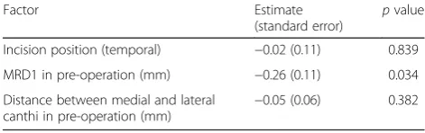
The multivariate regression analysis showed that the preoperative MRD1 was significantly associated with the reduction of MRD1 after surgery (p= 0.034, Table 2).
We examined the distributions of the absolute MRD1 and the change from pre-operation in MRD1. The two measures were basically normally distributed (p= 0.07 and
p= 0.66 by Shapiro-Wilk test, respectively). We added it as Additional file 2.
Discussion
Our data indicated that postoperative ptosis is rare fol-lowing phacoemulsification cataract surgery with either an upper or temporal 2.4-mm transconjunctival sclero-corneal incision. Furthermore, there was no significant difference between these two incision types. Interestingly the preoperative MRD1 was significantly associated with the reduction of MRD1 after surgery. Our findings sug-gest that the decision to perform phacoemulsification cataract surgery with an upper or temporal 2.4-mm transconjunctival sclerocorneal incision may be made based on the surgeon’s personal preference without in-fluencing the risk of postoperative ptosis.
Many factors may be involved in the development of postoperative ptosis. Kaplan et al. suggested that poten-tial causative factors may include local anesthesia, either through a volume effect or myotoxicity; the superior rec-tus bridle suture; the use of a lid speculum; the size and location of the incision; and upper eyelid edema [1]. They concluded that trauma to the superior rectus muscle by placement of a bridle suture was the most in-fluential factor in postoperative ptosis development. The use of a temporal sutureless incision reduces irritation beneath the upper lid, which is associated with inflam-mation and edema, and may cause ptosis [12]. With the recently developed technique of phacoemulsification surgery, the incision size and location are likely the most important factors in the development of postoperative ptosis. Our present study focused on the position of the small sutureless incision in phacoemulsification cataract surgery. In their prospective comparative study, Puvana-chandra et al. found a postoperative ptosis rate of 18 % in the extracapsular cataract extraction (ECCE) group and 0 % in the phacoemulsification group [9]. They defined ptosis as a decrease in the relative position of the upper lid of 2 mm or more compared to the preope-rative measurement, present 6 weeks after surgery. They
suggested that the principal factor influencing this dif-ference in ptosis rate was the smaller incision size in the phacoemulsification procedure (10 mm with ECCE compared to 3–4 mm with phacoemulsification).
Kawa et al. compared two groups that underwent pha-coemulsification without a bridle suture, but with either peribulbar or retrobulbar anesthesia [13]. Only one patient developed ptosis, and this low rate was attributed to not using a bridle suture. On the other hand, Patel et al. in-vestigated patients undergoing phacoemulsification under peribulbar anesthesia, and found no difference in ptosis rates between those operated using a superior incision and bridle suture and those with a temporal incision with no bridle suture [14]. Ptosis has been also reported after radial keratotomy and laser in situ keratomileusis [15–17], but these procedures are performed under top-ical anesthesia and with no bridle suture. In the present study, we did not place a bridle suture, and we used a fle-xible disposable lid speculum in all cases. Proposed mech-anisms of ptosis induction due to speculum use include traction on the superior rectus levator complex when a speculum is forced open, and damage to the levator apo-neurosis upon contraction of the orbicularis oculi against a rigid speculum. We anticipate that by eliminating as many possible causative factors as possible, we can greatly reduce the risk of postoperative ptosis.
Conclusions
Our present results indicate that ptosis following pha-coemulsification cataract surgery is rare when using either an upper or temporal 2.4-mm transconjunctival sclerocorneal incision, even applying our stricter defi-nition of postoperative ptosis. The longer preoperative MRD1 was significantly associated with the reduction of MRD1 after surgery. These findings suggest that the choice of whether to perform phacoemulsification cataract surgery with an upper or temporal 2.4-mm transcon-junctival sclerocorneal incision can be left to the surgeon without concern that either choice will influence ptosis risk. However we should take notice of postoperative ptosis in cases of longer preoperative MRD1.
Additional files
Additional file 1:Preoperative and postoperative MRD1 values, and preoperative distance between medial and lateral canthi. (XLSX 13 kb)
Additional file 2:Histograms of the absolute MRD1 and the change from pre-operation in MRD1, We examined the distributions of the absolute MRD1 (left) and the change from pre-operation in MRD1 (right). According to the figure, the two measures were basically normally distributed (p= 0.07 andp= 0.66 by Shapiro-Wilk test, respectively).
Abbreviations
CI, confidence interval; ECCE, extracapsular cataract extraction; MRD, marginal reflex distance; SD, standard deviation
Table 2Multivariate regression analysis for the change in MRD1
Factor Estimate
(standard error) p value
Incision position (temporal) −0.02 (0.11) 0.839
MRD1 in pre-operation (mm) −0.26 (0.11) 0.034
Distance between medial and lateral canthi in pre-operation (mm)
Acknowledgement
The authors received no grant support in reporting these clinical observations.
Funding
None.
Availability of data and materials
The dataset supporting the conclusions of this article is available in the repository: https://mynotebook.labarchives.com/share/MRD1%2520data/ MjIuMXwxNzE0NTYvMTcvVHJlZU5vZGUvNzc4NTE4NDM4fDU2LjE=.
Authors’contributions
RT was responsible for, collection of data, analysis and interpretation of results and wrote the first draft of the manuscript. KM participated in its design and supervised the study. MG performed the statistical analysis. NK was involved in data collection. MZ conceived the study. All authors reviewed and approved the final manuscript.
Competing interests
The authors declare that they have no competing interest.
Consent for publication
Not applicable.
Ethics approval and consent to participate
All patients provided informed consent prior to study entry, and the study protocol was approved by the internal review board of Aichi Medical University.
Author details
1Department of Ophthalmology, Aichi Medical University, Nagakute,
480-1195 Aichi, Japan.2Department of Clinical Trial and Clinical
Epidemiology, Faculty of Medicine, University of Tsukuba, Tsukuba, 305-8575 Ibaraki, Japan.
Received: 20 February 2016 Accepted: 22 June 2016
References
1. Kaplan LJ, Jaffe NS, Clayman HM. Ptosis and cataract surgery. A multivariant computer analysis of a prospective study. Ophthalmol. 1985;92:237–42. 2. Deady JP, Price NJ, Sutton GA. Ptosis following cataract and trabeculectomy
surgery. Br J Ophthalmol. 1989;73:283–5.
3. Loeffler M, Solomon LD, Renaud M. Postcataract extraction ptosis: effect of the bridle suture. J Cataract Refract Surg. 1990;16:501–4.
4. Singh SK, Sekhar GC, Gupta S. Etiology of ptosis after cataract surgery. J Cataract Refract Surg. 1997;23:1409–13.
5. Feibel RM, Custer PL, Gordon MO. Postcataract ptosis. A randomized, double-masked comparison of peribulbar and retrobulbar anesthesia. Ophthalmol. 1993;100:660–5.
6. Ropo A, Ruusuvaara P, Paloheimo M, Maunuksela EL, Nikki P. Periocular anaesthesia: technique, effectiveness and complications with special reference to postoperative ptosis. Acta Ophthalmol (Copenh). 1990;68:728–32. 7. Alpar JJ. Acquired ptosis following cataract and glaucoma surgery.
Glaucoma. 1982;4:66–8.
8. Altieri M, Truscott E, Kingston AE, Bertagno R, Altieri G. Ptosis secondary to anterior segment surgery and its repair in a two-year follow-up study. Ophthalmologica. 2005;219:129–35.
9. Puvanachandra N, Hustler A, Seah LL, Tyers AG. The incidence of ptosis following extracapsular and phacoemulsification surgery: comparison of two prospective studies and review of the literature. Orbit. 2010;29:321–3. 10. Malhotra R, Salam A, Then SY, Grieve AP. Visible iris sign as a predictor
of problems during and following anterior approach ptosis surgery. Eye (Lond). 2011;25:185–91.
11. Ropo A, Ruusuvaara P, Nikki P. Ptosis following periocular or general anaesthesia in cataract surgery. Acta Ophthalmol (Copenh). 1992;70:262–5. 12. Bernardino CR, Rubin PA. Ptosis after cataract surgery. Semin Ophthalmol.
2002;17:144–8.
13. Kawa P, Siwek M, Mańkowska A, Zagórski Z. Postoperative ptosis after cataract extraction: own material. Klin Oczna. 2000;102:25–8.
14. Patel JI, Blount M, Jones C. Surgical blepharoptosis–the bridle suture factor? Eye (Lond). 2002;16:535–7.
15. Carroll RP, Lindstrom RL. Blepharoptosis after radial keratotomy. Am J Ophthalmol. 1986;102:800.
16. Linberg JV, McDonald MB, Safir A, Googe JM. Ptosis following radial keratotomy. Performed using a rigid eyelid speculum. Ophthalmol. 1986;93:1509–12.
17. Cheng AC, Young AL, Law RW, Lam DS. Ptosis after laser in situ keratomileusis. J Cataract Refract Surg. 2004;30:1572–4.
• We accept pre-submission inquiries
• Our selector tool helps you to find the most relevant journal • We provide round the clock customer support
• Convenient online submission • Thorough peer review
• Inclusion in PubMed and all major indexing services • Maximum visibility for your research
Submit your manuscript at www.biomedcentral.com/submit


![9,10 Bis[3 (2 pyridylmethyl)imidazolium 1 ylmethyl]anthracene bis(hexafluorophosphate)](data:image/gif;base64,R0lGODlhAQABAIAAAP///wAAACH5BAEAAAAALAAAAAABAAEAAAICRAEAOw==)