R E S E A R C H
Open Access
Protein kinase C isoforms
α
,
δ
and
ε
are
differentially expressed in mouse ovaries at
different stages of postnatal development
Filiz Tepekoy, Ismail Ustunel and Gokhan Akkoyunlu
*Abstract
Background:The protein kinase C (PKC) is a family of serine/threonine kinases that consists of 12 different isoforms. Since PKC isoform expressions are known to be specific for different cell types and postnatal developmental stages, we aimed to determine immunolocalizations and protein expression levels of different PKC isoforms in pre-pubertal, pubertal and adult mouse ovaries.
Methods:Ovaries were obtained from postnatal day 1 (PND1) and PND7 of pre-pubertal, PND21 of pubertal and PND60 of adult mice. Immunolocalizations of PKCα, PKCδand PKCεisoforms were determined and immunostainings in different cellular components of all follicular stages were evaluated by H-Score. PKCα, PKCδand PKCεprotein expression levels were determined by Western blot. The bands were quantified via ImageJ software. The data obtained from H-Score and ImageJ evaluations were analyzed by ANOVA statistical test.
Results:PKCαimmunostainings were more intense in oocytes when compared to granulosa and theca cells at different follicular stages of all groups. The Western blot analysis revealed that PKCαexpression was significantly higher in PND60 adult ovaries. Conversely, PKCδimmunostainings were more intense in granulosa cells. According to the Western blot analysis, PKCδprotein expression was also higher in PND60 and significantly lower in PND1 ovaries. PKCεimmunostaining was more apparent in oocytes. PKCεprotein expression was significantly higher in adult PND60 and pubertal PND21 ovaries when compared to pre-pubertal PND7 and PND1 ovaries. Interestingly, PKCε immunostaining was significantly higher in primordial follicles, though PKCαand PKCδimmunostainings were more apparent in larger follicles. PKCαimmunostainings of corpora lutea (CL) were significantly higher when compared to follicles in PND60 ovaries.
Conclusions:This study demonstrates that PKCα, PKCδand PKCεisoforms are differentially expressed in particular cellular components of pre-pubertal, pubertal and adult mouse ovarian follicles. Therefore, we suggest that each PKC isoform has unique functions that are controlled by gonadotropin dependent mechanisms during follicular growth, oocyte maturation, ovulation and luteinization.
Keywords:Protein kinase C, Ovary, Postnatal development
Background
Main ovarian functions related to female fertility include folliculogenesis, oocyte maturation, ovulation and luteini-zation processes consecutively controlled by gonadotropin induced signal transduction pathways. The activation of these pathways shows difference between pre-pubertal,
pubertal and adult ovaries due to variation of gonado-tropin levels.
As reviewed by Richards J. et al. [1], particular signal-ing components show variable expression levels in ovar-ian follicles at different stages and corpus luteum (CL). Though, a number of signaling pathways are known to be specific for different stages of follicular development, less is known about protein kinase C (PKC) expression levels at particular follicular stages in pre-pubertal, pu-bertal or adult ovaries.
* Correspondence:[email protected]
Department of Histology and Embryology, Faculty of Medicine, Akdeniz University, 07070 Campus, Antalya, Turkey
PKC is a family of serine/threonine kinases that play essential roles in many signal transduction pathways [2,3]. PKC family consists of 12 different isoforms that show difference in terms of amino acid sequences of specific domains [4]. These isoforms are classified into 3 subtypes based on allosteric activators [5]: (a) conven-tional isoforms that are activated by Ca2+and diacylglyc-erol (DAG) (PKC α, β1, β2, and γ), (b) novel isoforms that are activated by DAG for activation (PKC μ,η, θ, ε and δ), and (c) atypical isoforms that are activated inde-pendently of Ca2+or DAG (PKCι,λandζ) [6]. Ca2+and DAG serve as allosteric activators of PKC as they bind to regulator domains of PKC for activation [7]. PKC iso-form expressions show specifity for the cell types and developmental stages [8]. PKC activation via specific hormones or phorbol ester leads to translocation of some of the PKCs to new subcellular sites where they phosphorylate their specific target proteins [9]. Earlier studies documented the importance of PKC in several physiological processes in ovary such as granulosa cell proliferation for follicular growth [10], oocyte matur-ation [11], ovulmatur-ation [6] and luteinizmatur-ation [12].
During follicular growth, controlled division of ham-ster granulosa cells is activated by a self-sustaining loop of PKC-MAPK-PLA2G4 (protein kinase C-mitogen acti-vated protein kinases-phospholipase A2 group IV family) triggered by FSH-EGF-EGFR kinase (follicle stimulating hormone- epidermal growth factor-epidermal growth fac-tor recepfac-tor kinase) pathway [10]. Self-sustaining loop in-cluding PKC is activated by FSH via triggering EGF and EGFR kinase respectively. EGFR kinase phosphorylates MAPK3/1 by sequential activation of RAF1 and MAP2K1. PKC is stimulated by PLA2G4 after MAPK3/1 activation and self-sustaining loop also becomes activated. After 2 h of exposure to FSH or EGF, self-sustaining loop becomes independent of the receptor kinase and sustains MAPK3/ 1 activity leading to cyclin dependent kinase 4 (CDK4) ac-tivation and DNA synthesis [10].
Isoform specifity of PKC family during follicular devel-opment is also underlined in earlier studies. PKCδis found to be associated with anti-apoptotic actions of basic fibro-blast growth factor (bFGF) sustaining rat granulosa cell viability [13]. bFGF’s anti-apoptotic activity is suggested to be controlled by maintaining [Ca2+]iwithin a physiological range [13]. PKC inhibitor chelerythrine chloride is shown to attenuate bFGF’s ability to regulate [Ca2+]iin granulosa cells [13]. Besides, PKCδ-specific inhibitor, rottlerin, abro-gates bFGF’s anti-apoptotic action whereas an activator of PKC, 12-O-Tetradecanoylphorbol-13-acetate (TPA-also known as phorbol 12-myristate 13-acetate [PMA]), prevents granulosa cell apoptosis [13]. Additionally, various PKC isoforms are expressed specifically at dif-ferent stages of mouse oocyte maturation process. The conventional PKC (cPKC) isoforms, PKCα, PKC-βI, and
PKC-βII are expressed in mouse oocytes at germinal vesicle (GV) and metaphase II (MII) stages. Treatment of GV oocytes with PKC activator TPA, resulted in PKCα accumulation at the plasma membrane, whereas treatment of MII oocytes with TPA leads to PKCβ accu-mulation in addition to PKCαaccumulation at the plasma membrane [14]. In addition to conventional isoforms, novel (δ) and atypical (λand ζ) isoforms are shown to be expressed in prophase I and MII stage mouse oocytes [15,16]. PKCδis located in the cytoplasm, associated with the spindle apparatus during the first meiotic division, whereas in MII stage oocytes PKCδ is found to be in a speckled pattern, associated with the chromosomes and upon oocyte activation, the protein is dephosphorylated and accumulates in the nuclei of early mouse embryos [11]. Global activation of PKC via PMA during bovine oo-cyte maturation is related to an acceleration of nuclear maturation [17]. In a follicle culture model, it is suggested that cPKC isoforms PKCαand PKCβI participate in FSH-induced meiotic resumption of mouse follicle enclosed oocytes, possibly by the activation of EGFR [18]. EGF and Amphiregulin are reported to reverse the inhibitory effects of PKCαandβI inhibitor Gö6976 on FSH-induced meiosis resumption indicating that PKCα and βI participate in FSH-induced oocyte meiotic resumption possibly by EGF and/or EGF-like factors [19]. EGF-like factors released from granulosa cells affect stimulation of ERK1/2MAPK dependent gene transcription related to ovulation in cu-mulus cells. The downstream positive signal that triggers germinal vesicle break-down (GVBD) is suggested to be related to reduced levels of cGMP transmission to the oo-cyte from cumulus cells. The reduction of cGMP trans-mission causes phosphodiesterase 3A (PDE3A) activation and thus cAMP is hydrolyzed by PDE3A which stops mei-otic arrest and results in resumption of meiosis. Though it is suggested that, the effects on cGMP may be dependent on activation of the EGF network [20,21], direct relation of this pathway with PKC remains unknown.
PKC is also related with control of ovulation. PKCζ, activation by luteinizing hormone (LH) or forskolin, re-sults in the induction of transcription factor nerve growth factor-induced protein B in cultured granulosa cells of pre-ovulatory follicles [12]. In primary mouse granulosa cells, PKC activation via PMA treatment leads to the induction of ovulatoryPrkg2gene through stimu-lation of extracellular signal-regulated kinase (ERK) [6].
the effect of adenosine triphosphate (ATP) by reducing the human chorionic gonadotropin (hCG)-induced cyclic adenosine monophosphate (cAMP) production in hu-man granulosa-luteal cells. The inhibitory action of ATP in hCG evoked cAMP production is reversed by global (Staurosporin), and selective (Bisindolylmaleimide I for PKCα, -β, and -γ), inhibitors of PKC [24].
PKC is included in the signal transduction pathways that control folliculogenesis, ovulation and luteinization under the control of specific hormones and growth fac-tors. Considering the fact that, the effectiveness of these hormones and growth factors at pre-pubertal, pubertal and adult ovaries shows variation, we hypothesize that, PKC is differentially expressed in mouse ovaries at dif-ferent stages of postnatal life and the expression levels of PKC isoforms in particular cell types of ovarian follicles at different stages and luteal cells of CL show variation which may be due to isoform specific functions of PKC in the ovary.
Methods
Animals and tissue processing
Female (n = 12, 3 for each group) Balb/C intact mice supplied by Animal Care and Usage Comittee of Akdeniz University were maintained under standard laboratory con-ditions (21 ± 1°C; ambient temperature; controlled light/ dark conditions, 14 L: 10D) and were given food and water ad libitum. The day of birth was designated as PND0 and the experimental protocol was approved by the Animal Ethics Committee of Akdeniz University, Turkey (2009.08.36).
Ovaries were harvested from postnatal days (PND) 1, 7, 21 and 60 female mice and left ovary samples were fixed by immersion in Bouin’s fixative (75 mL of satu-rated aqueous solution of picric acid [Sigma-Aldrich Co. LLC, Steinheim, Germany], 25 mL of formalin [Merck, NJ, USA] and 5 mL of glacial acetic acid [Sigma-Aldrich Co. LLC, Steinheim, Germany]) at room temperature for 4 hours. Then tissues were dehydrated through a graded series of ethanol, cleared with xylene and finally embedded in paraffin wax for immunohistochemical investigations. Right ovary samples were preserved in -80°C freezer until they were processed for protein extraction for Western blotting.
Immunohistochemistry
Paraffin-embedded samples were cut into 5μm sections and placed on superfrost ultra plus adhesion slides, (Thermo Sci., Rockford, IL, USA). After deparaffiniza-tion, the slides were boiled in citrate buffer (pH: 6.0) for 10 minutes for antigen retrieval and cooled for 20 minutes at room temperature. Then, the slides were immersed in 3% hydrogen peroxide for 20 minutes to block endogen-ous peroxidase. They were then incubated in a humidified
chamber with UltraV block (Lab-vision, Fremont, CA, USA) for 7 minutes at room temperature. Excess serum was drained and the slides were incubated with rabbit polyclonal primary antibodies PKCα (sc-208) PKCδ (sc-937) and PKCε (sc-214) antibodies (Santa Cruz, CA, USA) at 1:250 dilution overnight in a humidified chamber at 4°C. Negative controls were performed by replacing the primary antibody with phosphate buff-ered saline (PBS). The slides were washed three times for 5 minutes with PBS and then incubated with peroxidase-conjugated anti-rabbit secondary antibody (Vector Lab. Inc., Burlingame, CA, USA) at 1:500 dilution for 30 minutes at room temperature. Then the slides were washed with PBS and peroxidase activity was visualized with 3,3’ -Diaminobenzidine (DAB) (Sigma-Aldrich Co. LLC, Steinheim, Germany) for 3–5 minutes. The slides were counterstained with hematoxylin, dehydrated, mounted in entellan (Merck, NJ, USA) and examined by light microscopy. Since, immunohistochemical observations revealed only the localization of the proteins, semi-quantitative analysis of the observations performed in order to present differential amount of the proteins in specific cell types.
SDS polyacrylamide gel electrophoresis and Western blotting
USA) diluted 1:5000 for 1 h at room temperature. Immunolabeling was visualized using the chemilumin-escence based SuperSignal CL HRP Substrate System (Pierce, Rockford, IL, USA) and the membranes were exposed to Hyperfilm (AmershamPharmacia). β-Actin antibody (1:5000 dilution) (Abcam, Cambridge, UK) was used as an internal control for each blotting in order to confirm the equal loading of the samples. The bands were quantified using NIH image analysis soft-ware (ImageJ Version 1.36b, National Institutes of Health, Bethesda, MD, USA). During Western blot ex-periments, for all isoforms, multiple bands were de-tected within the predicted molecular weight range, however there was only one distinct band for all iso-forms. When analyzing Western blot bands, distinct bands were evaluated and the other insignificant bands were considered to be specific for different PKC iso-forms. Therefore, those insignificant bands were ex-cluded from the Western blot analysis.
Since this technique revealed the total amount of PKC isoforms in whole ovaries, differential expressions of these proteins in different cell types of the ovaries were evaluated by semi-quantitative analysis of immunohisto-chemical observations.
Histologic score (H-SCORE) and statistical analysis
The evaluations of the immunohistochemical labeling for PKCα, PKCδand PKCεin postnatal ovaries were ac-complished by utilizing H-SCORE as it has been used in previous studies [26]. Briefly, immunohistochemical la-belling for PKCα, PKCδ and PKCε was evaluated in a semiquantitative fashion by eye on a scale of 0 [absent] to 3 [most intense]. For each slide, an H-SCORE value was derived by summing the percentages of labelled cells at each labeling intensity, multiplied by the weighted in-tensity of the labeling, that is HSCORE =∑Pi(i + 1), where i defines the intensity score and Pi is the corre-sponding percentages of the cells labeled. For each slide, 10 different areas were evaluated using the light micro-scope at 400× magnification, and the percentage of the cells for each labeling intensity within these areas was determined at different times by two investigators (FT and GA) blinded to the source and type of the tissues. The average score of the two was used.
H-SCORE value for each follicle is obtained from the av-erages of H-SCORE values of oocytes and granulosa cells. The immunohistochemistry data from the H-SCORE and Western blotting data from ImageJ were analyzed with non-parametric ANOVA on ranks (Kruskal–Wallis test) and parametric One-way ANOVA, Holm Sidak method. The values were presented as mean ± SEM. Statistical cal-culations were performed using Sigma Stat for Windows, version 3.0 (Jandel Scientific Corp. San Rafael, CA, USA). Statistical significance was defined asP<0.05.
Results
Immunolocalization of PKCαin mouse ovaries
PKCα was detected in the cytoplasm of granulosa cells, theca cells and oocytes of all groups and luteal cells of PND60 ovaries (Figure 1). H-Score evaluation revealed that PKCαimmunostaining was intense in oocytes of all follicles (Figure 2A). Higher PKCα immunostainings in oocytes of primordial (PND60, PND7 and PND1), pri-mary (PND60, PND7 and PND1) and secondary follicles (PND60 and PND7) in adult and pre-pubertal ovaries were statistically significant. However, in PND21 ovaries, only oocytes of primordial follicles had significantly higher immunostaining of PKCα. Significantly higher immuno-staining of PKCα was detected in CL when compared to follicles at different stages of development in PND60 ovar-ies. Also, PKCαimmunostaining was decreased in primary and primordial follicles when compared to antral, pre-antral and secondary follicles in PND60 ovaries (Figures 1. a1-a7, 2B). In PND21 ovary, primordial and primary follicle PKCα immunostaining levels were significantly lower than the immunostaining levels in secondary and preantral follicles (Figures 1.b1-b4, 2B). In PND7 ovary, PKCα immunostaining level in primordial follicles was significantly lower than primary and secondary follicles (Figures 1.c1-c2, 2B). In PND1 ovary, though primordial follicle PKCα immunostaining level was higher than primary follicle (Figures 1.c3, 2B), this difference was not significant.
Immunolocalization of PKCδin mouse ovaries
Figure 1PKCαimmunostainings in postnatal mouse ovaries. (a1-a6)PND60 ovary primordial (a1), primary (a2), secondary (a3), pre-antral (a4), antral (a5) follicles and CL (a6).(b1-b3)PND21 ovary primordial and primary (b1), secondary (b2), pre-antral (b3) follicles.(c1-c3)PND7 ovary primordial and primary (c1), secondary (c2) follicles. PND1 ovary (c3). a7, b4 and c4 are representative negative control sections. Counterstain hematoxylin. Red arrows: Primordial follicles, Blue arrows: Primary follicles. Black arrow: Secondary follicle.
Figure 2H-Score evaluations of PKCαstainings in postnatal mouse ovaries in terms of follicular cells (A) and follicular stages (B).
Figure 3PKCδimmunostainings in postnatal mouse ovaries. (a1-a6)PND60 ovary primordial (a1), primary (a2), secondary (a3), pre-antral (a4), antral (a5) follicles and CL (a6).(b1-b3)PND21 ovary primordial and primary (b1), secondary (b2), pre-antral (b3) follicles.(c1-c3)PND7 ovary; primordial and primary (c1), secondary (c2) follicles, PND1 ovary (c3). a7, b4 and c4 are representative negative control sections. Counterstain hematoxylin. Red arrows: Primordial follicles, Blue arrows: Primary follicles.
Figure 4H-score evaluations of PKCδstainings in postnatal mouse ovaries in terms of follicular cells (A) and follicular stages (B).
Immunolocalization of PKCεin mouse ovaries
PKCε immunostaining was apparent only in the cyto-plasm of oocytes of all groups (Figure 5). H-Score evaluation revealed that PKCε immunostaining was significantly lower in granulosa cells and theca cells of all follicles and also luteal cells when compared to oocytes (Figure 6A). Interestingly, significantly higher immunostaining of PKCε was detected in primordial follicles in PND60 ovaries (Figures 5.a1-a7, 6B). In PND21 ovary, primordial and primary follicle PKCε immunostaining levels were significantly lower than the immunostaining levels in secondary and preantral follicles (Figure 5.b1-b4). In PND7 (Figures 5.c1-c2, 6B) and PND1 (Figures 5.c3, 6B) ovaries, there wasn’t any statistical difference in terms of PKCε immunostain-ing levels between follicles though primordial follicles had the highest immunostaining level of PKCε in both groups.
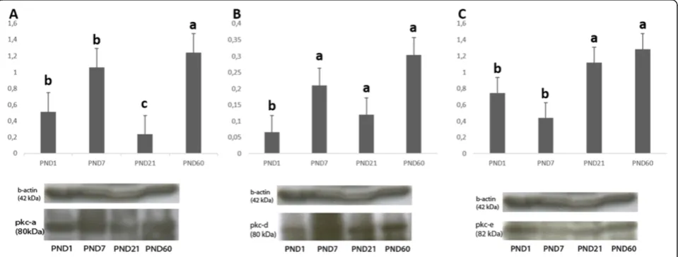
Western blot analysis of PKCα, PKCδand PKCεin mouse ovaries
PKCαprotein expression level was detected to be signifi-cantly higher in PND60 ovaries. PKCα expression in PND7 and PND1 was also significantly higher when compared to PND21 ovaries. The lowest PKCα protein expression was observed in PND21 ovaries (Figure 7A).
PKCδ expression was significantly lower in PND1 ovaries when compared to other groups. PND60 had the highest expression level of PKCδ(Figure 7B).
PKCεexpression level was significantly higher in PND60 and PND21 ovaries when compared to pre-pubertal PND7 and PND1 ovaries (Figure 7C).
Discussion
This study demonstrates that different PKC isoforms are differentially expressed in particular cellular components
of ovarian follicles of pre-pubertal, pubertal and adult mouse ovaries.
Data obtained from H-Score evaluation of immunohis-tochemistry findings revealed that PKCαexpression was more apparent in oocytes of all follicles in pre-pubertal, pubertal and adult ovaries, though it was also expressed in granulosa cells. Interestingly, in adult and pre-pubertal ovaries, intense immunostaining of PKCα in oocytes was statistically significant in primordial (PND60, PND7 and PND1), primary (PND60, PND7 and PND1) and secondary follicles (PND60 and PND7), whereas in PND21 ovaries only oocytes of primordial follicles had significantly higher immunostaining level of PKCα. These findings support the idea that PKCαexpression in oocytes of larger follicles may have low significance due to granulosa-oocyte interactions
during initiation of hormone dependent follicular growth [27,28]. PKCαimmunostaining was shown to be higher in larger follicles in all groups which might be related to pro-liferation of granulosa cells and development of oocytes. Only in PND1 ovary, primordial follicles had higher immu-nostaining level of PKCαwhich was not statistically signifi-cant. PKCαprotein expression showed the highest level in PND60 ovaries according to our Western blot experiments which might be due to its higher expression in larger folli-cles and CL.
Oocyte maturation of preovulatory follicles is induced by the signals from the extra-follicular environment dur-ing the LH surge and these signals are transmitted by cumulus cells to the oocyte through gap junctions as reviewed by Huang, Z. et al. [29]. Once the signal for
Figure 6H-Score evaluations of PKCεstainings in postnatal mouse ovaries in terms of follicular cells (A) and follicular stages (B).
Different letters mark statistical significance (p < 0.05). (One way anova, Holm Sidak method).
cAMP reduction is received by cumulus cells, cAMP levels also decrease in oocytes through the action of PDE3A and results in protein kinase A (PKA) inactivation. Consequently, inactivating phosphorylation of CDC25b (cell division cycle 25b) by PKA is inhibited and active CDC25b removes inactivating phosphates from CDK1 of maturation promoting factor (MPF). Then MPF becomes active and maturation progresses [30]. While cumulus cells transmit signals to control oocyte maturation, oocyte secretes factors that promote follicle growth such as growth differentiation factor 9 (GDF9) and bone morpho-genetic protein 15 (BMP15) [31].
The resumption of meiosis and entry into metaphase I (MI) is inhibited in oocytes arrested at prophase I upon PKC activation [16,32] whereas in Xenopus, mouse and rat MII oocytes, PKC activation promotes entry into interphase [33-35]. However, activation of PKC during bovine oocyte maturation is reported to induce GVBD [17]. In the present study, higher PKCαimmunostaining level in oocytes of smaller follicles supports the idea that PKCα might be critical for meiotic arrest of oocytes ra-ther than meiotic resumption.
According to the findings of the current study, another conventional PKC isoform, PKCδ was found to be expressed highly in granulosa cells of all follicles in pre-pubertal, pubertal and adult ovaries. Interestingly, intense immunostaining of PKCδin granulosa cells compared to oocytes was significant only in adult and pubertal ovaries. This might be due to altered gonadotropin levels and re-lated signal transduction pathways during and after pu-berty [28]. PKCδ immunostaining was also higher in larger follicles of all groups. Analysis of the Western blot bands showed that PKCδ had an increased expression in PND60 ovaries whereas PND1 ovaries had the lowest PKCδ expression level. These results might be related to higher expression of PKCδin larger follicles.
The expression of PKCα and PKCδ in granulosa cells of all follicles might be related to their importance in granulosa cell proliferation and viability. The controlled division of hamster granulosa cells has already been found to be activated by a self-sustaining loop of PKC and MAPK pathway [10]. Though PKC is found to be included in activation of DNA synthesis through induc-tion of MAPK pathway, it remains unknown which PKC isoform specifically participates in this process. PLA2G4 which takes part in DNA synthesis activation loop as a PKC activator [10], catalyzes phosphatidylinositol 4, 5 bisphosphate (PIP2) into diacylglycerol (DAG) and inosi-tol 1, 4, 5-triphosphate (IP3). Since these catalysis prod-ucts stand for the activation of conventional and novel PKC; atypical PKC might be excluded from these activa-tion loop. Thus, the effect of PKC activators in DNA synthesis might be considered for detection of specific PKC isoforms participated in this process.
Inhibitor of phospholipase C (PLC), that hydrolyses PIP2 is found to significantly reduce PGV (simian virus infected porcine granulosa cell line) cell proliferation. Moreover, inhibitor of phosphatidic acid phosphatase (PAP), that converts phosphatidic acid (PA) to DAG, also significantly reduces PGV cell numbers. PMA restores PGV cell proliferation that is reduced by both PLC and PAP [36]. cAMP independent growth promoting effects of FSH are shown to be activated by calcium ion and MAPK-dependent pathways [37]. These pathways inMAPK-dependent of cAMP but dependent of Ca2+and MAPK might include PKC. However, specific PKC isoforms included in these signaling pathways remain unknown.
Research focused on isoform specifity of PKC is very restricted. PKCδ is suggested to be related with anti-apoptotic actions of bFGF in rat granulosa cells [13]. The results of the current study showing that PKCδ is highly expressed in granulosa cells, also support the idea that PKCδ might specifically be critical for inhibition of granulosa cell apoptosis during pre-pubertal, pubertal and adult stages. PKCδ was also found to have specific functions during mouse oocyte maturation [11]. Accord-ing to our results, PKCδmight also have important roles in granulosa cells. In a recent study, it is suggested that cPKC isoforms PKCα and PKCβI participate in FSH-induced meiotic resumption of mouse follicle enclosed oocytes in follicle culture model [18]. Thus, specific PKC isoforms might be critical for oocyte maturation process. Once, specific PKC isoforms responsible for progression and arrest of oocyte maturation is determined, the mecha-nisms lying behind oocyte maturation defects can be bet-ter understood as well as in vitro maturation of oocytes for treatment and preservation of fertility can be manipu-lated by the use of specific chemical activators or inhibi-tors of these isoforms.
PKCε was found to be expressed at a significantly higher level in oocytes of all follicles in pre-pubertal, pu-bertal and adult ovaries. However, unlike PKCα and PKCδ, PKCεexpression was found to be higher in prim-ordial follicles of adult and pre-pubertal ovaries. In PND21 pubertal ovaries, PKCεexpression was higher in secondary and pre-antral follicles when compared to primordial and primary follicles. This might be due to differential hormonal levels and related signaling during puberty [28]. PKCε might be critical for primordial fol-licle survival which is related with balance of oocyte sur-vival and death [38]. The Western blot analysis revealed that PKCεprotein expression level was higher in PND60 ovaries when compared to other groups. This might be due to increased number of larger oocytes and elevated expression levels of PKCε in primordial follicles of PND60 ovaries.
activation of DNA synthesis of granulosa cells through in-duction of MAPK [10]. There is also evidence that the luteolysis process triggered by prostaglandin F2α (PGF2α) is also under the control of PKC through the activation of Raf/MEK1/ERK1 and ERK2 pathway [22].
PGF2αactivates PLC through its plasma membrane G-protein-coupled receptor and this activation causes PIP2 hydrolysis and as a result IP3 and DAG accumulation as well as mobilized intracellular Ca2+ stimulation [39]. Calcium is required to support progesterone (P4) syn-thesis in bovine luteal cells and LH increases IP3, and [Ca2+]i in bovine luteal cells and in porcine granulosa cells leading to activation of PKC [40]. When PKCε is inhibited with PKCε-specific inhibitors, the PGF2α – in-duced rise in [Ca2+]ii is decreased in luteal cells and con-sequently the ability of PGF2αto inhibit LH-stimulated P4 secretion becomes restricted leading to inadequate exten-sion of the luteal life span [41]. On the other hand, inhib-ition of specific PKC isoform resulting in high P4 level can compensate for luteal phase defects characterized by low P4 levels [42].
Though it is documented that PKCε is related to pro-gesterone synthesis/secretion in luteal cells in earlier studies [23], in the current study PKCα expression was found to be significantly higher in CL compared to the other follicles. These results indicate that PKCα might be important for luteolysis process and luteal functions. PKCδ and PKCε expressions in luteal cells of CL was found to be lower than developing follicles. PKCα must also be studied in terms of its possible functions in luteal cells. Though it is found that PKC family is associated with the MAPK signaling cascade in luteal cells [22], it is still unclear which PKC isoform is specifically related to these signaling pathways in CL.
PKC is found to be participated in the induction process of ovulatory Prkg2 gene through stimulation of ERK, but not JNK [6]. Thus, PKC induces particular genes through interaction with specific proteins. These specific target proteins might be stimulated by specific PKC isoforms. Since, in the current study PKCα and PKCδ expressions were found to be more significant in granulosa cells, signaling pathways related to ovulatory genes might be related to these isoforms.
Though isoform specific PKC antibodies can detect mul-tiple isotypes to some extent, based on our observations in Western blot experiments, we concluded that the anti-bodies are notably specific for the studied isoforms for Western blot as well as immunohistochemistry.
In addition to our findings through immunohisto-chemistry and Western blotting, the existence of mRNA of specific PKC isoforms in different cells of the ovary can be revealed by application of q-PCR on isolated cells from follicles at specific developmental stages without any concern about the amount of specimen. Besides,
proteomics technology can clearly reveal the structure and function of different PKC isoforms on isolated cells of the ovary.
Conclusions
We conclude that PKCα, PKCδ and PKCεisoforms show different expression profiles in mouse ovary at pre-pubertal, pubertal and adult stages. Thus, considering the differential expression levels of these isoforms in specific cellular components of the ovary, we suggest that these isoforms have particular roles in ovarian function. The iso-form specifity of PKC family must be further studied to re-veal undiscovered mechanisms of follicular development, oocyte maturation, ovulation and luteal cell functions. The proof of PKC isoform specifity for particular ovarian functions will allow the use of isoform specific inhibitors or activators to compensate ovarian dysfunction which is one of the major causes of female infertility.
Competing interests
The authors declare that they have no competing interests.
Authors’contributions
FT carried out tissue processing, immunohistochemistry and Western blot experiments, performed the statistical analysis. IU and GA conceived of the study, and participated in its design and coordination and helped to draft the manuscript. All authors read and approved the final manuscript.
Acknowledgements
This work was supported by The Scientific Research Projects Coordination Unit of Akdeniz University (Project Number: 2009.02.0122.010).
Received: 14 July 2014 Accepted: 29 November 2014
References
1. Richards JS, Russell DL, Ochsner S, Hsieh M, Doyle KH, Falender AE, Lo YK, Sharma SC:Novel signaling pathways that control ovarian follicular development, ovulation, and luteinization.Recent Prog Horm Res2002,
57:195–220.
2. Nishizuka Y:Intracellular signaling by hydrolysis of phospholipids and activation of protein kinase C.Science1992,258:607–614.
3. Hug H, Sarre TF:Protein kinase C isoenzymes: divergence in signal transduction?Biochem J1993,291(Pt 2):329–343.
4. Gomberts BDKI, Tatham ER:Signal Transduction.MA, USA: Academic; 2009. 5. Corbalan-Garcia S, Gomez-Fernandez JC:Protein kinase C regulatory
domains: the art of decoding many different signals in membranes.
Biochim Biophys Acta2006,1761:633–654.
6. Sriraman V, Modi SR, Bodenburg Y, Denner LA, Urban RJ:Identification of ERK and JNK as signaling mediators on protein kinase C activation in cultured granulosa cells.Mol Cell Endocrinol2008,294:52–60. 7. Dekker LV:Protein Kinase C.NY, USA: Springer; 2004.
8. Aderem A:The MARCKS family of protein kinase-C substrates.
Biochem Soc Trans1995,23:587–591.
9. Baron CB, Cunningham M, Strauss JF 3rd, Coburn RF:Pharmacomechanical coupling in smooth muscle may involve phosphatidylinositol
metabolism.Proc Natl Acad Sci U S A1984,81:6899–6903.
10. Yang P, Roy SK:A novel mechanism of FSH regulation of DNA synthesis in the granulosa cells of hamster preantral follicles: involvement of a protein kinase C-mediated MAP kinase 3/1 self-activation loop.
Biol Reprod2006,75:149–157.
11. Viveiros MM, O’Brien M, Wigglesworth K, Eppig JJ:Characterization of protein kinase C-delta in mouse oocytes throughout meiotic maturation and following egg activation.Biol Reprod2003,69:1494–1499.
12. Park JI, Kim SG, Chun JS, Seo YM, Jeon MJ, Ohba M, Kim HJ, Chun SY:
forskolin-induced NGFI-B expression in preovulatory granulosa cells of rat ovary.Mol Cell Endocrinol2007,270:79–86.
13. Peluso JJ, Pappalardo A, Fernandez G:Basic fibroblast growth factor maintains calcium homeostasis and granulosa cell viability by stimulating calcium efflux via a PKC delta-dependent pathway.
Endocrinology2001,142:4203–4211.
14. Luria A, Tennenbaum T, Sun QY, Rubinstein S, Breitbart H:Differential localization of conventional protein kinase C isoforms during mouse oocyte development.Biol Reprod2000,62:1564–1570.
15. Gangeswaran R, Jones KT:Unique protein kinase C profile in mouse oocytes: lack of calcium-dependent conventional isoforms suggested by rtPCR and Western blotting.FEBS Lett1997,412:309–312.
16. Downs SM, Cottom J, Hunzicker-Dunn M:Protein kinase C and meiotic regulation in isolated mouse oocytes.Mol Reprod Dev2001,58:101–115. 17. Mondadori RG, Neves JP, Goncalves PB:Protein kinase C (PKC) role in
bovine oocyte maturation and early embryo development.Anim Reprod Sci2008,107:20–29.
18. Wang J, Chen Q, Zhou J, Wen J, Bian F, Li G, Mu X, Han Y, Xia G, Zhang M:
Specific protein kinase C isoforms alpha and betaI are involved in follicle-stimulating hormone-induced mouse follicle-enclosed oocytes meiotic resumption.PLoS One2012,7:e45043.
19. Zhang M, Ouyang H, Xia G:The signal pathway of gonadotrophins-induced mammalian oocyte meiotic resumption.Mol Hum Reprod2009,
15:399–409.
20. Norris RP, Freudzon M, Mehlmann LM, Cowan AE, Simon AM, Paul DL, Lampe PD, Jaffe LA:Luteinizing hormone causes MAP kinase-dependent phosphorylation and closure of connexin 43 gap junctions in mouse ovarian follicles: one of two paths to meiotic resumption.
Development2008,135:3229–3238.
21. Vaccari S, Weeks JL 2nd, Hsieh M, Menniti FS, Conti M:Cyclic GMP signaling is involved in the luteinizing hormone-dependent meiotic maturation of mouse oocytes.Biol Reprod2009,81:595–604. 22. Chen DB, Westfall SD, Fong HW, Roberson MS, Davis JS:Prostaglandin
F2alpha stimulates the Raf/MEK1/mitogen-activated protein kinase signaling cascade in bovine luteal cells.Endocrinology1998,139:3876–3885. 23. Goravanahally MP, Sen A, Inskeep EK, Flores JA:PKC epsilon and an
increase in intracellular calcium concentration are necessary for PGF2 alpha to inhibit LH-stimulated progesterone secretion in cultured bovine steroidogenic luteal cells.Reprod Biol Endocrinol2007,5:37.
24. Tai CJ, Kang SK, Choi KC, Tzeng CR, Leung PC:Antigonadotropic action of adenosine triphosphate in human granulosa-luteal cells: involvement of protein kinase Calpha.J Clin Endocrinol Metab2001,86:3237–3242. 25. Lowry OH, Rosebrough NJ, Farr AL, Randall RJ:Protein measurement with
the Folin phenol reagent.J Biol Chem1951,193:265–275.
26. Akkoyunlu G, Erdogru T, Seval Y, Ustunel I, Koksal T, Usta MF, Baykara M, Demir R:Immunolocalization of glial cell-derived neurotrophic factor (GDNF) and its receptor GFR-alpha1 in varicocele-induced rat testis.
Acta Histochem2007,109:130–137.
27. Canipari R:Oocyte–granulosa cell interactions.Hum Reprod Update2000,
6:279–289.
28. Fauci ASKD, Braunwald E, Hauser SL, Longo DL, Jameson JL, Loscalzo J:The Female Reproductive System: Infertility and Contraception.InHarrison’s Principles of Internal Medicine.17th edition. New York City: The McGraw-Hill Companies; 2008:2530–2652.
29. Huang Z, Wells D:The human oocyte and cumulus cells relationship: new insights from the cumulus cell transcriptome.Mol Hum Reprod2010,
16:715–725.
30. Conti M, Andersen CB, Richard F, Mehats C, Chun SY, Horner K, Jin C, Tsafriri A:Role of cyclic nucleotide signaling in oocyte maturation.Mol Cell Endocrinol2002,187:153–159.
31. Gilchrist RB, Lane M, Thompson JG:Oocyte-secreted factors: regulators of cumulus cell function and oocyte quality.Hum Reprod Update2008,
14:159–177.
32. Lefevre B, Pesty A, Koziak K, Testart J:Protein kinase C modulators influence meiosis kinetics but not fertilizability of mouse oocytes.J Exp Zool1992,264:206–213.
33. Bement WM, Capco DG:Parallel pathways of cell cycle control during Xenopus egg activation.Proc Natl Acad Sci U S A1991,88:5172–5176. 34. Colonna R, Tatone C, Francione A, Rosati F, Callaini G, Corda D, Di Francesco
L:Protein kinase C is required for the disappearance of MPF upon artificial activation in mouse eggs.Mol Reprod Dev1997,48:292–299.
35. Eliyahu E, Shalgi R:A role for protein kinase C during rat egg activation.
Biol Reprod2002,67:189–195.
36. Lin MT:Establishment of an immortalized porcine granulosa cell line (PGV) and the study on the potential mechanisms of PGV cell proliferation.Keio J Med2005,54:29–38.
37. Babu PS, Krishnamurthy H, Chedrese PJ, Sairam MR:Activation of extracellular-regulated kinase pathways in ovarian granulosa cells by the novel growth factor type 1 follicle-stimulating hormone receptor. Role in hormone signaling and cell proliferation.J Biol Chem2000,
275:27615–27626.
38. McLaughlin EA, McIver SC:Awakening the oocyte: controlling primordial follicle development.Reproduction2009,137:1–11.
39. Davis JS, Alila HW, West LA, Corradino RA, Hansel W:Acute effects of prostaglandin F2 alpha on inositol phospholipid hydrolysis in the large and small cells of the bovine corpus luteum.Mol Cell Endocrinol1988,
58:43–50.
40. Flores JA, Aguirre C, Sharma OP, Veldhuis JD:Luteinizing hormone (LH) stimulates both intracellular calcium ion ([Ca2+]i) mobilization and transmembrane cation influx in single ovarian (granulosa) cells: recruitment as a cellular mechanism of LH-[Ca2+]i dose response.
Endocrinology1998,139:3606–3612.
41. Sen A, Choudhary E, Inskeep EK, Flores JA:Effects of selective protein kinase c isozymes in prostaglandin2alpha-induced Ca2+ signaling and luteinizing hormone-induced progesterone accumulation in the mid-phase bovine corpus luteum.Biol Reprod2005,72:976–984. 42. Jordan J, Craig K, Clifton DK, Soules MR:Luteal phase defect: the
sensitivity and specificity of diagnostic methods in common clinical use.
Fertil Steril1994,62:54–62.
doi:10.1186/s13048-014-0117-z
Cite this article as:Tepekoyet al.:Protein kinase C isoformsα,δandε are differentially expressed in mouse ovaries at different stages of postnatal development.Journal of Ovarian Research20147:117.
Submit your next manuscript to BioMed Central and take full advantage of:
• Convenient online submission
• Thorough peer review
• No space constraints or color figure charges
• Immediate publication on acceptance
• Inclusion in PubMed, CAS, Scopus and Google Scholar
• Research which is freely available for redistribution