R E V I E W
Open Access
EBV-positive mucocutaneous ulcers: a
presentation of two cases and a brief
literature review
Karolinne Correia Wanderlei
1*, Danielle Carvalho Quintella
2, Tullia Cuzzi
3, Denize D
’
Azambuja Ramos
2,
José Carlos Morais
2, Mário Romañach
4and Cristiane Bedran Milito
2Abstract
Mucocutaneous ulcers associated with the Epstein Barr virus constitute an EBV-induced B-cell lymphoproliferative disorder first described in 2010 by Stefan D. Dojcinov et al. These lesions can occur in association with a spectrum of immunosuppressive conditions, including primary immune deficiency, Human Immunodeficiency Virus (HIV) infection, post-transplantation and the use of methotrexate or tumor necrosis factor-alpha (TNF-a) antagonists. Patients clinically present with slowly developing indurated cutaneous and/or mucosal ulcers, especially in the oropharynx. Histopathology reveals circumscribed ulcers containing a mixture of lymphocytes, plasma cells, histiocytes, eosinophils and large transformed cells resembling Hodgkin and Reed-Sternberg cells. The adjacent squamous epithelium presents reactive nuclear atypia and pseudoepitheliomatous hyperplasia. The large
transformed cells show positivity for CD20, CD30, Oct-2, PAX5 and EBV. These cells are also positive for MUM1, yet lack CD10 expression, with absent or focal positivity for BCL6. Despite the presence of highly atypical cells, the clinical course is indolent, without progression to disseminated disease. We report herein two cases of diagnosed EBV-positive mucocutaneous ulcers to add to the relatively few cases previously described in the literature.
Keywords:Epstein Barr Virus, EBV positive mucocutaneous ulcer, Immunosuppression, Lymphoproliferative
Resumo
A úlcera mucocutânea associada ao Vírus Epstein Barr é uma doença linfoproliferativa indolente de linfócitos B que foi descrita em 2010 por Stefan D. Dojcinov e cols. Pode ocorrer em associação com etiologias imunossupressoras, incluindo deficiência imune primária, infecção pelo vírus da Imunodeficiência Humana (HIV), pacientes pós transplantes e em uso de metotrexato ou antagonistas TNF-alfa. Os pacientes apresentam quadro clínico de úlcera de surgimento insidioso, com localização cutânea ou de mucosas, preferencialmente de orofaringe. Os achados histopatológicos da lesão revelam áreas ulceradas com infiltrado polimórfico de linfócitos, plasmócitos, histiócitos e eosinófilos em meio a células grandes, pleomórficas que lembram células de Hodgkin e de Reed-Sternberg. O epitélio adjacente geralmente mostra núcleos atípicos e hiperplasia pseudoepiteliomatosa. As células grandes pleomórficas mostram positividade para CD20, CD30, Oct-2, PAX5 e EBV. Estas células podem ser também positivas para MUM1, focalmente positivas para Bcl-6 e não costumam expressar CD10. Apesar da presença de células muito atípicas, o curso clínico é indolente, sem progressão para doença disseminada, sendo essencial diferenciá-la de doenças linfoproliferativas malignas. Descrevemos dois casos de pacientes com diagnóstico de úlcera mucocutânea associada ao vírus Epstein Barr (EBV) para adicionar aos poucos casos já descritos na literatura.
© The Author(s). 2019Open AccessThis article is distributed under the terms of the Creative Commons Attribution 4.0 International License (http://creativecommons.org/licenses/by/4.0/), which permits unrestricted use, distribution, and reproduction in any medium, provided you give appropriate credit to the original author(s) and the source, provide a link to the Creative Commons license, and indicate if changes were made. The Creative Commons Public Domain Dedication waiver (http://creativecommons.org/publicdomain/zero/1.0/) applies to the data made available in this article, unless otherwise stated. * Correspondence:karolinnecw@hotmail.com
Background
Mucocutaneous ulcers positive for the Epstein Barr Virus (EBVMCU) are a newly recognized clinicopatho-logical entity in the 2017 revision to the World Health Organization diagnostic criteria. (Swerdlow et al., 2017) This condition was described as a lymphoproliferative lesion associated with isolated skin or mucosal ulcers in elderly or immunosuppressed patients. Since 2010, 53 cases have been published. (Dojcinov et al.,2010; Au et al.,2006; Kalantzis et al.,1997; Deeming et al.,2005; Del Pozo et al., 2001; Kazlow et al., 2003; Nalesnik et al.,
1988; Warner et al., 2008; Lawrence & Dahl, 1984; Hashizume et al.,2012; Au et al.,2011; Kleinman et al.,
2014; Moran et al., 2015; Yamakawa et al., 2014; Attard et al., 2012; Matnani & Peker, 2014; McGinness et al.,
2012; Di Napoli et al.,2011; Hart et al.,2014; Kanemitsu et al., 2015; Sadiku et al., 2012; Magalhaes et al., 2015; Soni et al.,2014; Mendes et al.,n.d.; Roberts et al.,2016) Ninety percent of the adult population become in-fected with the Epstein Barr virus via the oral route, (Aldridge et al.,2017) and most infections occur early in life. In adulthood, EBV can persistently infect B-cells. The viral genes can upregulate a variety of cellular anti-gens and pathways, such as NF-kappaB. Physiologically, the proliferation of B cells induced by EBV infection is generally controlled by the immune system. In patients suffering from various causes of immunosuppression (IS), EBV has been associated with B-cell lymphoprolif-erative disorders (LPD). (Dojcinov et al.,2010)
EBV-LPD is associated with many etiologies, including pri-mary immunodeficiency, HIV infection, post-transplantation (Gru & Jaffe,2016), immunosenescence owing to aging and iatrogenic causes, such as methotrexate and TNF-alfa antag-onists. The spectrum of age-related EBV-positive lymphopro-liferative disorders was first described by Oyama and colleagues, and can occur in elderly patients without a his-tory of immunosuppression. (Oyama et al.,2003)
EBVMCU was described, in 2010, as localized sharply circumscribed ulcerative lesions, typically solitary (83%), that can occur in the oropharynx (52%), skin (29%) or gastrointestinal tract (19% - 40% colon, 30% esophagus, 20% rectum and 10% terminal ileum) (Roberts et al.,
2016). It occurs more commonly in women, around a mean age of 77. Isolated regional lymphadenopathy can accompany these ulcers, yet usually in the absence of systemic findings. (Gru & Jaffe, 2016) The diagnosis of EBVMCU requires a combination of clinical, morpho-logical and immunophenotypic parameters.
The histopathologic features of EBVMCU usually consist of a well-circumscribed ulcer with a polymorphous infil-trate containing histiocytes, eosinophils and plasma cells, large pleomorphic blasts resembling Hodgkin Reed Stern-berg (HRS)-like cells, numerous medium-sized T-cells, plasmacytoid apoptotic bodies, as well as angioinvasion and
necrosis. The large pleomorphic blast cells express CD20, CD30, CD15, PAX5, OCT2, MUM1, BOB1 and CD45, with a background of lymphocytes positive for CD3, CD4 and CD8. Reduction or absence of CD20 expression is observed in 33% of cases. These large atypical cells are positive for the latent membrane protein-1 of EBV (LMP-1). The pres-ence of a high Ki67 proliferative index does not exclude the diagnosis of EBVMCU. (Gratzinger & Jaffe,2016)
Although considered a self-limited disorder, monoclonal-ity studies investigating immunoglobulin and T-cell recep-tor (TCR) gene rearrangements by PCR-based genotyping showed monoclonal immunoglobulin heavy chain or kappa light chain gene rearrangement and monoclonal TCR gene rearrangement in some cases. (Dojcinov et al.,2010; Au et al.,2006; Moran et al.,2015; McGinness et al.,2012; Hart et al.,2014; Kanemitsu et al.,2015; Roberts et al.,2016; Gru & Jaffe, 2016; Gratzinger & Jaffe,2016; Stojanov & Woo,
2015; Asano et al.,2009; Shimoyama et al.,2009). Patients with EBVMCU present typically undetectable EBV DNA in peripheral blood, in contrast to many other types of EBV-associated LPDs. (Hart et al.,2014)
Typically, EBVMCU presents an indolent course. While no treatment guidelines exist, management tends to be conservative and, in cases related to the use of munosuppressants, the withdrawal or decrease in im-munosuppressant dosages is common. Nearly two-thirds of immunosuppressive-associated cases evolved to complete clinical remission exclusively by dosage reduc-tion, with a median time to lesion resolution of four weeks (range: 2-12 weeks). A lack of response to im-munosuppressant dosage reduction or withdrawal within three months should prompt the reevaluation of an EBVMCU diagnosis. (Hujoel et al.,2018)
The differential diagnosis of EBVMCU includes sec-ondary involvement by Classic Hodgkin Lymphoma (cHL), diffuse large B-cell lymphoma (DLBCL) associ-ated or not with EBV, primary cutaneous anaplastic large cell lymphoma, lymphomatous granulomatosis (LyG) and aggressive Posttransplant Lymphoproliferative Dis-ease (PTLD). (Gru & Jaffe, 2016) Distinctions based solely on pathological findings can prove extremely diffi-cult due to considerable overlap in morphology and immunophenotype; consequently, making correlations with clinical parameters is essential.
Main text
Patient characteristics and clinical course
We report two cases of EBVMCU compromising the oral mucosa and skin. Both patients were over 65 years and one was undergoing immunosuppressive treatment.
yellowish center containing fibrin. The other lesion mea-sured 2 cm and presented scarring aspects. His laboratory evaluation was negative for HIV, anti-HBsAg and HCV antibodies. Previous pathological history consisted of splenectomy due to hemorrhaging of unknown etiology.
The second patient, a 74-year-old female, presented to her dentist with fever and a painful ulcer on her lower right alveolar ridge. She had a history of Sjögren's dis-ease and had been taking methotrexate for 18 months. Laboratory examinations were negative for anti-HIV, anti-HBsAg, anti-HCV, VDRL, EBV (IgM), CMV (IgM) and HSV.
In both cases, lesions of the oral mucosa and skin regressed spontaneously without treatment.
Histology
The histopathological examination of the first patient re-vealed a sharply circumscribed cutaneous ulcer extend-ing to the hypodermis, with large and pleomorphic mononucleate or binucleate cells resembling Hodgkin/ Reed-Sternberg cells, associated with an inflammatory background of small lymphocytes, as well as some eosin-ophils, plasma cells and histiocytes. Apoptotic cells and foci of necrosis were also observed (Fig.1).
The histopathological examination of the second patient revealed a necrotic mucosal ulcer and angioinvasion by large cells, some with Hodgkin/Reed Sternberg-like fea-tures, in an inflammatory background of small lympho-cytes and rare eosinophils (Fig.2).
Immunohistochemistry
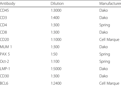
Immunohistochemistry was performed in paraffin sec-tions using monoclonal antibodies against CD30, LMP-1/EBV, CD45, CD20, CD3, CD4, CD8, PAX5, OCT2, MUM1 and BCL6 (Table 1). Sections were dewaxed in xylene and rehydrated using serial concen-trations of ethanol. Heat-mediated antigen retrieval was performed, and endogenous peroxidase activity was blocked with 5% alcoholic hydrogen peroxide for 30 mi-nutes. Slides were then incubated overnight with the pri-mary antibodies. After pripri-mary antibody binding was detected by the EnVision® + Dual Link/Peroxidase sys-tem (Dako, Carpenteria, Ca, USA), sections were coun-terstained using Harris haematoxylin. (Table1).
Immunohistochemical analysis of the first patient depicted large pleomorphic cells positive for CD30, OCT-2, CD20, MUM-1, CD45 and EBV (LMP1). The base of the ulcer showed a dense rim of small lympho-cytes positive for CD3, CD4, CD8 and CD45. CD8 posi-tive T-lymphocytes were more abundant than CD4. (Tables2and3) (Fig.1).
Immunohistochemical analysis of the second patient demonstrated large pleomorphic cells positive for CD30, OCT-2, CD20, MUM-1, CD45, PAX5 and EBV (LMP1).
The base of the ulcer showed small lymphocytes positive for CD3, CD4 and CD8, with some cells positive for OCT-2, CD45 and EBV (LMP1). CD8 positive T-lymphocytes were more abundant compared to CD4. (Tables2and3) (Fig.2).
Discussion/conclusion
EBV-positive mucocutaneous ulcers have been recently described, and are characterized by an ulcerated lesion not associated with lymphadenopathy, hepatosplenome-galy or bone marrow involvement. This condition is usu-ally associated with immunodeficiency. The majority of cases present an indolent course and can regress spon-taneously. When associated with immunosuppressive therapy, treatment should be discontinued and/or asso-ciated with rituximab. Despite the fact that 30% of cases present clonal rearrangement of immunoglobulin and/or clonal T rearrangement, the prognosis is generally excel-lent. Clonality has also been observed in both poly-morphic and monopoly-morphic cases of PTLD. (Swerdlow et al.,2017; Dojcinov et al.,2010; Au et al.,2006; Kalant-zis et al., 1997; Nalesnik et al., 1988; Di Napoli et al.,
2011; Gru & Jaffe,2016)
The distinct localization of EBV-positive mucocutane-ous ulcers is not fully understood. It has been suggested that this process might be a consequence of reduced immunosurveillance against EBV in sites rich in EBV-infected B-cells, such as the Waldeyer ring. It has also been speculated that EBV-positive mucocutaneous ulcers may result from chronic irritation, leading to de-creased immune resistance and the localized prolifera-tion of EBV-infected B-cells. (McGinness et al.,2012)
The histopathological aspects of EBVMCU may present challenges to pathologists, as the presence of large pleo-morphic cells around necrotic foci are suggestive of malig-nant lymphoproliferative processes. Morphological findings
Table 1Immunohistochemistry–Primary Antibodies
Antibody Dilution Manufacturer
CD45 1:3000 Dako
CD3 1:400 Dako
CD4 1:300 Spring
CD8 1:300 Dako
CD20 1:1000 Cell Marque
MUM 1 1:300 Dako
PAX 5 1:50 Spring
Oct-2 1:100 Spring
LMP-1 1:5000 Dako
CD30 1:300 Dako
BCL6 1:2400 Cell Marque
may be suggestive of a diagnosis of cHL, DLBCL associated or not with EBV, anaplastic large cell lymphoma (ALCL) or LyG. Immunophenotyping can also share similarities with these diseases, especially cHL. HRS-like cells typically ex-press CD30, PAX5, LMP1/EBV and, sometimes, CD15. Some immunohistochemical findings could prove helpful in performing differential diagnosis. Firstly, the inflammatory
background should contain abundant CD8-positive T-cells and a large number of EBV-positive plasmacytoid apoptotic cells, neither of which is common in cHL. Another import-ant feature not typically seen in cLH but usually found in EBVMCU is positivity for CD45 and CD20 in large pleo-morphic cells. Furthermore, LMP1/EBV expression is usu-ally detected in small cells, immunoblasts and large
Fig. 1Patient 1:aCutaneous lesion showing a well-circumscribed ulcer with a dense rim of lymphocytes at the base (25X).bandcLarge and pleomorphic Hodgkin/Reed-Sternberg-like cells in an inflammatory background (B:200X; C:400X). These atypical cells present immunoreactivity for CD30dCD20eand LMP1/EBVf. (D, E, F: 400X)
HRS-like cells. Finally, it is important to note that since Hodgkin’s Lymphoma is essentially a lymph node disease, extra-nodal involvement is generally secondary. (Swerdlow et al.,2017; Ohata et al.,2017)
Another important disease to consider in the differential diagnosis of EBVMCU is LyG, a similarly EBV-driven B-cell LPD with angiocentric and angiodestructive fea-tures. LyG is rare, also commonly associated with im-munosuppression, and is characterized by a background of small T-lymphocytes, with variable numbers of large atypical immunoblast-like cells, admixed with HRS-cells. Cells are positive for LMP1/EBV, CD20 and CD30, but usually not for CD15. Differential diagnosis relies on clin-ical presentation, as LyG typclin-ically compromises the lung, sometimes in association with other organs, more fre-quently the central nervous system (Swerdlow et al.,2017; Song et al.,2015).
EBV-positive DLBCL is another important differential diagnosis. These tumors occur in apparently immuno-competent individuals, usually older than 50 years of age, and carry a poorer prognosis when compared with
EBV-negative DLBCL. Presentation occurs at extra nodal sites in 70% of patients and can involve the skin. Morpho-logically, large lymphoid cells resembling centroblasts, immunoblasts or HRS-cells are present. The background is reactive with lymphocytes, plasma cells and histiocytes. Neoplastic cells express B-cell antigens, CD30 and EBV. (Swerdlow et al.,2017; Goodlad et al.,2017).
In conclusion, EBVMCU poses a diagnostic challenge to the pathologist due to its histopathological similarity with some EBV-associated malignant lymphoprolifera-tive disorders. A combination of clinical history, mor-phological findings and immunohistochemical features are crucial to achieving a definitive diagnosis, as well as to selecting appropriate therapy for affected patients.
Abbreviations
ALCL:Anaplastic Large Cell Lymphoma; cHL: classic Hodgkin Lymphoma; DLBCL: Diffuse Large B-cell Lymphoma; EBV: Epstein Barr Virus; LPD: EBV-induced B cell lymphoproliferative disorder; EBVMCU: EBV-positive mucocutaneous ulcer; HIV: Human Immunodeficiency Virus; HRS: Hodgkin and Reed-Sternberg; IS: Immunosuppression; LPD: Lymphoproliferative disorders; LyG: Lymphomatous Granulomatosis; PTLD: Posttransplant Lymphoproliferative Disease; TCR: T-cell receptor
Acknowledgements
Not applicable.
Funding
The authors contributed with the funding of the research.
Availability of data and materials
Not applicable.
Authors’contributions
Authors KCW and CBM were major contributors in writing the manuscript. Authors CBM, JCM and DDA performed histological examinations and diagnosis in the two cases. KCW accompanied the histological examination of each case. Authors TC and DCQ identified the first patient and provided figures depicting histological examinations. Author MR identified the second patient. Authors KCW and CBM wrote the manuscript. DCQ and CBM revised the final manuscript. All authors read and approved the final manuscript.
Ethics approval and consent to participate
Not applicable.
Consent for publication
Not applicable.
Competing interests
The authors declare that they have no competing interests.
Publisher’s Note
Springer Nature remains neutral with regard to jurisdictional claims in published maps and institutional affiliations.
Author details
1Serviço de Anatomia Patológica, Hospital Universitário Clementino Fraga Filho, Universidade Federal do Rio de Janeiro, Rio de Janeiro, Brazil. 2Departamento de Patologia, Faculdade de Medicina, Universidade Federal do Rio de Janeiro, Rio de Janeiro, Brazil.3Serviço de Anatomia Patológica, Instituto Nacional de Infectologia, FIOCRUZ, Rio de Janeiro, Brazil.4Patologia Oral da Faculdade de Odontologia da, Universidade Federal do Rio de Janeiro, Rio de Janeiro, Brazil.
Table 2Summary of Immunophenotype of B-cell Blasts, including Cells with HRS-like Features
Antibody Patient 1 Patient 2
OCT-2 + +
CD30 + +
CD20 + +
CD3 – –
CD8 – –
CD4 – –
MUM1 + +
EBV (LMP-1) + +
CD10 – –
CD45 + +
PAX5 – +
Table 3Summary of Immunophenotype of Background Small Lymphocytes
Antibody Patient 1 Patient 2
OCT-2 – + some cells
CD30 – –
CD20 – + rare cells
CD3 + +
CD8 + +
CD4 + +
MUM1 – –
EBV (LMP-1) – +
CD45 + +
Received: 2 December 2018 Accepted: 18 April 2019
References
Aldridge T et al (2017) Epstein-Barr-virus-related mucocutaneous ulceration that mimics oral squamous cell carcinoma: the importance of recognising this new condition. J Oral Maxillofac Surg 55(4):418–419.https://doi.org/10.1016/j. bjoms.2017.01.003
Asano N, Yamamoto K, Tamaru J et al (2009) Age-related Epstein-Barr virus (EBV) associated B-cell lymphoproliferative disorders: comparison with EBV positive classic Hodgkin lymphoma in elderly patients. Blood 113:2629–2636 Attard AA, Praveen P, Dunn PJS, James GJ (2012) Epstein–Barr virus-positive
mucocutaneous ulcer of the oral cavity: the importance of having a detailed clinical history to reach a correct diagnosis. Oral Surg Oral Med Oral Pathol Oral Radiol 114:e37–e39
Au WY, Loong F, Wan TSK, Tong ACK (2011) Multi-focal EBV-mucocutaneous ulcer heralding late-onset T-cell immunodeficiency in a women with lupus erythematosus. Int J Hematol 94:501–502
Au WY, Ma ES, Choy C, Chung LP, Fung TK, Liang R, Kwong YL (2006) Therapy related lymphomas in patients with autoimmune diseases after treatment with disease modifying anti-rheumatic drugs. Am J Hematol 81:5–11 Deeming GM, Collingwood J, Pemberton MN (2005) Methotrexate and oral
ulceration. Br Dent J 198:83–85
Del Pozo J, Martinez W, Garcia-Silva J, Almagro M, Pena-Penabad C, Fonseca E (2001) Cutaneous ulceration as a sign of methotrexate toxicity. Eur J Dermatol 11:450–452
Di Napoli A, Glubettini M, Duranti E, Ferrari A, Guglielmi C, Uccini S, Ruco L (2011) Iatrogenic EBV-positive lymphoproliferative disorder with features of EBV+ mucocutaneous ulcer: evidence for concomitant TCRγ/IGH rearrangements in the Hodgkin-like neoplastic cells. Virchows Arch 458:631–636
Dojcinov SD et al (2010) EBV positive Mucocutaneous ulcer–a study of 26 cases associated with various sources of immunosuppression. Am J Surg Pathol 34(3):405–417
Goodlad JR et al (2017) Epstein-Barr virus-associated lymphoproliferative disorders in the skin. Surg Pathol Clin 10(2):429–453.https://doi.org/10.1016/j. path.2017.01.001. Epub 2017 Mar 18
Gratzinger D, Jaffe ES (2016) Mucocutaneous ulcer: a mimic of EBV+ diffuse large B cell lymphoma in the immunodeficiency setting. Leuk Lymphoma. 57(8): 1982–1983.https://doi.org/10.3109/10428194.2016.1166492
Gru AA, Jaffe ES (2017) Cutaneous EBV-related lymphoproliferative disorders. Semin Diagn Pathol 34(1):60–75.https://doi.org/10.1053/j.semdp.2016.11.003. Epub 2016 Dec 7.
Hart M, Thakral B, Yohe S, Balfour HH Jr, Sing C, Spears M, McKenna RW (2014) EBV-positive mucocutaneous ulcer in organ transplant recipients. A localized indolent posttransplant lymphoproliferative disorder. Am J Surg Pathol 38(11):1522–1529
Hashizume H, Uchiyama I, Kawamura T, Suda T, Takigawa M, Tokura Y (2012) Epstein–Barr virus-positive mucocutaneous ulcers as a manifestation of methotrexate-associated B-cell lymphoproliferative disorders. Acta DermVenereol 92:276–277
Hujoel IA et al (2018) Epstein-Barr virus-positive Mucocutaneous ulcer in an immunosuppressed patient. ACG case reports journal.https://doi.org/10. 14309/crj.2018.32
Kalantzis A, Marshman Z, Falconer DT, Morgan PR, Odell EW (1997) Oral effects of low-dose methotrexate treatment. Cancer Surv 30:233–248
Kanemitsu M, John D, Lim A, Jaffe ES, Aoki J (2015) Clonal Epstein–Barr viruspositive mucocutaneous ulcer mimicking a mature B-cell lymphoma in a patient with mycophenolate-induphoma. Leuk Lymphoma 56(6):1908–10. https://doi.org/10.3109/10428194.2014.982646. Epub 2015 Jan 21 Kazlow DW, Federgrun D, Kurtin S, Lebwohl MG (2003) Cutaneous ulceration
caused by methotrexate. J Am Acad Dermatol 49:S197–S198
Kleinman S, Jhaveri D, Caimi P, Cameron R, Lemonovich T, Meyerson H, Hostoffer R, Tcheurekdjian H (2014) A rare presentation of EBV+ mucocutaneous ulcer that led to a diagnosis of hypogammaglobulinemia. J Allergy Clin Immunol Pract 2(6):810–812
Lawrence CM, Dahl MG (1984) Two patterns of skin ulceration induced by methotrexate in patients with psoriasis. J Am Acad Dermatol 11:1059–1065 Magalhaes M, Ghorab Z, Morneault J, Akinfolarin J, Bradley G (2015) Age-related Epstein–Barr virus-positive mucocutaneous ulcer: a case report. Clin Case Rep 3(7):531–534
Matnani R, Peker D (2014) Azathioprine induced, Epstein–Barr Virus-positive mucocutaneous ulcer arising in perianal fistula and abscess associated with Crohn’s disease. J Crohns Colitis 8:1747–1748
McGinness JL, Spicknall KE, Mutasim DF (2012) Azathioprine-induced EBV positive mucocutaneous ulcer. J Cutan Pathol 39:377–381
Mendes LST, et al. Epstein Barr virus positive mucocutaneous ulcer in a background of Crohn’s disease and Waldenstrom macroglobulinemia: a case report highlighting diagnostic pitfalls; doi:https://doi.org/10.1111/his.13420 Moran NR, Webster B, Lee KM, Trotman J, Kwan YL, Napoli J, Leong RW (2015)
Epstein Barr virus-positive mucocutaneous ulcer of the colon associated Hodgkin lymphoma in Crohn’s disease. World J Gastroenterol 21(19):6072– 6076
Nalesnik MA, Jaffe R, Starzl TE, Demetris AJ, Porter K, Burnhan JA, Makowka L, Ho M et al (1988) The pathology of posttransplant lymphoproliferativedisorders occurring in the setting of cyclosporine A-prednisone immunosuppression. Am J Pathol 133:173–192
Ohata Y, Tatsuzawa A, Ohyama Y, Ichikawa A et al (2017) A distinctive subgroup of oral EBV + B-cell neoplasm with polymorphous features is potential identical to EBV + mucocutaneous ulcer. Hum Pathol 69:129–139
Oyama T, Ichimura K et al (2003) Senile EBV+ B-cell lymphoproliferative disorders: a clinicopathologic study of 22 patients. Am J Surg Pathol 27(1):16–26 Roberts TK, Chen X, Liao JJ (2016) Diagnostic and therapeutic challenges of EBV
positive mucocutaneous ulcer: a case report and systematic review of the literature. Exp Hematol Oncol 5:13
Sadiku S, Kurshumliu F, Krasniqi X, Brovina A, Kryeziu E, Rrudhani I, Meqa K, Gashi-Luci L, Merz H (2012) Age-related Epstein–Barr virus-positive cutaneous ulcer arising after a self-limited subcutaneous abscess: a case report. J Med Case Rep 6:288–292
Shimoyama Y, Asano N, Kojima M et al (2009) Age-related EBV-associated B-cell lymphoproliferative disorders: diagnostic approach to a newly recognized clinicopathological entity. Pathol Int 59:835–843
Song JY, Pittaluga S, Dunleavy K et al (2015) Lymphomathoid granulomatosis - a single institution experience. Am J Surg Pathol 39:141–156
Soni S, Mercer R, Pattani K, Magill J (2014) Epstein–Barr virus positive mucocutaneous ulcer: a rare lesion presenting as a large lower lip mass. Poster presentation from the University of Central Florida college of medicine
Stojanov IJ, Woo SB (2015) Human papillomavirus and Epstein-Barr virus associated conditions of the oral mucosa. Semin Diagn Pathol 32:3–11 Swerdlow SH et al (2017) WHO classification of Tumours of Haematopoietic and
lymphoid tissues (revised 4th edition). IARC, Lyon
Warner J, Brown A, Whitmore SE, Cowan DA (2008) Mucocutaneous ulcerations secondary to methotrexate. Cutis. 81:413–416