Engeholmet al. BMC Neurology2014,14:118 http://www.biomedcentral.com/1471-2377/14/118
C A S E R E P O R T
Open Access
A novel mutation in LRSAM1 causes axonal
Charcot-Marie-Tooth disease with dominant
inheritance
Maik Engeholm
1,2*, Julia Sekler
1,2, David C Schöndorf
1,2,3, Vineet Arora
1,2,3, Jens Schittenhelm
4,
Saskia Biskup
1,5, Caroline Schell
1and Thomas Gasser
1,2Abstract
Background: Charcot-Marie-Tooth disease (CMT) refers to a heterogeneous group of genetic motor and sensory neuropathies. According to the primary site of damage, a distinction is made between demyelinating and axonal forms (CMT1 and 2, respectively, when inherited as an autosomal dominant trait). Leucine-rich repeat and sterile alpha motif-containing protein 1 (LRSAM1) is a ubiquitin-protein ligase with a role in sorting internalised cell-surface receptor proteins. So far, mutations in the LRSAM1 gene have been shown to cause axonal CMT in three different families and can confer either dominant or recessive transmission of the disease.
Case presentation: We have identified a novel mutation in LRSAM1 in a small family with dominant axonal CMT. Electrophysiological studies show evidence of a sensory axonal neuropathy and are interesting in so far as giant motor unit action potentials (MUAPs) are present on needle electromyography (EMG), while motor nerve conduction studies including compound motor action potential (CMAP) amplitudes are completely normal. The underlying mutation c.2046+1G>T results in the loss of a splice donor site and the inclusion of 63 additional base pairs of intronic DNA into the aberrantly spliced transcript. This disrupts the catalytically active RING (Really Interesting New Gene) domain of LRSAM1.
Conclusions: Our findings suggest that, beyond the typical length-dependent degeneration of motor axons, damage of cell bodies in the anterior horn might play a role in LRSAM1-associated neuropathies. Moreover, in conjunction with other data in the literature, our results support a model, by which disruption of the C-terminal RING domain confers dominant negative properties to LRSAM1.
Keywords: Axonal CMT, LRSAM1, Anterior horn cell disease, Splice site mutation, RING domain, Exome sequencing
Background
CMT comprises a clinically and genetically heterogeneous group of inherited motor and sensory neuropathies [1]. With an overall prevalence of approximately 1 in 2,500 individuals, CMT is the most common genetic disorder of the PNS [2]. In a majority of cases, CMT is transmitted as an autosomal dominant trait and is further classified as CMT1 or CMT2 according to the primary site of damage
*Correspondence: maik.engeholm@uni-tuebingen.de
1Department of Neurology and Hertie Institute for Clinical Brain Research, Hoppe-Seyler-Str. 3, 72076 Tübingen, Germany
2German Center for Neurodegenerative Diseases (DZNE), Otfried-Müller-Str. 27, 72076 Tübingen, Germany
Full list of author information is available at the end of the article
(demyelinating and axonal, respectively) [1,3]. In other families, CMT is transmitted as an X-linked or autosomal recessive trait. These latter cases are commonly classified as CMT4 when they show a demyelinating phenotype, while axonal forms are referred to as autosomal reces-sive CMT2 (AR-CMT2) [4]. Since the identification of a duplication of the peripheral myelin protein 22 locus as the cause of CMT1A more than 20 years ago [5,6], muta-tions in more than 60 genes implicated in a variety of different cellular functions have been associated with var-ious forms of CMT [3]. Beyond their relevance for clinical classification, these genes identify molecules and molec-ular pathways that play a primary role for the structural and functional integrity of PNS neurons and their myelin
sheath, respectively, and are potential targets for future therapeutic interventions.
Three different mutations in the LRSAM1 gene have been shown to cause axonal CMT in humans. Guernsey et al. [7] reported a large, multiply consanguineous family from Eastern Canada, in which axonal CMT was inherited as an autosomal recessive trait. The clinical presentation included moderate weakness and wasting, predominantly affecting distal lower limb muscles, with an onset in early adulthood. Needle EMG revealed signs of den-ervation and reinnden-ervation, and sensory nerve action potentials (SNAPs) were reduced or absent. Homozygos-ity mapping yielded a splice site mutation c.1913-1G>A in the LRSAM1 gene giving rise to a premature stop codon 20 bp inside the penultimate exon. Subsequently, two further mutations in LRSAM1 were identified in a Dutch and a Sardinian family with dominant axonal CMT [8,9]. In both studies, the clinical and electrophys-iological findings were very similar to that reported by Guernsey et al. [7]. Both mutations, p.Leu708ArgfsX28
and p.Ala683ProfsX3, disrupt the RING domain
of LRSAM1.
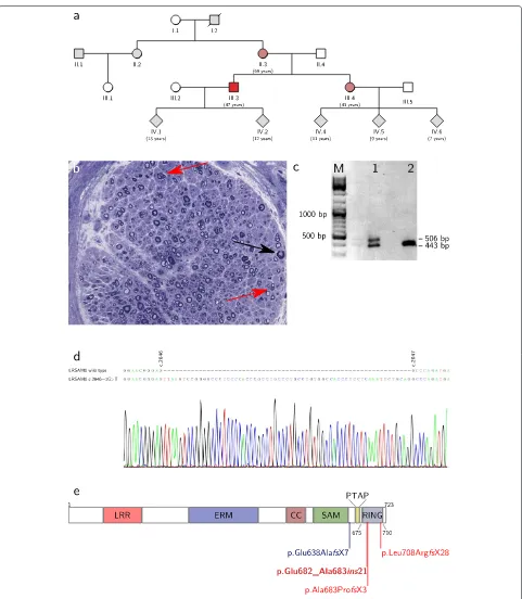
LRSAM1 is a E3 ubiquitin-protein ligase highly con-served throughout vertebrate evolution [10]. Alternative splicing gives rise to three different isoforms in humans, the largest of which consists of 723 amino acids and har-bours an N-terminal leucine-rich repeat domain, an ezrin-radixin-moezin domain, a coiled-coil region, a sterile alpha motif domain and a C-terminal C3HC4-type RING finger domain (Figure 1e). In human and mouse, LRSAM1 is highly expressed in motor neurons of the spinal cord and cell bodies of sensory neurons of dorsal root gan-glia [8,11]. Moreover, some expression is observed in the central nervous system [10,11]. In a cell culture system, LRSAM1 has been shown to interact with and medi-ate monoubiquitination of the Tumour susceptibility gene 101 protein (TSG101) [10]. TSG101 is a component of the ESCRT (Endosomal Sorting Complexes Required for Transport)-1 complex, which is involved in the sorting of endocytic ubiquitinated cargoes into lumenal vesicles of late endosomes [12]. Upon LRSAM1-mediated ubiq-uitination, TSG101 relocalises from these multivesicular bodies to a detergent-soluble compartment and loses its ability to direct internalised receptor proteins to the lyso-some [10]. In a mouse model, loss of LRSAM1 results in decreased motor NCVs and motor axon counts follow-ing acrylamide challenge, while neuromuscular function is otherwise unimpaired [11].
Here, we report a novel mutation in LRSAM1 in a small family with autosomal dominant axonal CMT. We present an in-depth electrophysiological examination of three affected individuals and characterise the functional consequences of the splice site mutation at the level of the transcript. Our results provide new insights into the
pathomechanism of LRSAM1-associated neuropathies, both at the cellular and molecular level.
Case presentation
We present a family with three members suffering from a predominantly sensory neuropathy with fascic-ulations and hyper-CK-aemia but without evidence of motor axonal neuropathy in nerve conduction studies (Figure 1a). The 47-years old index patient complained of a reduced sensation for touch on his feet, for instance when trying on shoes, which he had first noticed more than 20 years ago. Over the past few years, these deficits had gradually spread over his lower legs and recently also reached his finger tips. For several years he had noticed an unsteady gait, especially when walking in the dark, and he reported long-standing fasciculations of his calf and thigh muscles as well as crampi affecting both the leg and hand muscles. Over the past year, he had observed a reduction in size of his calf muscles. He did not complain of weak-ness, paresthesias or vegetative symptoms. His 43-years old sister and his 69-years old mother had very similar complaints with an age of onset between 20 and 40 years.
Clinical examination of the index patient showed fasci-culations of proximal leg muscles. The intrinsic foot and hand muscles appeared slender, but no other signs of atro-phy or skeletal deformities were observed. There was no motor weakness. Deep tendon reflexes were normal in the upper extremities but absent in the lower extrem-ities. Sensation for touch, vibration, heat and pain was markedly reduced in the lower legs, and tandem gait was mildly impaired. While clinical examination of the sis-ter gave very similar results, in the mother an additional grade 4/5 weakness of ankle dorsiflexion and intrinsic hand muscles was present. Laboratory testing revealed a moderate hyper-CK-aemia in all three individuals (maxi-mum 480 U/l in the mother), while other parameters were normal. Electrodiagnostic studies yielded similar findings in all three individuals (Table 1). Firstly, sensory nerve conduction studies showed reduced SNAPs in upper and lower extremity nerves. Secondly, while there was no evi-dence of acute denervation on needle examination, giant MUAPs were observed both in proximal and distal mus-cles. Thirdly, motor nerve conduction studies including CMAPs were completely normal. A biopsy of the sural nerve in the index patient showed a chronic neuropathy with predominantly axonal damage (Figure 1b).
Engeholmet al. BMC Neurology2014,14:118 Page 3 of 7 http://www.biomedcentral.com/1471-2377/14/118
al.
BMC
Neurology
2014,
14
:118
Page
4
o
f
7
Table 1 Electrophysiological studies
Index Sister Mother
Nerve CMAP/mV mNCV/ms−1 CMAP/mV mNCV/ms−1 CMAP/mV mNCV/ms−1
N. ulnaris [4.0 mV / 50 ms−1] 23 68 19 61 15 57
N. tibialis [5.0 mV / 40 ms−1] 11 51 24 51 6.4 40
N. peronaeus [4.0 mV / 41 ms−1] 9.9 47 n.d. n.d. n.d. n.d.
Index Sister Mother
Nerve SNAP/μV sNCV/ms−1 SNAP/μV sNCV/ms−1 SNAP/μV sNCV/ms−1
N. radialis [16μV / 55 ms−1] 8.8 67 15 55 6.1 58
N. suralis [3.8μV / 39 ms−1] 1.6 50 2.7 43 Ø n.a.
Index Sister Mother
Muscle ASA MUAP/mV ASA MUAP/mV ASA MUAP/mV
M. deltoideus [1.5 mV] Ø 2.0 Ø 2.7 Ø 6.4
M. IOD1 [2.3 mV] Ø 7.5 n.d. n.d. Ø 12
M. vastus medialis [2.0 mV] Ø 16 Ø 28 Ø 28
M. tibialis anterior [1.6 mV] Ø 4.3 n.d. n.d. n.d. n.d.
Engeholmet al. BMC Neurology2014,14:118 Page 5 of 7 http://www.biomedcentral.com/1471-2377/14/118
mutation in LRSAM1 c.2046+1G>T, which could be con-firmed by Sanger sequencing and was also present in the sister and mother. Other family members were not avail-able for testing. The mutation destroys the splice donor site downstream of exon 25. To test if the mutant allele is expressed and to map the alternative splice donor site, RT-PCR was performed on RNA isolated from blood using primers located in exon 23 and 26 (LRSAM1_c2424_fw: acctgctgagccaaatgagc and LRSAM1_c2803_rv: tcagctgct gtggtagatgc). In a healthy control subject, this yielded a single product of 443 bp, as expected. In the index patient, however, a second slightly larger band was observed (Figure 1c). Sanger sequencing showed that this second band corresponds to an aberrantly spliced LRSAM1 tran-script from the mutant allele, in which an alternative splice donor site 63 bp downstream of the mutated site is used (Figure 1d). This results in the insertion of 21 additional amino acids into the LRSAM1 protein leading to a dis-ruption of the RING domain (Figure 1e and Table 2). This mutation is predicted to be deleterious by PROVEAN [13] with high confidence (PROVEAN score of -19.2).
Conclusions
We report a small family with autosomal dominant axonal CMT associated with a novel mutation in LRSAM1. In the affected individuals sensory deficits including impaired sensation of touch and disturbed proprioception dom-inate the clinical picture. Motor symptoms are present as well but are limited to fasciculations and crampi in the younger two individuals, while only the 70-years old mother showed distal predominant weakness. The onset of symptoms was reported between the second and fourth decade in all individuals, and the clinical course is relatively mild and benign. The underlying mutation in the LRSAM1 gene destroys the splice donor site at the 3’-end of exon 25. Instead, an alternative splice site 63 bp further downstream is used. This results in the insertion of 21 amino acids into the RING domain of LRSAM1 most likely rendering this domain catalytically inactive (Table 2). All in all, the clinical features present in our patients are very similar to those reported for the three other families. CMT associated with mutations in LRSAM1 is now referred to as CMT2P, irrespective of its (dominant or recessive) mode of inheritance [1,3].
We have observed rather distinctive electrodiagnos-tic findings in our family, which in some respect differ from those generally reported in patients with axonal CMT. Despite clear motor involvement on clinical and laboratory examination, including distal predominant weakness in the mother and fasciculations and crampi in all three individuals, motor nerve conduction stud-ies (including CMAP amplitudes) were entirely normal (Table 1). At the same time, needle EMG showed giant MUAPs both in proximal and distal muscles of all indi-viduals. Such giant potentials are frequently observed in slowly progressive forms of anterior horn cell dis-ease. The finding might be taken as an indication that in the patients reported here cellular damage affects not only the axon in a typical length-dependent man-ner but also the cell body of the lower motor neuron. Consistent with this idea, denervation of paraspinal mus-cles has been documented in one family with LRSAM1-associated CMT [7]. The normal CMAP amplitudes, similar to giant MUAPs, may be considered the result of extensive axonal regeneration in LRSAM1-associated neuropathies. However, all in all, further detailed reports of electrophysiological and histopathological findings in genetically defined neuropathies are needed to address the question of how well this type of study can pro-vide information regarding the subcellular site of dam-age or the kinetics of degenerative and regenerative processes.
An interesting feature of LRSAM1 is that different mutations in the same gene cause either recessive or dominant forms of CMT. A similar behaviour has been observed for a number of other disease-associated genes, and the underlying molecular mechanisms are subject of intensive research [14-17]. In the case of LRSAM1, the mutation described by Guernsey et al. [7] is likely to be a null mutation due to a stop codon in the penultimate exon of the aberrantly spliced transcript. The stop codon is normally found within the very last exon of a gene, and its premature occurrence in any other exon can trig-ger degradation of the mRNA by a mechanism referred to as nonsense-mediated decay [18]. Consistent with this notion, Guernsey et al. [7] have failed to detect LRSAM1 protein in blood cells of homozygous mutation carriers (Table 2).
Table 2 Comparison of LRSAM1 mutations
DNA Protein Expression RING domain Transmission Reference
c.1913-1G>A p.Glu638AlafsX7 Ø n.a. Recessive Guernsey et al. 2010 [7]
c.2122_2123insGC p.Leu708ArgfsX28 n.d. Disrupted Dominant Weterman et al. 2012 [8]
c.2047-1G>A p.Ala683ProfsX3 + Disrupted Dominant Nicolaou et al. 2013 [9]
c.2046+1G>T p.Glu682_Ala683ins21 n.d. Disrupted Dominant Herein
The absence of clinical symptoms in heterozygous car-riers of the null mutation described by Guernsey et al. [7] suggests that the other three mutations reported by Weterman et al. [8], Nicolaou et al. [9] and herein exert a dominant negative effect, rather than lead to haploin-sufficiency. Consistent with this idea, the mutant protein could be detected in blood cells of heterozygous muta-tion carriers in one family [9]. It is tempting to speculate that such a dominant negative effect results immediately from the disruption of the RING domain, where all three mutations occur (Table 2). Artificially engineered muta-tions inactivating the RING domain of LRSAM1 not only fail to confer ubiquitination, but at the same time show an increased physical association with the substrate TSG101 [10]. This appears to reinforce the endosomal localisa-tion of TSG101 and enhance its sorting activity, possibly through blocking its ubiquitination by wild-type LRSAM1 or other ubiquitin ligases. It is conceivable that a similar type of blocking interaction occurs between catalytically inactive mutants of LRSAM1 and other physiological sub-strates.
Further studies are needed to investigate if the misregu-lation of ESCRT complexes in LRSAM1 mutation carriers is responsible for the CMT phenotype or if other yet unknown functions of LRSAM1 also play a role. In this context, it is worth mentioning that the product of another CMT2-associated gene, small integral membrane pro-tein of lysosome/late endosome (SIMPLE), also interacts with various components of ESCRT complexes including TSG101 [19]. Note however, that in cell culture experi-ments SIMPLE and LRSAM1 (as well as the respective disease-associated mutations) have opposite effects on the function of ESCRT complexes: While SIMPLE promotes the assembly of endosomal ESCRT complexes and thereby accelerates lysosomal degradation of activated growth fac-tor recepfac-tors, LRSAM1-dependent ubiquitination causes the disassembly of these sorting complexes resulting in prolonged signalling of the activated receptors [10,20]. It is conceivable that a tightly controlled balance between the two processes is a prerequisite for an optimal reg-ulation of neurotrophic signalling in PNS neurons. The use of patient-derived induced pluripotent stem cells [21], which can be differentiated into motor neurons [22], will offer the opportunity to study these processes in a more authentic context than previous cell culture experiments.
Finally, our study illustrates the power of targeted exome analyses as a diagnostic procedure especially for patients with neurodegenerative disorders. As a first step, the approach involves whole exome sequencing using standard next-generation sequencing technologies. Sub-sequent data analysis, however, is confined to genes that have previously been associated with the clinically sus-pected diagnosis. In our case, this group comprised 46 genes at the time of examination. In our experience,
this strategy is far more efficient than are conventional sequencing panels or the successive sequencing of individ-ual candidate genes, especially when dealing with genet-ically diverse diseases such as CMT [1,3]. In addition, it provides a high degree of flexibility, as it is possible to re-assess the original exome data at a later stage in case new genes are added to a list of candidate genes or new symptoms appear in the course of a disease indicating a different clinical diagnosis. Our case illustrates that by this method it is possible to identify novel heterozygous dis-ease causing mutations, as long as they occur within a gene that belongs to a suitable group of candidate genes defined by clinical considerations.
Consent
Written informed consent was obtained from the patient for publication of this Case report and any accompany-ing images. A copy of the written consent is available for review by the Editor of this journal.
Abbreviations
CMAP: Compound motor action potentials; CMT: Charcot-Marie-Tooth disease; EMG: Electromyography; ESCRT: Endosomal sorting complexes required for transport; LRSAM1: Leucine-rich repeat and sterile alpha motif-containing protein 1; MUAP: Motor unit action potentials; NCV: Nerve conduction velocities; PNS: Peripheral nervous system; RING: Really interesting new gene; SIMPLE: Small integral membrane protein of lysosome/late endosome; SNAP: Sensory nerve action potential; TSG101: Tumour susceptibility gene 101 protein.
Competing interests
The authors declare that they have no competing interests.
Authors’ contributions
ME, CS, SB and TG saw the patient and diagnosed CMT; CS performed electrophysiological studies; JSch performed histopathological studies; SB performed exome sequencing; JS, DCS, VA, ME and TG performed molecular biology studies. ME wrote the manuscript with contributions from all other authors. All authors read and approved the final version of the manuscript.
Acknowledgements
The authors would like to acknowledge support by Deutsche
Forschungsgemeinschaft and Open Access Publishing Fund of Tübingen University to cover publication costs of this article.
Author details
1Department of Neurology and Hertie Institute for Clinical Brain Research, Hoppe-Seyler-Str. 3, 72076 Tübingen, Germany.2German Center for Neurodegenerative Diseases (DZNE), Otfried-Müller-Str. 27, 72076 Tübingen, Germany.3Graduate School of Cellular & Molecular Neuroscience, Österbegstr. 3, 72074 Tübingen, Germany.4Division of Neuropathology, Calwerstr. 3, 72076 Tübingen, Germany.5CeGaT GmbH, Paul-Ehrlich-Str. 17, 72076 Tübingen, Germany.
Received: 14 April 2014 Accepted: 30 May 2014 Published: 3 June 2014
References
Engeholmet al. BMC Neurology2014,14:118 Page 7 of 7 http://www.biomedcentral.com/1471-2377/14/118
2. Braathen GJ, Sand JC, Lobato A, HÃÿyer H, Russell MB:Genetic epidemiology of charcot-marie-tooth in the general population. Eur J Neurol2011,18(1):39–48.
3. Rossor AM, Polke JM, Houlden H, Reilly MM:Clinical implications of genetic advances in charcot-marie-tooth disease.Nat Rev Neurol
2013,9(10):562–571.
4. Tazir M, Bellatache M, Nouioua S, Vallat J-M:Autosomal recessive charcot-marie-tooth disease: from genes to phenotypes.J Peripher Nerv Syst2013,18(2):113–129.
5. Lupski JR, de Oca-Luna RM, Slaugenhaupt S, Pentao L, Guzzetta V, Trask BJ, Saucedo-Cardenas O, Barker DF, Killian JM, Garcia CA, Chakravarti A, Patel PI:Dna duplication associated with charcot-marie-tooth disease type 1a.Cell1991,66(2):219–232.
6. Raeymaekers P, Timmerman V, Nelis E, Jonghe P. D, Hoogendijk JE, Baas F, Barker DF, Martin JJ, Visser MD, Bolhuis PA:Duplication in chromosome 17p11.2 in charcot-marie-tooth neuropathy type 1a (cmt 1a). the hmsn collaborative research group.Neuromuscul Disord1991, 1(2):93–97.
7. Guernsey DL, Jiang H, Bedard K, Evans SC, Ferguson M, Matsuoka M, Macgillivray C, Nightingale M, Perry S, Rideout AL, Orr A, Ludman M, Skidmore DL, Benstead T, Samuels ME:Mutation in the gene encoding ubiquitin ligase lrsam1 in patients with charcot-marie-tooth disease.PLoS Genet2010,6(8):e1001081.
8. Weterman MAJ, Sorrentino V, Kasher PR, Jakobs ME, van Engelen BGM, Fluiter K, de issel MB, Sizarov A, Nürnberg G, Nürnberg P, Zelcer N, Schelhaas HJ, Baas F:A frameshift mutation in lrsam1 is responsible for a dominant hereditary polyneuropathy.Hum Mol Genet2012, 21(2):358–370.
9. Nicolaou P, Cianchetti C, Minaidou A, Marrosu G, Zamba-Papanicolaou E, Middleton L, Christodoulou K:A novel lrsam1 mutation is associated with autosomal dominant axonal charcot-marie-tooth disease.Eur J Hum Genet2013,21(2):190–194.
10. Amit I, Yakir L, Katz M, Zwang Y, Marmor M. D, Citri A, Shtiegman K, Alroy I, Tuvia S, Reiss Y, Roubini E, Cohen M, Wides R, Bacharach E, Schubert U, Yarden Y:Tal, a tsg101-specific e3 ubiquitin ligase, regulates receptor endocytosis and retrovirus budding.Genes Dev2004, 18(14):1737–1752.
11. Bogdanik LP, Sleigh JN, Tian C, Samuels ME, Bedard K, Seburn KL, Burgess RW:Loss of the e3 ubiquitin ligase lrsam1 sensitizes peripheral axons to degeneration in a mouse model of charcot-marie-tooth disease.Dis Model Mech2013,6(3):780–792.
12. Hurley JH:Escrt complexes and the biogenesis of multivesicular bodies.Curr Opin Cell Biol2008,20(1):4–11.
13. Choi Y, Sims GE, Murphy S, Miller JR, Chan AP:Predicting the functional effect of amino acid substitutions and indels.PLoS One2012, 7(10):46688.
14. Rees MI, Andrew M, Jawad S, Owen MJ:Evidence for recessive as well as dominant forms of startle disease (hyperekplexia) caused by mutations in the alpha 1 subunit of the inhibitory glycine receptor. Hum Mol Genet1994,3(12):2175–2179.
15. Zhang J, George AL, Griggs RC, Fouad GT, Roberts J, Kwieci ´nski H, Connolly AM, Ptácek LJ:Mutations in the human skeletal muscle chloride channel gene (clcn1) associated with dominant and recessive myotonia congenita.Neurology1996,47(4):993–998. 16. Barletta MRD, Ricci E, Galluzzi G, Tonali P, Mora M, Morandi L, Romorini A,
Voit T, Orstavik KH, Merlini L, Trevisan C, Biancalana V,
Housmanowa-Petrusewicz I, Bione S, Ricotti R, Schwartz K, Bonne G, Toniolo D:Different mutations in the lmna gene cause autosomal dominant and autosomal recessive emery-dreifuss muscular dystrophy.Am J Hum Genet2000,66(4):1407–1412.
17. Houlden H, Laura M, Vrièze FW-D, Blake J, Wood N, Reilly MM:Mutations in the hsp27 (hspb1) gene cause dominant, recessive, and sporadic distal hmn/cmt type 2.Neurology2008,71(21):1660–1668.
18. Kervestin S, Jacobson A:Nmd: a multifaceted response to premature translational termination.Nat Rev Mol Cell Biol2012,13(11):700–712. 19. Shirk AJ, Anderson SK, Hashemi SH, Chance PF, Bennett CL:Simple
interacts with nedd4 and tsg101: evidence for a role in lysosomal sorting and implications for charcot-marie-tooth disease.J Neurosci Res2005,82(1):43–50.
20. Lee SM, Chin L-S, Li L:Charcot-marie-tooth disease-linked protein simple functions with the escrt machinery in endosomal trafficking. J Cell Biol2012,199(5):799–816.
21. Bellin M, Marchetto MC, Gage FH, Mummery CL:Induced pluripotent stem cells: the new patient?Nat Rev Mol Cell Biol2012,13(11):713–726. 22. Ebert AD, Yu J, Rose FF, Mattis VB, Lorson CL, Thomson JA, Svendsen CN:
Induced pluripotent stem cells from a spinal muscular atrophy patient.Nature2009,457(7227):277–280.
doi:10.1186/1471-2377-14-118
Cite this article as:Engeholmet al.:A novel mutation in LRSAM1 causes axonal Charcot-Marie-Tooth disease with dominant inheritance.
BMC Neurology201414:118.
Submit your next manuscript to BioMed Central and take full advantage of:
• Convenient online submission
• Thorough peer review
• No space constraints or color figure charges
• Immediate publication on acceptance
• Inclusion in PubMed, CAS, Scopus and Google Scholar
• Research which is freely available for redistribution