ANTIFUNGAL ACTIVITY of PROPOLIS SAMPLES COLLECTED
from DIFFERENT GEOGRAPHICAL REGIONS of TURKEY
AGAINST TWO FOOD-RELATED MOLDS,
Aspergillus versicolor
and
Penicillium aurantiogriseum
Abstract
The aim of this study was to investigate antifungal activities of 10 propolis samples collected from various geographical regions of Turkey against 2 important food-related mycotoxin producer molds, Aspergillus versicolor and Penicillium aurantiogriseum. Chemical compositions of ethyl alcohol extracts of the propolis (EEP) samples were determined by gas chromatography coupled to mass spectrometry. Antifungal activities of EEP samples were tested at 3 different concentrations of 1%, 5% and 10% (v/v) by determination percentage of mycelial growth inhibition of the mold strains on Potato Dextrose Agar. Main organic components of EEP samples were flavonoids, aromatic alcohols, aromatic acid esters, aliphatic acid esters, aromatic acids, alcohols and aromatic hydrocarbons. All EEP samples at 10% concentration showed 100% inhibition on mycelial growth of both mold strains. Antifungal activities of EEP samples against the mold strains were variable at 1% and 5% concentrations. The results indicated that a higher EEP concentration was necessary for the inhibitory effect on mycelial growth of P. auantiogriseum comparing with A. versicolor. Percentage of flavonoids in the effective propolis samples was considerably higher than the others. These results indicated that propolis samples had a marked antifungal action against both mold strains depending on EEP concentration and chemical composition.
Keywords: Propolis, antifungal activity, food related molds, Aspergillus versicolor, Penicillium aurantiogriseum
Ayhan Temiz*1, Ayla Şener Mumcu2, Aslı Özkök Tüylü3, Kadriye Sorkun3, Bekir Salih4
1Hacettepe University, Faculty of Engineering, Department of Food Engineering, Ankara, Turkey 2Republic of Turkey Ministry of Food, Agriculture and Livestock, Bodrum District
Directorate, Mu¤la Provincial Control Directorate, Mu¤la, Turkey
3Hacettepe University, Faculty of Science, Department of Biology, Ankara, Turkey 4Hacettepe University, Faculty of Science, Department of Chemistry, Ankara, Turkey
Received / Gelifl tarihi : 27.01.2013 Kabul tarihi/ Accepted:05.03.2013
INTRODUCTION
Molds are very common filamentous fungi and well adapted to use a wide range of substrates as their carbon, nitrogen and energy source in the environment. The growth of many molds is difficult to control because of their ability to metabolize many substances by their complex enzyme systems. Molds are important in food products because they can cause spoilages and produce mycotoxins, and are also used for flavor development in certain foods such as Roquefort and Camembert cheeses. Molds growth on food products can result in economic losses because of spoilage of foods and also may cause important diseases if mycotoxins are produced. Mycotoxins are secondary metabolites produced by certain filamentous fungi. They cause a toxic response, termed as mycotoxicosis, when ingested by humans and animals. The mycotoxigenic fungi related with food products belong mainly to three genera: Aspergillus, Fusarium and Penicillium(1).
Chemical food preservatives have been used for centuries to prevent bacterial and fungal growth
on foods. Sodium benzoate, potassium sorbate and their mixtures are commonly used chemical preservatives with a broad-spectrum activity against yeasts and molds and are generally considered safe and well accepted world-wide (2). However, the application of natural preservatives with antimicrobial properties to control microbial growth on food products has been subject of concern in recent years and might provide an alternative to the chemical preservatives (2-5). Propolis is a natural resinous bee product collected by honeybee workers from leaf buds, twigs, trunk wounds and trees. The bees pack the propolis on their hind legs, and carry it back to their colony, where it is combined with beeswax and used by worker "hive" bees as a sealant and sterilant in the colony nest (6-8). Propolis has gained popularity as an alternative medicine or food for health protection and disease prevention (9-13). Antimicrobial properties of propolis have been known for many years. Antimicrobial activity of propolis against human pathogenic bacteria, fungi and viruses has been extensively investigated (6,
TÜRKİYE’NİN FARKLI COĞRAFİK BÖLGELERİNDEN TOPLANAN
PROPOLİS ÖRNEKLERİNİN
Aspergillus versicolor
VE
Penicillium
aurantiogriseum
’E KARŞI ANTİFUNGAL AKTİVİTELERİ
Özet
Bu çal›flman›n amac›, Türkiye’nin çeflitli co¤rafik bölgelerinden toplanan 10 propolis örne¤inin mikotoksin üreticisi olan, g›da kaynakl› iki küf (Aspergillus versicolorve Penicillium aurantiogriseum) sufluna karfl› antifungal aktivitelerini araflt›rmakt›r. Propolis örneklerinin etil alkol ekstraktlar›n›n (EEP) kimyasal kompozisyonlar› kütle spektorometresine birlefltirilmifl gaz kromatografisi ile belirlenmifltir. EEP örneklerinin antifungal aktiviteleri %1, %5 ve %10 olacak flekilde üç farkl› konsantrasyonda test edilmifltir. EEP örneklerinin antifungal aktiviteleri, incelenen küf suflunun Patates Dekstroz Agar besiyeri yüzeyindeki misel gelifliminde meydana gelen inhibisyonun yüzdesi fleklinde belirlenmifltir. EEP örneklerinin ana bileflenleri flavonoitler, aromatik alkoller, aromatik asit esterler, alifatik asit esterler, aromatik asitler, alkoller ve aromatik hidrokarbonlar olarak tespit edilmifltir. EEP örneklerinin tümü %10 konsantrasyonda her iki küf suflunun misel geliflimi üzerinde %100 inhibisyon sa¤lam›flt›r. EEP örneklerinin iki küf suflu üzerindeki antifungal etkisi %1 ve %5 konsantrasyonlar›nda de¤iflkenlik göstermifltir. Araflt›rma sonuçlar›, P. aurantiogriseumsuflunun misel geliflimi üzerinde inhibisyon etkisi yaratmas› için A. versicolor’a k›yasla daha yüksek bir EEP konsantrasyonuna gereksinim oldu¤una iflaret etmektedir. Küf sufllar› üzerinde etkin olan propolis örneklerinin flavonoit yüzdesi di¤er bileflenlerin yüzdesinden oldukça daha yüksektir. Bu sonuçlar, propolis örneklerinin EEP konsantrasyonuna ve kimyasal kompozisyonuna ba¤l› olarak iki küf suflu üzerinde de belirgin kuvvette bir antifungal etki yaratt›¤›na iflaret etmektedir.
Anahtar Kelimeler: Propolis, antifungal aktivite, g›da kaynakl› küfler,Aspergillus versicolor, Penicillium aurantiogriseum
14-31). However, a few in vitro and in vivo studies have been conducted on the antimicrobial activity of propolis against food-borne bacteria (32) and food-borne or plant-origin fungi (33-36). The efficacy of propolis in extending of the storage life of fruits and in controlling fungal decay in different fruits during storage has been investigated by several authors (37, 38). Antifungal effect of propolis was also studied in controlling fungal growth on the surface of cheese during ripening (39). Aspergillus versicolorand Penicillium aurantiogriseum are common molds which related to food products and can cause health problems in humans because of their mycotoxin producing properties. A. versicolor is the heavy producers of sterigmatocystin, its toxicity refers primarily to liver and kidney (39, 40). P. aurantiogriseum is another mold species found in food products and food plants environment. It is common in stored cereals and a potential producer of mycotoxins, notably ochratoxin A and citrinin, which cause nephrotoxicity in humans (41). The aim of the present study was to determine antifungal activities of 10 propolis samples collected from various geographical regions of Turkey against 2 important food-related mycotoxin producer molds, A. versicolorand P. aurantiogriseum.
MATERIALS and METHODS
Propolis Samples
Ten propolis samples belonged to Apis mellifera colonies were collected from different regions of Turkey. The samples were collected using propolis traps and stored in the freezer until further processing. Geographical regions and some other properties of the propolis samples are listed in Table 1.
Propolis sample was hardened in a freezer and ground in a handy grinder. Then 100 g of the sample was dissolved in 300 mL of 96% ethanol. This mixture was incubated for 4 weeks at 30° C in a tightly closed bottle with periodically stirring. After incubation, supernatant was filtered twice with Whatman No. 4 then with No. 1 filter papers. The final filtered solution (concentrated EEP, Table 1) was diluted by 1:10 ratio (w/v) with 96% ethanol and this solution was called EEP. For the analysis of gas chromatography coupled to mass spectrometry (GC-MS), a portion of the EEP was evaporated to dryness. Then about 5 mg of residue was mixed with 75 µl of dry pyridine and 50 µl bis(trimethylsilyl) trifluoroacetamide, heated at 80° C for 20 min and then the final supernatant was analyzed by GC-MS (29, 31).
GC-MS analysis
A GC 6890N from Hewlett-Packard (Palo Alto, CA, USA) coupled with mass detector (MS5973, Hewlett-Packard) was used for the analysis of the diluted EEP samples. Experimental conditi-ons of GC-MS system were as follows: a DB 5MS column (30 m x 0.25 mm and 0.25 µm of film thickness) was used and flow rate of mobile phase (He) was set at 0.7 mL/min. In the gas chromatography part, temperature was kept at 50° C for 1 min. After this period, the temperature was increased to 150° C with a 10° C/min heating ramp and then kept at 150° C for 2 min. Finally, temperature was increased to 280° C with a 20° C/min heating ramp and then kept at 280° C for 30 min (29, 31).
Mold strains and preparation of spore suspension
Aspergillus versicolor 200853 and Penicillium aurantiogriseum501588 isolated from the Turkish Cashar cheese were obtained from TUBITAK
Table 1. Propolis samples tested in the study
Propolis sample location Symbol of EEP Concentrated EEP solution (0.01 g/mL)
Artvin / Camili ARC 0.25
Bart›n BA 0.37
Bursa Tahtaköprü BURT 0.33
Erzincan / Kemaliye ERK 0.33
Karakavaz / Zonguldak KAZON 0.43
K›r›kkale / Sulakyurt KIS 0.25
Tekirda¤ TEK 0.35
Tunceli TUN 0.25
(The Scientific and Technological Research Council of Turkey) Marmara Research Center, Turkey. These two mold strains were used to investigate antifungal activity of EEP samples. The spore suspensions of them were prepared as follows (42). The test mold strain was grown on the slope surface of Potato Dextrose Agar (PDA) (Merck, Darmstadt, Germany) medium at 25° C for up to 7 days. The mycelium of the test strain was suspended in 10 mL of sterile saline containing 0.05% (w/v) Tween 80. After dispersing the fungal clumps, the suspension was filtered through a filter paper and centrifuged at 3000 x g for 15 min. The washing procedure was repeated two times and then spore suspension was prepared in saline containing 0.05% (w/v) Tween 80. The spore counting was carried out by using a Thoma counting chamber. The spore densities of the suspensions were 2.80x10⁶and 3.18x10⁶spores/ mL for A. versicolor200853 and P. aurantiogriseum 501588, respectively.
Antifungal Activity
The test EEP sample was added to sterile PDA at about 48 °C to final concentrations of 1%, 5% and 10% (mL/mL). EEP-free sterile PDA was used as control. PDA containing 0.1% and 0.01% sodium benzoate (Na-B) was used as a positive chemical antifungal agent control to compare antifungal effects of EEP samples. They were poured into sterile Petri dishes. Ten µL of the spore suspension was inoculated at the centre of the agar plate. Plates were incubated at 25° C for 7 days and the radial growth of mycelia (colony diameter, mm) was measured. The percentage of the growth inhibition was calculated from mean values of 3
replicates by using the following equation (34, 43, 44);
Percentage of mycelial growth inhibition=C-T/C x100 where C is an average of 3 replicates of mycelial growth (mm) of controls and T is an average of 3 replicates of mycelial growth (mm) of EEP samples.
RESULTS AND DISCUSSION
In this study, the antifungal activities of 10 different EEP samples were tested at 3 different concentrations of 1%, 5% and 10% against two food-related mycotoxin producer molds, A. versicolorand P. aurantiogriseum. The antifungal activities of EEP samples were evaluated according to the inhibition on myclial growth of the mold strains. Percentage inhibition values of EEP samples against A. versicolorand P. aurantiogriseum are presented in Table 2 and 3, respectively. The results showed that all EEP samples at the highest concentration (10%) had a strong fungicidal effect on both mold strains, i.e. no mycelial growth was observed after 7 d incubation of the inoculated medium. However, EEP samples at lower concentrations (1% and 5%) had variable inhibitory action on the mycelial growth of the mold strains.
The most effective EEP samples against A. versicolor were BA and ERK with a complete inhibition (100%) on mycelial growth at both 1% and 5% concentraitons. TEK, KAZON, YA and BURT also showed complete inhibition against this mold strain at 5% concentration. While TEK showed
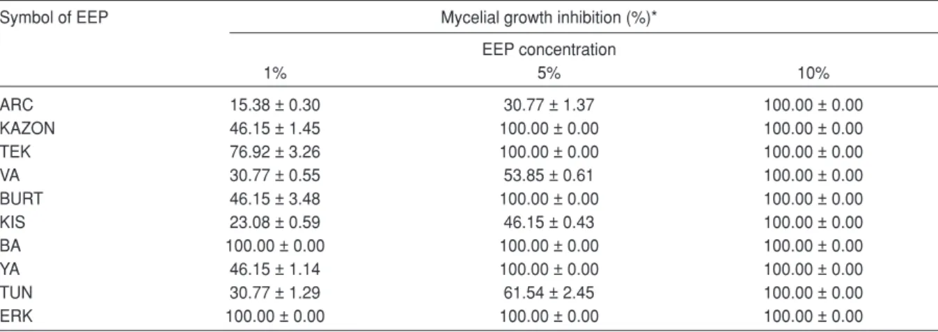
Table 2. Antifungal activity of EEP samples against Aspergillus versicolor200853
Symbol of EEP Mycelial growth inhibition (%)*
EEP concentration 1% 5% 10% ARC 15.38 ± 0.30 30.77 ± 1.37 100.00 ± 0.00 KAZON 46.15 ± 1.45 100.00 ± 0.00 100.00 ± 0.00 TEK 76.92 ± 3.26 100.00 ± 0.00 100.00 ± 0.00 VA 30.77 ± 0.55 53.85 ± 0.61 100.00 ± 0.00 BURT 46.15 ± 3.48 100.00 ± 0.00 100.00 ± 0.00 KIS 23.08 ± 0.59 46.15 ± 0.43 100.00 ± 0.00 BA 100.00 ± 0.00 100.00 ± 0.00 100.00 ± 0.00 YA 46.15 ± 1.14 100.00 ± 0.00 100.00 ± 0.00 TUN 30.77 ± 1.29 61.54 ± 2.45 100.00 ± 0.00 ERK 100.00 ± 0.00 100.00 ± 0.00 100.00 ± 0.00
76.92% inhibition on the mycelial growth of A. versicolor at 1% concentration, KAZON, YA and BURT provided 46.15% inhibition at this concentration. The lowest antifungal effect was obtained with ARC sample by 15.38% inhibition at 1% concentration and 30.77% inhbition at 5% concentration. Sodium benzoate (Na-B), a positive chemical antifungal agent, at 0.01% and 0.1% concentrations showed 15.38% and 46.15% inhibition on the mycelial growth of A. versicolor, respectively.
The most effective EEP sample against P. auantiogriseum was KAZON with a complete inhibition on mycelial growth at 5% concentraiton. BURT, TEK and BA at 5% concentration had relatively lower inhibitory effect on the mycelial growth of P. aurantiogriseumthan that of KAZON. While BURT showed 92.59% inhibition, TEK and
BA showed 88.89% inhibition on the mycelial growth of this mold strain. KAZON, BURT, TEK and BA at the concentration of 1% produced 55.56% inhibition on mycelial growth of P. aurantiogriseum. None of EEP samples at 1% concentration provided a complete inhibition on mycelial growth of this mold strain. ARC had again the lowest antifungal activity against P. aurantiogriseum as that of A. versicolor. ARC exihibited 25.93% inhibition 1% and 51.85% inhibition at 5%. Na-B, a positive chemical antifungal agent, at 0.01% and 0.1% concentrations, showed 3.70% and 25.30% inhibition on the mycelial growth of P. aurantiogriseum, respectively. From the GC-MS analysis, seven organic components were dominantly detected in the EEP samples (Table 4). Flavonoids, aromatic alcohols and aromatic acid esters were the shared
Table 3. Antifungal activity of EEP samples against Penicillium aurantiogriseum501588
Symbol of EEP Mycelial growth inhibition (%)*
EEP concentration 1% 5% 10% ARC 25.93 ± 0.72 51.85 ± 0.70 100.00 ± 0.00 KAZON 55.56 ± 1.55 100.00 ± 0.00 100.00 ± 0.00 TEK 55.56 ± 0.78 88.89 ± 1.58 100.00 ± 0.00 VA 48.15 ± 0.34 62.96 ± 0.61 100.00 ± 0.00 BURT 55.56 ± 1.02 92.59 ± 3.33 100.00 ± 0.00 KIS 48.15 ± 1.30 62.96 ± 2.09 100.00 ± 0.00 BA 55.56 ± 0.93 88.89 ± 1.47 100.00 ± 0.00 YA 55.56 ± 1.36 74.07 ± 1.15 100.00 ± 0.00 TUN 55.56 ± 2.07 66.67 ± 0.98 100.00 ± 0.00 ERK 48.15 ± 0.29 66.67 ± 0.80 100.00 ± 0.00
*: Average value of three replicates
Table 4. Percentage of the main types of compounds in EEP samples identified by GC-MS
Sample Aromatic Alcohols Aromatic Flavonoids Aromatic Aliphatic Aromatic The others
alcohols acids (flavones, acid esters acid esters hydrocarbons (aliphatic ketones,
flavanones, aromatic ketones,
flavonols) terpenes, bee wax,
vitamin E and aliphatic acids) ARC 11.45 -* - 1.34 33.72 20.5 6.07 -KAZON 8.03 1.14 - 46.01 0.53 - - -TEK 1.86 1.71 3.35 53.54 7.09 0.68 0.72 -VA 3.11 1.12 1.64 27.30 7.92 3.01 0.97 9.57 BURT 8.91 1.30 2.71 49.48 1.82 0.7 - 8.27 KIS 4.75 - - 22.54 2.14 - - 41.81 BA 10.02 1.12 8.55 42.64 0.57 - 1.07 13.82 YA 5.57 1.92 - 36.87 1.94 0.57 1.13 1.72 TUN 5.58 - - 23.12 1.48 1.10 - 0.87 ERK 8.06 - 2.18 40.04 3.15 1.90 - 2.18 -*: Not determined
organic components found in all EEP samples with different levels. Flavonoids were found to be the most abundant organic components of all EEP samples, except ARC.
The results of the study showed that EEP samples had strong antifungal effect on both food-borne mycotoxin producer molds, A. versicolor and P. auantiogriseum, depending on EEP concentration. All EEP samples at the highest concentration (10%) had fungicidal effect on both mold strains. The EEP samples of KAZON, BURT, TEK, BA, ERK and YA had high antifungal activity against these two mold strains at 1% and 5% concentrations. However, only BA and ERK had a complete inhibition on the mycelial growth of A. versicolor at 1% and 5% concentrations. These EEP samples at these 2 concentrations had only limited inhibitory effect on the mycelial growth of P. auantiogriseum. KAZON, BURT, TEK, BA, ERK and YA had a complete inhibition on the mycelial growth of A. versicolor at 5% concentration wheras only KAZON showed a complete inhibition on the mycelial growth of P. auantiogriseum at the same concentration. These results indicated that a higher EEP concentration was necessary for the inhibitory effect on the mycelial growth of P. auantiogriseumcomparing with A. versicolor (Table 2 and 3). It may be said that A. versicolor is more sensitive against EEP samples than P. auantiogriseum. It is clear that the antifungal effect of EEP samples displays variations depending on the mold genus and species. These results are in good agreement with the results of previous studies on the antifungal effect of propolis against food-borne or plant-origin molds. It was stated that the application of 1%, 5%, and 10% concentrations of ethanol-extracted propolis inhibited in vitro growth of Penicillium digitatum (35, 36) and limited the growth of Botrytis cinerea on strawberry (33). Özcan showed that treatment with 4% water-extracted propolis resulted in more than 50% inhibition of some plant pathogens, including P. digitatum and B. cinerea, in vitro (45). The application of 2% and 5% concentrations of methanol-extracted propolis inhibited in vitro growth of Alternaria alternataand Fusarium oxysporium f.sp. melonis. However, F. oxysporium was found to be more sensitive than A. alternata(34).
EEP samples had various degrees of antifungal acitivities. It is thought that antifungal activities of
EEPs is related with the chemical compositions of propolis samples. In fact, there was a clear correlation between the amount of flavonoids and the antifungal effect of EEP samples tested in the present study. EEP samples, which had higher antifungal activities on the test mold strains contained considerably higher flavonoids content than the other EEP samples. TEK had the highest flavonoids content with 53.54%. ARC, which showed the lowest antifungal activity had the lowest flavonoids with 1.34% among the EEP samples. Flavonoids are well known for their antibacterial, antifungal and antiviral action and are thought to be responsible for the antimicrobial properties of propolis (18, 22, 32, 46).
Antifungal effect of propolis in controlling fungal growth in different fruits during storage and was studied by several authors (37, 38). It was stated that the application of 5% and 10% concentrations of EEP extended the storage life of Fremont mandarins, as compared to untreated control fruits (38). Treatment with EEP was also effective in preventing fungal decay in cherries stored for 4 weeks, but adversely affected sensory quality and stem color (37). Özdemir et al. studied to determine the effect of EEP samples at the concentrations of 1%, 5% and 10% on the storage life of Star Ruby grapefruit (38). They showed that the treatment with 5% EEP was effective in preventing fungal decay in grapefruits. However, EEP treatment adversely affected sensory quality (appearance and taste) in all of the fruits treated with 10% EEP. On the other hand, Aly and Elewa investigated to determine the efficacy of different concentrations of aqueous propolis extract against the growth of A. versicolor as well as biosynthesis of sterigmatocystin during ripening of Egyptian Ras cheese (39). They determined that mold growth and toxin production were completely inhibited at the highest concentration 1000 part per million (ppm), while the lower concentrations 250 and 500 ppm exhibited definite fungistatic activity during 90 days of ripening.
In the present study, Na-B, a positive chemical antifungal agent, at 0.01% and 0.1% concentrations caused lower inhibitory effects on the mycelial growth of A. versicolorand P. auantiogriseum comparing with EEP samples. However, Na-B at both concentrations had more inhibitory effect on the mycelial growth of A. versicolorthan that of P. auantiogriseum.
As a result, EEP samples have strong antifungal effect on both food-borne molds, A. versicolor and P. auantiogriseum, if an appropriate EEP concentration is used. The results of the present study and the other studies on the inhibitory effect of propolis against food-borne and plant-origin bacteria and fungi (32-39) indicated that propolis preparations might provide a superior and safe alternative to the chemical preservatives and they can be used as a natural preservative to control bacterial and fungal growth on/in foods. It is thought that the possible sensory quality problems of the foods due to EEP using could be overcome by adjusting the effective EEP concentration.
REFERENCES
1. Sweeney MJ, Dobson ADW. 1998. Mycotoxin production by Aspergillus, Fusarium and Penicillium species. Int J Food Microbiol, 43: 141-158.
2. Fleet G. 1992. Spoilage yeasts. Crit Rev Biotechnol, 12: 1-14.
3. Fuglsang C, Johnson G, Christgau S. 1995. Antimicrobial enzymes: Applications and future potential in food industry. Trends Food Sci Technol, 6 (2): 390-399.
4. Brul E, Coote P. 1997. Novel antifungal strategies of potential use to the food industry. Food Chem Toxicol, 35 (4): 720-726.
5. Varanda E, Monti R, Tavares DC. 1999. Inhibitory effect of propolis and bee venom on the mutagenicity of some direct- and indirect-acting mutagens. J Carcinog, 19 (4): 403-419.
6. Ghisalberti EL. 1979. Propolis: A Review. Bee World, 60: 59-84.
7. Crane E. 1990. Bees and Beekeeping: Science, Practice and World Resources. Cornstock Publishing, Ithaca, NY., USA, 614 p.
8. Sorkun K, Suer B, Salih B. 2001. Determination of chemical composition of Turkish propolis. Z Naturforsch, 56: 666-668.
9. Greenaway W, Scaybrook T, Whatley FR. 1990. The composition and plant origin of propolis. Mycologia, 72 (2): 107-118.
10. Burdock GA. 1998. Review of the biological properties and toxicity of bee propolis (propolis). Food Chem Toxicol, 36: 347-363.
11. Cafarchia C, De Laurentis N, Milillo MA, Losacco V, Puccini V. 1999. Antifungal activity of Apulia region propolis. Parassitologia, 4 (12): 5870-5880.
12. Wohrl S. 2003. Patch testing in children, adults, and the elderly: influence of propolis on age and sex.J Dermatol, 46 (2): 119-123., 18 (1): 6-9. 13. Tan-No K, Nakajima T, Shoji T, Nakagawasai O, Niijima F, Ishikawa M, Endo Y, Sato T, Satoh S, Tadano T. 2006. Anti-inflammatory effect of propolis through inhibition of nitric oxide production on carrageenin-induced mouse paw edema. Biol Pharm Bull, 29 (1): 96-99.
14. Lindenfelser LA. 1967. Antimicrobial activity of propolis. Am Bee J, 107 (3): 90-92, 130-131. 15. Grange JM, Davey RW. 1990. Antibacterial properties of propolis (bee glue). J Roy Soc Med, 83: 159-160.
16. Dobrowolski JW, Vohora SB, Sharma K, Shah SA, Naqvi SAH, Dandiya PC. 1991. Antibacterial, antifungal,antiamoebic, anti inflammatory and antipyretic studies on propolis bee products. J Ethnopharmacol, 35: 77-82.
17. Kujumgiev A, Bankova V, Ignatova A, Popov S. 1993. Antibacterial activity of propolis, some of its components and their analogs. Pharmazie, 48: 785-786.
18. Marcucci MC. 1995. Propolis: chemical composition, biological properties and therapeutic activity. Apidologie, 26: 83–99.
19. Kujumgiev A, Tsvetkova I, Serkedjieva Y, Bankova V, Christov R, Popov S. 1999. Antibacterial, antifungal and antiviral activity of propolis of different geographic origin. J Ethnopharmacol, 64: 235-240.
20. Nivea M, Isla M, Cudmani NG, Vattuone MA, Sampietro AR.1999. Screning of antibacterial activity of Amaicha del Valle (Tucuman, Argentina) propolis.J. Ethnopharmacol, 68: 97-102.
21. Hegazi AG, Abd El Hady FK, Abd Allah FA. 2000. Chemical composition and antimicrobial activity of European propolis. Z Naturforsch C, 55: 70-75.
22. Sforcin JM, Fernandes Jr. A, Lopes CAM, Bankova V, Funari SRC. 2000. Seasonal effect on Brazilian propolis antibacterial activity. J Ethnopharmacol, 73: 243-249.
23. Velikova M, Bankova V, Sorkun K, Houcine S, Tsvetkova I, Kujumgiev A. 2000. Propolis from Mediterranean region: Chemical composition and antimicrobial activity. Z Naturforsch C, 55: 790-793.
24. Keskin N, Haz›r S., Baser KH, Kürkçüo¤lu M. 2001. Antibacterial activity and chemical composition of Turkish propolis. Z Naturforsch C, 56: 1112-1115.
25. Ota C, Unterkircher C, Fantinato V, Shimizu MT. 2001. Antifungal activity of propolis on different species of Candida. Mycoses, 44: 375-378. 26. Abd El Hady, FK, Hegazi AG. 2002. Egyptian propolis: 2. Chemical composition, antiviral and antimicrobial activities of East Nile Delta propolis. Z Naturforsch C, 57: 386-394.
27. Kartal M, Y›ld›z S, Kaya S, Kurucu S, Topçu G. 2003. Antimicrobial activity of propolis samples from two different regions of Anatolia. J Ethnopharmacol, 86: 69-73.
28. Erdem GB, Ölmez S. 2004. Inhibitory effect of Bursa Propolis on dental caries formation in rats inoculated with Streptococcus sobrinus. Turk J Zool, 28: 29-36.
29. Cihangir N, Sorkun K, Salih B. 2005. Chemical composition and antibacterial activities of propolis collected from different regions of Turkey. Hacet J Biol Chem, 34: 59-67.
30. Silici S, Koç NA, Ayangil D, Çankaya S. 2005. Antifungal activities of propolis collected by different races of honeybees against yeasts isolated from patients with superficial mycoses. J Pharmacol Sci, 99: 39-44.
31. Uzel A, Sorkun K, Önça¤ Ö, Ço¤ullu D, Gençay Ö, Salih B. 2005. Chemical compositions and antimicrobial activities of four different Anatolian propolis samples. Microbiol Res, 160: 189-195.
32. Temiz A, fiener A, Tüylü AÖ, Sorkun K, Salih B. 2011. Antibacterial activity of bee propolis samples from different geographical regions of Turkey against two foodborne pathogens, SalmonellaEnteritidis and Listeria monocytogenes. Turk J Biol, 35 (4): 503-511.
33. La Torre A, Imbroglini G, Guccione M. 1990. Action of propolis based preparations against Botrytis cinerea Pers. of strawberry. First Observation Agric, 6: 169-177.
34. Özcan M, Ünver A, Ceylan DA, Yetiflir R. 2004. Inhibitory effect of pollen and propolis extracts. Nahrung, 48 (3): 188– 194.
35. Soylu EM, Özdemir AE, Ertürk E, fiahinler N. 2004. Antifungal activity of propolis against Penicillium digitatum, causal agent of green mold of citrus fruits. Proceedings of the First European Conference of Apidology ‘EurBee’, Udine, Italy; pp. 160.
36. Soylu EM, Özdemir AE, Ertürk E, fiahinler N, Soylu S. 2008. Chemical composition and antifungal activity of propolis against Penicillium digitatum. Asian J Chem, 20: 4823-4830.
37. Çand›r EE, Özdemir AE, Soylu EM, fiahinler N, Gül A. 2009. Effects of propolis on storage of sweet cherry cv. Aksehir Napolyon. Asian J Chem, 21: 2659-2666.
38. Özdemir AE, Çand›r EE, Kaplank›ran M, Soylu EM, fiahinler N, Gül, A. 2010. The effects of ethanol-dissolved propolis on the storage of grapefruit cv. Star Ruby. Turk J Agric For, 34: 155-162. 39. Aly SA, Elewa NA. 2007. The effect of Egyptian honeybee propolis on the growth of Aspergillus versicolor and sterigmatocystin biosynthesis in Ras cheese. J Dairy Res, 74: 74-78.
40. Engelhart S, Loock A, Skutlarek D, Sagunski H, Lommel A, Färber H, Exneri M. 2002. Occurrence of toxigenic Aspergillus versicolor isolates and sterigmatocystin in carpet dust from damp indoor environments. Appl Environ Microb, 68 (8): 3886-3890.
41. Northolt MD, Soentoro PSS. 1988. Fungal Growth on Foodstuffs Related to Mycotoxin Contamination. In: Introduction to Food Borne Fungi, Samson RA, van Reenen-Hoekstra ES (ed), Centraalbureau voor Schimmelcultures, Baarn, The Netherland; pp. 231-238.
42. Sato J, Goto K, Nanjo F, Kawai S, Murata K. 2000. Antifungal activity of plant extracts against Arthrinium sacchari and Chaetomium funicola. J Biosci Bioeng, 90 (4): 442-446.
43. Quiroga EN, Sampietro AR, Vattuone MA. 2001. Screening antifungal activities of selected medicinal plants. J Ethnopharmacol, 74, 89-96. 44. Çakir A, Kordali S, K›l›ç H, Kaya E. 2005. Antifungal properties of essential oil and crude extracts of Hypericum linarioides Bosse. Biochem Syst Ecol, 33: 245-256.
45. Özcan M. 1999. Antifungal properties of propolis. Grasas Aceites, 50: 395-398.
46. Hemández NMR, Bemal KC. 1990. Efecto antibiótico del propóleo frente a cepas de Staphylococcus aureus origen clinico humano. Rev Cubana Farm, 24: 45-50.