UNIVERSITI PUTRA MALAYSIA
REGULATING THE DEGREE OF CONTRAST ENHANCEMENT IN
GLOBAL HISTOGRAM EQUALIZATION-BASED METHOD FOR
GRAYSCALE PHOTO PROCESSING
CHEN SOONG DER
REGULATING THE DEGREE OF CONTRAST ENHANCEMENT IN
GLOBAL HISTOGRAM EQUALIZATION-BASED METHOD FOR
GRAYSCALE PHOTO PROCESSING
CHEN SOONG DER
DOCTOR OF PHILOSOPHY
UNIVERSITI PUTRA MALAYSIA
REGULATING THE DEGREE OF CONTRAST ENHANCEMENT IN GLOBAL HISTOGRAM EQUALIZATION-BASED METHOD FOR GRAYSCALE PHOTO
PROCESSING
By
CHEN SOONG DER
Thesis Submitted to the School of Graduate Studies, Universiti Putra Malaysia, in Fulfilment of the Requirements for the degree of Doctor of Philosophy
DEDICATION
This thesis is dedicated to my parents whose selfless sacrifices and dedications have made it possible for me to reach this stage of my studies.
iii
Abstract of thesis presented to the Senate of the Universiti Putra Malaysia in fulfilment of the requirement for the degree of Doctor of Philosophy
REGULATING THE DEGREE OF CONTRAST ENHANCEMENT IN GLOBAL HISTOGRAM EQUALIZATION-BASED METHOD FOR GRAYSCALE PHOTO
PROCESSING
By
CHEN SOONG DER November, 2007
Chairman: Associate Professor Abdul Rahman Ramli, PhD Faculty: Engineering
Global Histogram equalization (GHE) is a popular image contrast enhancement method. However, it is rarely used on photo processing because it tends to create noise-artifacts, especially in simple-structure-image. A few GHE-based methods have been proposed to address this issue but whether they are noise-artifacts-proof remains questionable. This is because the methods are fully automatic and the evaluation conducted was not comprehensive.
A novel automatic GHE-based method called Minimum Mean Brightness Error Bi-Histogram Equalization (MMBEBHE) has been proposed in this thesis. It has been evaluated thoroughly together with the existing automatic methods. The results have proven that none of the automatic GHE-based methods is noise-artifacts-proof. The
conclusion has motivated author to look into scalable GHE-based methods that allows user to regulate the degree of contrast enhancement.
A novel scalable GHE-based method called Recursive Mean-Separate Histogram Equalization (RMSHE) has been proposed in this thesis. It has been evaluated thoroughly together with other two existing scalable methods - Clip Limited Adaptive HE (CLAHE) and Stark’s Adaptive HE (StarkAHE). The results of separate evaluations consistently showed that none of the three methods could effectively enhance the contrast of simple-structure-image without creating any noise-artifacts.
Another novel scalable GHE-based method called Scalable Global Histogram Equalization with Selective Enhancement (SGHESE) has been developed then to overcome the limitation of the existing methods. Evaluation results showed that SGHESE could enhance the image’s contrast effectively without creating any noise-artifacts. The results of subjective evaluation involving human observer also showed that the preference level of SGHESE was significantly higher compared to those of other methods.
Finally, the thesis recommends extending the study of SGHESE to color image processing because majority of the images nowadays are color images.
v
Abstrak tesis yang dikemukakan kepada Senat Univeriti Putra Malaysia sebagai memenuhi keperluan untuk ijazah Doktor Falsafah
PENYESUAIAN TAHAP PENINGKATAN KEJELASAN IMEJ DALAM TEKNIK YANG BERASASKAN PENYAMAAN HISTOGRAM GLOBAL BAGI
PEMPROSESAN FOTO HITAM-PUTIH
Oleh
CHEN SOONG DER November 2007
Pengerusi: Profesor Madya Abdul Rahman Ramli, PhD Fakulti: Kejuruteraan
Penyamaan Histogram Global (GHE) adalah satu kaedah yang popular bagi meningkatkan kejelasan imej. Namun, ia jarang digunakan dalam pemprosesan foto kerana ia sering mendatangkan kesan hingar, terutama sekali dalam memproses foto-berstruktur-mudah. Beberapa kaedah yang berasaskan GHE telah dicadangkan sebelum ini, namun ia masih menjadi satu persoalan samada masalah kesan hingar telah diselesaikan. Ini kerana kaedah tersebut adalah automatik dan kajiannya tidak dijalankan secara menyeluruh.
Satu kaedah baru yang digelar Penyamaan Dwi-Histogram dengan Putra Ralat Kecerahan Minima (MMBEBHE) telah dibentangkan dalam tesis ini. Kaedah ini telah dikaji secara teliti bersama dengan kaedah automatik yang lain. Kajian telah membuktikan bahawa
tiada satu pun daripada kaedah automatik tersebut dapat menyelesaikan masalah kesan hingar secara menyeluruh. Keputusan yang sedemikian telah memberi motivasi supaya mengkaji kaedah berskala yang boleh dilaraskan tahap peningkatan-kejelasan-imej.
Satu kaedah berskala baru yang digelar Penyamaan Histogram secara Pembahagian-Purata Berulang (RMSHE) telah dibentangkan dalam tesis ini. Ia telah dikaji secara teliti bersama dua kaedah berskala yang lain – Penyamaan Histogram Tempatan Berhad (CLAHE) dan Penyamaan Histogram Tempatan (StarkAHE). Keputusan daripada kajian berasingan secara konsistennya menunjukkan bahawa, tiada satu pun daripada kaedah berskala tersebut dapat meningkatkan kejelasan imej dengan berkesan tanpa mendatangkan kesan hingar.
Lanjutan daripada kajian di atas, satu lagi kaedah berskala baru yang digelar Penyamaan Histogram Berskala Terpilih (SGHESE) telah dicadangkan bagi mengatasi kelemahan kaedah berskala yang sedia ada. Kajian menunjukkan SGHESE berupaya meningkatkan kejelasan imej dengan berkesan tanpa mendatangkan kesan hingar. Kajian subjektif yang melibatkan pemerhatian manusia juga menunjukkan tahap kegemaran pemerhati terhadap SGHESE adalah jauh lebih tinggi berbanding kaedah-kaedah yang lain.
Dalam mengakhiri tesis ini, pengarang telah mengesyorkan supaya melanjutkan kajian terhadap SGHESE dalam pemprosesan imej berwarna kerana kebanyakan imej sekarang adalah imej berwarna.
vii
ACKNOWLEDGEMENTS
I am very grateful to my supervisor Associate Professor Dr. Abdul Rahman Ramli for the technical and moral support he provided throughout my study. He has opened my mind to research in the field of image enhancement. I am also grateful to other member of the dissertation committee – Professor Mohd Khazani Abdullah for his support and feedback in many ways. I had the opportunity to work with them and it has helped my research work immensely. I also wish to thank all those who have volunteered to participate in the subjective evaluation conducted as part of the research work of this thesis. Last but not least, I wish to express my appreciation to College of IT, Universiti Tenaga Nasional that has supported my studies in many ways.
I certify that an Examination Committee has met on 28th Nov 2007 to conduct the final examination of Mr. Chen Soong Der on his PhD thesis entitled " REGULATING THE DEGREE OF CONTRAST ENHANCEMENT IN GLOBAL HISTOGRAM EQUALIZATION-BASED METHOD FOR GRAYSCALE PHOTO PROCESSING" in accordance with Universiti Pertanian Malaysia (Higher Degree) Act 1980 and Universiti Pertanian Malaysia (Higher Degree) Regulations 1981. The Committee recommends that the candidate be awarded the relevant degree. Members of the Examination Committee are as follows:
Abdul Rashid Mohamed Shariff, PhD
Associate Professor Faculty of Engineering Universiti Putra Malaysia (Chairman)
Mohammad Hamiruce Marhaban, PhD
Lecturer
Faculty of Engineering Universiti Putra Malaysia (Internal Examiner)
Abdul Aziz Jaafar, PhD
Lecturer
Faculty of Engineering Universiti Putra Malaysia (Internal Examiner)
Kasmiran Jumari, PhD
Professor
Faculty of Engineering
Universiti Kebangsaan Malaysia (External Examiner)
________________________________ HASANAH MOHD. GHAZALI, PhD
Professor and Deputy Dean School of Graduate Studies Universiti Putra Malaysia Date: 29 January 2008
ix
This thesis was submitted to the Senate of Universiti Putra Malaysia and has been accepted as fulfilment of the requirement for the degree of Doctor of Philosophy. The members of the Supervisory Committee were as follows:
Abdul Rahman Ramli, PhD Associate Professor
Faculty of Engineering Universiti Putra Malaysia (Chairman)
Mohd. Khazani Abdullah, PhD Professor
Faculty of Engineering Universiti Putra Malaysia (Member)
______________________ AINI IDERIS, PhD
Professor and Dean
School of Graduate Studies Universiti Putra Malaysia Date:
DECLARATION
I hereby declare that the thesis is based on my original work except for quotations and citations, which have been duly acknowledged. I also declare that it has not been previously or concurrently submitted for any other degree at Universiti Putra Malaysia or other institutions.
______________________________
CHEN SOONG DER
xi TABLE OF CONTENTS DEDICATION ii ABSTRACT iii ABSTRAK v ACKNOWLEDGEMENTS vii APPROVAL viii DECLARATION x
LIST OF TABLES xiv
LIST OF FIGURES xv GLOSSARY OF TERMS xx CHAPTER 1 INTRODUCTION 1 1.1 Background 2 1.2 Problem Statement 3 1.3 Motivations 4 1.4 Objectives 5
1.5 Thesis Scope and Limitation 5
1.6 Evaluation Methodology 7
1.7 Contributions 8
1.8 Thesis Organization 9
2 LITERATURE REVIEW 11
2.1 Classification of Contrast Enhancement Methods 11
2.1.1 Intensity-based Methods 11
2.1.2 Feature-based Methods 13
2.1.3 Intensity-based vs. Feature-based Methods 14
2.1.4 Global vs. Adaptive Intensity-based Methods 15
2.2 Application of HE 16 2.2.1 HE in Medical Imaging 16 2.2.2 HE in Satellite Imaging 21 2.2.3 HE in Microscopic Imaging 24 2.2.4 HE in Computer Vision 26 2.2.5 HE in Image Processing 34 2.3 Implementation of HE 37
2.4 HE in Color Image Processing 39
2.5 Variants of HE 41
2.5.1 Variants of AHE 41
2.5.2 Variants of GHE 53
2.6 Conclusion 57
3 A STUDY ON AUTOMATIC GHE-BASED METHODS 60
3.1 Introduction 60
3.2 Minimum Mean-Brightness Error Bi-Histogram Equalization 60
3.2.1 Algorithm of MMBEBHE 61
3.2.2 Fast Algorithm of MMBEBHE 61
3.3 Evaluation Methodologies 64
3.3.1 Test Images 64
3.3.2 Algorithm Implementation 65
3.3.3 Noise-artifacts Proof Test 66
3.3.4 Background Contrast Change Measurement 66
3.4 Results and Discussions 68
3.5 Conclusions 70
4 A STUDY ON SCALABLE GHE-BASED METHODS 72
4.1 Introduction 72
4.2 Recursive Mean-Separate Histogram Equalization 73
4.2.1 Algorithm of RMSHE 73
4.2.2 Mathematical Analysis on RMSHE 76
4.3 CLAHE and StarkAHE 80
4.4 Evaluation Methodologies 80
4.4.1 Test Images 81
4.4.2 Algorithm Implementation 81
4.4.3 Objective Evaluation 83
4.4.3.1 Main-object Contrast Change Measurement 83
4.4.3.2 Main-object Contrast Recovery Rate 84
4.4.3.3 Structural-Similarity Index Measurement 85
4.4.4 Selection of Parameters’ Value 86
4.5 Results and Discussions 86
4.5.1 RMSHE 87
4.5.2 CLAHE 89
4.5.3 StarkAHE 92
4.6 Conclusions 94
5 A STUDY ON NOVEL SCALABLE GLOBAL HISTOGRAM
EQUALIZATION WITH SELECTIVE ENHANCEMENT 95
5.1 Introduction 95
5.2 Analysis of Limitation in CLAHE and StarkAHE 95
5.3 Scalable Global Histogram Equalization with Selective
Enhancement (SGHESE) 103 5.3.1 Philosophy of SGHESE 103 5.3.2 Design of SGHESE 106 5.3.3 Algorithm of SGHESE 111 5.4 Evaluation Methodologies 111 5.4.1 Test Images 111 5.4.2 Algorithm Implementation 112 5.4.3 Objective Evaluation 113
xiii
5.4.4 Subjective Evaluation 113
5.4.5 Selection of Parameters’ Value 115
5.5 Results and Discussions 115
5.5.1 Objective Evaluation 116
5.5.2 Subjective Evaluation 118
5.6 Conclusions 122
6 CONCLUSIONS AND RECOMMENDATIONS 123
6.1 Introduction 123
6.2 Summary of Research Findings 123
6.2.1 Study on Automatic GHE-based Methods 123
6.2.2 Study on Scalable GHE-based Methods 124
6.2.3 Study on SGHESE 125
6.3 Recommendation for Future Research 126
6.4 Summary of Contributions 126
BIBLIOGRAGHY 128
APPENDICES 140
BIODATA OF THE AUTHOR 152
LIST OF TABLES
Table Page
3.1 The results of visual inspection for noise-artifacts in the output images of various automatic GHE-based methods
68
3.2 BCGP of the output images of various automatic GHE-based
methods
69
4.1 Value of parameters chosen to produce the respective output
images of RMSHE, CLAHE and StarkAHE
87
4.2 MCRR of RMSHE’s output image of RMSHE 88
4.3 MCRR of CLAHE’s output images 90
4.4 MCRR of StarkAHE’s output image 93
5.1 Value of parameters chosen to produce the output images of
SGHESE
116
xv
LIST OF FIGURES
Figure Page
1.1 Worldwide revenue from digital photo prints, 2002-2008 1
1.2a Original image, Girl 2
1.2b After contrast enhancement 2
1.3 Result of GHE 3
2.1 A brain MRI image (Baudraa et al., 2000) 16
2.2 A portal image of a pelvis of a patient (Fielding et) 17
2.3 Tomography slices of teeth (Chai-U-Dom et al.) 17
2.4 Gastric sonogram image (Fu et al., 2000) 17
2.5 Cardiovascular MRI (Fu et al., 2000) 18
2.6 Cephalogram (Sanei et al., 1999) 18
2.7 Neural image (Blomstrand et al., 1999) 18
2.8 Sample medical image before (left) and after (right) HE (Pizer et al., 2003)
19
2.9 Sample cardiovascular image before (left) and after (right) HE (Fu et al., 2000)
19
2.10 Sample brain MRI image before (left) and after (right) HE
(Baudraa et al., 2000)
20
2.11 Sample gastric sonogram image before (left) and after (right) HE (Fu et al., 2000)
20
2.12 Sample cephalogram image before (left) and after (right) HE (Sanei et al., 1999)
20
2.13 (Left) Original image (Right) Image after HE 21
2.14 Original (left) and HE enhanced (right) satellite image (Wan et al., 2002)
2.15 Satellite image after lineament (white lines) detection (Raghavan et al., 1995)
23
2.16 Gray scale microscopic image and its histogram (Szmaja,
1998)
25
2.17 Image in fig. 2.16 after enhanced by HE (Szmaja, 1998) 26
2.18 Sample outputs of face detection algorithm developed by Heisele et al.(2003)
27
2.19 Eye image before (left column) and after (right column) HE (Li et al., 2001)
28
2.20 Process flow of face verification system developed by Bengio et. al. (2002)
28
2.21 (Left) Original image. (Right) After HE was applied to the
bounded skin blobs. (Koh et al., 2002)
29
2.22 Sample face image before (left) and after (right) HE (Ayinde and Yang, 2002)
29
2.23 Ridge structures - an important feature in fingerprint matching (Greenberg et al., 2000)
31
2.24 Fingerprint image before (left) and after (right) histogram
equalization. (Greenberg et al., 2000)
31
2.25 Process flow-chart of the algorithm to build panoramic image mosaic developed by Kim et al. (2003)
33
2.26 Sample radar image before (left) and after (right) HE (Olheoft, 2000)
35
2.27 Fringes images showing different mode shape of vibrating
surface of computer hard disk. (Kumar et al., 2001)
36
2.28 Combination of sub-blocks 45
2.29 Processes of CLAHE 48
3.1 GUI of the window-based application used to test GHE,
BBHE, DSIHE, Multi-peak HE and MMBEBHE
66
xvii
3.3 Log10(BCGP) of the output images 70
4.1 Histogram before and after GHE 75
4.2 Histogram before and after equalization with two
segments
75
4.3 Histogram before and after equalization with four
segments
75
4.4 Histogram before and after RMSHE with r=0 (equivalent
to GHE)
76
4.5 Histogram before and after RMSHE with r=1 (equivalent
to BBHE)
77
4.6 Histogram before and after RMSHE with r=2 78
4.7 GUI of window application used evaluate RMSHE 82
4.8 GUI of window application used evaluate CLAHE 82
4.9 GUI of window application used evaluate StarkAHE 83
4.10 MCGP of the RMSHE’s output image vs. the MCGP of
their respective original image
88
4.11 SSIM_CRI vs. SSIM_ORI of RMSHE’s output images 89
4.12 MCGP of the CLAHE’s output image vs. the MCGP of
their respective original image
90
4.13 SSIM_CRI vs. SSIM_ORI of CLAHE’s output images 91
4.14 MCGP of the StarkAHE’s output image vs. the MCGP of
their respective original image
92
4.15 SSIM_CRI vs. SSIM_ORI of StarkAHE’s output images 94
5.1 Original image of clock 96
5.2 Histogram of original image clock 96
5.4 Image clock before (left) and after (right) conventional GHE
98
5.5 Histogram of image clock after conventional GHE 98
5.6 Gray level transformation function of CLAHE, clip limit = 0.4
99
5.7 Image clock before (left) and after (right) applying CLAHE, clip = 0.4
100
5.8 Histogram of image clock after applying CLAHE, clip limit = 0.4
101
5.9 Gray level transformation function of StarkAHE,
=0.65, = 1
102
5.10 Image clock before (left) and after (right)
StarkAHE,=0.65,=1
102
5.11 Histogram of image clock after applying
StarkAHE,=0.65,=1
103
5.12 Gray level transformation function proposed by this thesis 104
5.13 Image clock before (left) and after (right) transformed by
the function proposed by the thesis
105
5.14 Histogram of image clock after transformed by function proposed by the thesis
105
5.15 Transformation function after stretching with clipping at upper end
108
5.16 Transformation function after stretching with clipping at lower end
109
5.17 Transformation function after sliding upward 109
5.18 Transformation function after sliding downward 110
5.19 GUI of window application used to evaluate SGHESE 112
5.20 GUI used for the subjective evaluation 113
5.21 MCGP of the SGHESE’s output images vs. the MCGP of
respective original image
xix
5.22 Readings of SSIM_CRI vs. SSIM_ORI of SGHESE’s
output images
118
5.23 Distribution of the observers’ races 119
5.24 Distribution of the observers in group 1,2 and 3 120
5.25 OPL of the output images from all GHE-based methods in
study
121
5.26 OPL of the output images from all GHE-based methods in
study
121
GLOSSARY OF TERMS
AHE Adaptive Histogram Equalization
AMBE Absolute Mean Brightness Error
BBHE Brightness Preserving Bi-Histogram Equalization
BMA Block Matching Algorithm
CDF Cumulative Density Function
CLAHE Clip Limited Adaptive Histogram Equalization
CRT Cathode Ray Tube
CT Computed Tomography
DSIHE Dualistic Sub-Image Histogram Equalization
DSPI Digital Speckle Pattern Interferometry
FPGA Field Programmable Gate Array
GHE Global Histogram Equalization
GOES Geostationary Operational Environmental Satellite
GUI Graphical User Interface
HE Histogram Equalization
LPF Low Pass Filter
LRM Local Range Modification
MaxBE Maximum Brightness Error
MIDAG Medical Image Display and Analysis Group
MLE Multi Level Histogram Equalization
MMBEBHE Minimum Mean Brightness Error Bi-Histogram Equalization
xxi
MSE Mean Square Error
PC Personal Computer
PCA Principal Component Analysis
PDF Probability Density Function
POSHE Partially Overlapped Sub-Block HE
PSNR Peak Signal to Noise Ratio
RMSHE Recursive Mean-Separate Histogram Equalization
SEM Scanning Electron Microscope
SGHESE Scalable Global Histogram Equalization with Selective Enhancement
SIMD Single Instruction Multiple Data
SSIM Structural- Similarity-Based Image Assessment
1
CHAPTER 1
INTRODUCTION
Figure 1.1 shows graph of the forecasted worldwide revenue from digital photo prints, 2002-2008 (Lyra Research Inc, 2004). The graph indicates that digital photo processing is getting more important as the demand for it is expanding rapidly. There are a few types of image processing methods used to process digital photo. One of them is image enhancement. Image enhancement aims to improve the detect-ability of important image details or objects by man or machine (Shapiro and Stockman, 2001). Contrast enhancement, image sharpening and image smoothing are among the most common type of enhancement (Gonzalez and Woods, 2002).
2
1.1 Background
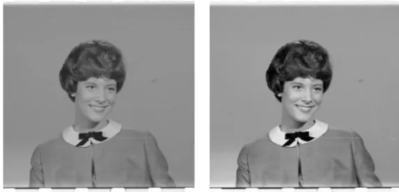
The purpose of image contrast enhancement is to increase the visibility of an image. Figure 1.2a and 1.2b show an image before and after contrast enhancement. Notice
that the image shows better visibility after contrast enhancement. Many methods have been proposed and they can be generally classified into two main categories: intensity-based methods and feature-based methods (Zhu et al., 1999).
In feature-based methods, the ways to extract the feature components to be enhanced must be based on the knowledge about these features. So feature-based methods are often used in special applications such detecting tumor in medical imaging. On the other hand, the intensity-based methods are more general. They are widely used in the preprocessing of various types of image. One of the very popular intensity-based methods is histogram equalization (HE) (Zhu et al., 1999). Intensity-based methods can be further classified into two main categories: global and adaptive methods. In global methods, a single transformation of the image gray levels is applied to the
Figure 1.2b: after contrast enhancement