Reference document on exposure to
metallic mercury and the development of
symptoms with emphasis on neurological
and neuropsychological diseases or
complaints
ByJesper Baelum Heidi Pöckel
Department of Occupational and Environmental Medicine Odense University Hospital
DK-5000 Odense November 2007
Contents
CONTENTS 3 PREFACE 6
DANSK RESUMÉ 7
EKSPONERING FOR KVIKSØLV I TANDPLEJE-ERHVERVET 8
HELBREDSEFFEKTER AF KVIKSØLV I TANDPLEJE-ERHVERVET 9
SYGDOMME LANG TID EFTER OPHØR AF EKSPONERING 11
EFFEKTER PÅ FORPLANTNINGSEVNEN 12
OPSUMMERING, EVIDENS FOR ÅRSAG 13
BEHOV FOR VIDEN OG YDERLIGERE FORSKNING 14
1 INTRODUCTION 15
1.1 METHODS OF THE REVIEW 16
1.2 A SHORT DESCRIPTION OF THE UPTAKE, DISTRIBUTION, AND EXCRETION
OF INORGANIC MERCURY 17
1.2.1 Inhalation 17
1.2.2 Gastrointestinal uptake 18
1.2.3 Dermal uptake 19
1.3 BIOLOGICAL INDICES OF EXPOSURE 19
1.4 DENTAL AMALGAM 21
1.4.1 Procedures for the use of amalgam in dentistry. 21
1.4.2 Mixing of amalgam 22
1.4.3 Condensation of amalgam 22
1.4.4 Polishing and removal of amalgam 22
1.4.5 Waste removal 23
1.5 EXPOSURE TO MERCURY BY AMALGAM IN OWN TEETH 23
1.6 CLINICAL TOXICOLOGY OF INORGANIC MERCURY 23
1.7 PERMISSIBLE EXPOSURE LEVELS 25
2 EXPOSURE IN DENTISTRY 26
2.1 MERCURY IN AIR IN RELATION TO THE TYPE OF CLINIC AND PROCEDURES26
2.1.1 Older studies, observations before 1990 26
2.1.2 Studies later than 1990 28
2.2 URINARY MERCURY IN DENTAL PERSONNEL 29
2.3 HG IN BLOOD 34
2.4 HG IN HAIR AND NAILS 35
2.5 GENDER DIFFERENCE IN THE BIOLOGICAL MEASUREMENTS 35
2.6 DISCUSSION AND CONCLUSION 35
2.6.1 Mercury in workroom air. 35
2.6.2 Mercury in urine 36
2.7 TABLES FOR CHAPTER 2 38
3 STUDIES OF SYMPTOMS AND NEUROPSYCHOLOGICAL
PERFORMANCE IN DENTISTRY 43
3.1 STUDIES OF DELAYED NEUROPSYCHOLOGICAL EFFECTS IN DENTAL
PERSONNEL 43
3.2 NEUROPSYCHOLOGICAL MEASUREMENTS IN DENTISTRY DURING
EXPOSURE 50
3.3 DISCUSSION AND CONCLUSION 54
3.4 TABLES FOR CHAPTER 3 56
4 EFFECTS OF MERCURY ON NEUROPSYCHOLOGICAL
PERFORMANCE AND SYMPTOMS 61
4.1 CRITICAL REVIEW AND METAANALYSES 61
4.2 STUDIES OF THE EFFECT OF PAST EXPOSURE 64
4.3 INDIVIDUAL RISK FACTORS 65
4.3.1 Confounding and bias 66
4.4 CONCLUSIONS 66
5 REPROTOXIC EFFECTS 68
5.1 DEFINITION OF REPRODUCTIVE OUTCOMES 68
5.2 SPONTANEOUS ABORTION/MISCARRIAGE 69
5.3 FECUNDABILITY/INFERTILITY 70
5.4 CONGENITAL MALFORMATIONS 71
5.5 LOW BIRTH WEIGHT 71
5.6 STILLBIRTHS AND PERINATAL DEATHS 72
5.7 NEUROPSYCHOLOGICAL DEVELOPMENT IN OFFSPRING 72
5.8 MALE EXPOSURE AND EFFECTS IN OFFSPRING 72
5.9 DISCUSSION AND CONCLUSION 73
5.10 TABLE FOR CHAPTER 5 74
6 CONCLUSIONS 80
6.1 EXPOSURE TO MERCURY IN DENTISTRY. 80
6.2 HEALTH EFFECTS OF MERCURY IN DENTISTRY 82
6.3 DISEASES LONG TIME AFTER CESSATION OF EXPOSURE 83
6.4 EFFECTS ON REPRODUCTION 84
6.5 IN SUMMARY, EVIDENCE OF CAUSAL ASSOCIATIONS 84
6.6 NEED FOR KNOWLEDGE OR FURTHER RESEARCH 85
7 REFERENCES 87
ADDENDA
1: The notice about the reference document. 1a. Notice in Danish
1.b. Notice translated to English 2: Description of selected neuropsychological tests 3: Extended tables for chapter 2
Table 2-1 Air levels of mercury in relation to different technologies/locations in dentistry. Table 2-2 Urinary mercury levels in relation to different technolgies in dentistry. Table 2-3 Urinary mercury levels in relation to occupation in dentistry. Table 2-4 Mercury levels in blood in relation to occupation in dentistry. Table 2-5 Mercury levels in blood in relation to occupation in dentistry.
Table 2-6 Mercury levels in head/pubic hair in relation to occupation, different technologies, etc. in dentistry.
Table 2-7 Mercury levels in fingernails/toenails in relation to occupation, type of technology, etc. in dentistry.
4: Extended tables for chapter 5
Table 5-1 Measurements of mercury levels in women occupationally exposed to mercury and their offspring.
Table 5-2 The rate of spontaneous abortion/miscarriage in women occupationally exposed to mercury.
Table 5-3 The fecundability (probability of conception each menstrual cycle) of women Table 5-4 The rate of infertility in women occupationally exposed to mercury
Table 5-5 The rate of menstrual disorders in women occupationally exposed to mercury. Table 5-6 The rate of congenital abnormalities/malformations in children of women occupationally exposed to mercury
Table 5-7 The rate of women occupationally exposed to mercury, giving birth to low birth weight infants.
Table 5-8 The rate of women occupationally exposed to mercury, giving birth to infants suffering from perinatal death.
Table 5-9 Neuropsychological development in children of women occupationally exposed to mercury
.
Preface
The present report is a result of a task set by the Danish Health and Safety Research Fund by an announcement made in May 2006 for a reference document on the health effects of metallic mercury. Department of Occupational and Environmental Medicine got the contract and the work has been done by the authors.
The reference document has been reviewed by professor Lars Barregard, Department of Occupational and Environmental Medicine, Sahlgrenska University Hospital and Academy, Gothenburg, Sweden and Dr. Andreas Seeber, former affiliated at the Institute for Occupational Physiology at the University of Dortmund, Germany. A quality review board, professor Svend Sabroe, Institute of Public Health, University of Aarhus, Denmark and professor Staffan Skerfving, Department of Occupational and Environmental Medicine, Lund University Hospital, Sweden has together with the authors and reviewers made comments on the report on a seminar held in Copenhagen October 12. Afterwards corrections and additions have been made and the revised report has been c submitted to the Danish Health and Safety Research Fund.
Odense November, 09, 2007.
Dansk resumé
Dette referencedokument er baseret på en gennemgang af den videnskabelige litteratur om helbredseffekter af udsættelse for metallisk kviksølv. Baggrunden for dokumentet er et ønske fra Arbejdsmiljøforskningsfonden om
”Referencedokumentet skal på baggrund af en, primært epidemiologisk baseret, gennemgang af de væsentligste internationale undersøgelsesresultater på området sammenfatte og vurdere medicinsk viden, som er af særlig relevans til belysning af de eventuelle årsagssammenhænge mellem udsættelse for metallisk kviksølv, herunder særligt påvirkning på lavdosis-niveau igennem længere tid indenfor tandplejeregi, og udvikling af sygdomme/gener, herunder særligt sygdomme/gener af neurologisk og/eller neuropsykologisk karakter.”
Opslaget til referencedokumentet indeholdt en række detaljerede spørgsmål om forhold vedrørende eksponering for metallisk kviksølv relateret til tandplejearbejde, eksponering ved forskellige arbejdsprocesser gennem de sidste 50 år, biologiske mål for eksponering, sandsynlige følger af mangeårigt arbejde med kviksølvholdig amalgam, specielt om muligheden for udvikling af symptomer eller egentlig sygdom flere år efter ophør af eksponeringen. Et særligt ønske har været at vurdere
betydningen af gravides udsættelse for metallisk kviksølv for forløbet af graviditeten og for barnets sundhed.
Opslaget med de detaljerede spørgsmål kan ses i det vedlagte bilag 1a. Anledningen er en debat om mulige senfølger af eksponering for kviksølv hos tandklinikassistenter tilbage i tiden med udvikling af gener af
neurologisk/neuropsykologisk karakter. Debatten blev udløst af en norsk rapport fra 2005 som beskrev en overhyppighed af en række symptomer hos
tandklinikassistenter i sammenligning med en gruppe sygehjælpere.
Referencedokumentet er opdelt i i alt 6 kapitler 1. Indledning med beskrivelse af metallisk kviksølvs generelle optagelse og helbredseffekter samt en kort beskrivelse af amalgam og dens håndtering i tandplejen. 2: Eksponering for kviksølv i tandplejen. 3. Helbredseffekter af kviksølv i tandplejen. 4. Kviksølvs virkning på den
neuropsykologiske funktion. 5) Effekten på forplantningsevnen. 6) Konklusioner
Eksponering for kviksølv i tandpleje-erhvervet
Eksponeringen for metallisk kviksølv i tandplejeerhvervet er blevet fulgt i forskellige lande. især i Norge og til en vis grad i Sverige. Der er foretaget en del målinger i 1960erne, men fra ca. 1975 har der i Norge været et omfattende måleprogram af af kviksølv i urin, som anses som det bedste mål for en persons udsættelse. I Danmark er der kun foretaget få og usystematiske målinger.
Det gennemsnitlige niveau af kviksølv i urinen hos ansatte i tandplejen var i
perioden frem til 1969 160-320 nmol/l med betragtelige individuelle variationer idet ca. 10% var over 500 nmol/l. Omkring 1980 faldt det norske niveau til omkring 43-50 nmol/l baseret på et stort antal målinger og faldt yderligere til 22 nmol/l i perioden1990-2000. Senere viste britiske og amerikanske undersøgelser samme kviksølvsniveauer eller lavere. Bemærkelsesværdigt viste svenske undersøgelser fra 1970-90 værdier generelt 50% lavere end de norske målinger i samme periode. Den aktuelle biologiske grænseværdi er i de fleste europæiske lande 200 nmol/l, der arbejds med forskellige lavere såkaldte aktionsværdier.
Niveauerne for tandlæger og tandklinikassistenter var på næsten samme niveau, mens den noget mindre gruppe af tandteknikere frem til 1984 havde værdier 100% højere end de to øvrige grupper.
Nogle af forskellene i mængden af kviksølv i urin skyldtes arbejdsmetoder og indretningen af klinikkerne. Mængden af kviksølv i urinen øgedes med antallet af fyldninger med en variation på 27 til 48 nmol/l (80%) mens trægulve øgede mængden med ca. 30% i sammenligning med fliser eller linoleum, mens andre karakteristika af klinikkerne var af mindre vigtighed i disse ældre studier. Der kunne ikke findes systematiske forskelle mellem mænd og kvinder, som ikke skyldtes forskelle i arbejdsfunktionen.
Den umiddelbare eksponering målt som koncentrationen af kviksølv i luften i indåndingszonen kunne give en bedre beskrivelse af kilderne til eksponering ved forskellige arbejdsmetoder i tandpleje-erhvervet. Et antal ældre studier fra 1957 til 1980 fandt, at hovedkilden til inhaleret kviksølv med eksponeringer så højt som 2 mg/m3, men for det meste omkring 0.15 mg/m3,var når gamle fyldninger skulle
bores ud. Dette var væsentligt højere end i forbindelse med isætning af fyldninger. Anvendelsen af sug nedsatte denne eksponering ved udboring af plomber med en faktor 10-50. den aktuelle grænseværdi i Danmark er 0.025 mg/m3.
På grund af forureningen omkring tandlægestolen var kviksølvskoncentrationerne i
luften højere end i andre områder i klinikkerne. Dog var koncentrationen i området omkring amalgam blandingsmaskinerne og affaldsbeholderne på samme niveau som omkring tandlægestolene. Ventilation begrænsede koncentrationerne noget, men forskelle i arbejdsmetoderne havde større betydning.
Af de forskellige metoder til fremstilling af amalgam gav manuel blanding, for eksempel i morter anledning til højere værdier end ved brug af lukkede systemer mens brugen af præfabrikerede kapsler nedsatte antallet af høje koncentrationer yderligere. Antallet af rapporterede spild er blevet sammenstillet med
koncentrationen af kviksølv i urin, men en direkte årsagssammenhæng er ikke blevet dokumenteret. En direkte effekt er mulig, men spild kan også være en indikator for dårlige hygiejniske tiltag generelt.
Brugen af kobberamalgam har haft særlig bevågenhed. Under opvarmningen af kobber amalgam kunne høje koncentrationer af kviksølv måles i luften i korte perioder, men vendte tilbage til baggrundsniveauet efter få minutter. Der er imidlertid foretaget meget få målinger, og betydningen af kobberamalgam på kviksølvsmængden i urin er ikke rapporteret.
Eksponering for metallisk kviksølv sker hovedsagelig ved indånding. Optagelse af kviksølv gennem huden er kun blevet undersøgt i begrænset grad. Den er sandsynligvis kun få procent af inhalationen og vil vise sig i kviksølvsniveauet i urinen. Den mulige hudoptagelse ved blanding og modellering af amalgam i hænderne må derfor ikke anses som et større skjult problem.
Helbredseffekter af kviksølv i tandpleje-erhvervet
Akut kviksølvsforgiftning er karakteriseret ved neurologiske og neuropsykologiske symptomer og problemet har været i hvilken grad disse effekter kan detekteres som følge af eksponering i lavere niveauer. Der er rapporteret meget få reelle
kviksølvsforgiftninger i tandpleje-erhvervet og symptomer på forgiftning har været meget uspecifikke. Derfor er effekterne af eksponering for kviksølv hovedsageligt blevet baseret på at vise mindre forringelser i forskellige neuropsykologiske
funktioner, ved sammenligning med ikke-eksponerede personer. En række af disse studier er blevet udført i tandplejeerhvervet, enten ved at se på relationen til mængden af kviksølv i urinen eller ved sammenligning med eksterne kontrol grupper.
Tre større studier er af stor interesse. I en undersøgelse fra Singapore havde
tandlæger, som var eksponeret for kviksølv svarende til urinniveauer på omkring 125
nmol/l forringede præstationer i et antal funktioner sammenlignet med
kontrolgruppen. Ved opdeling af tandlægerne i en høj- og laveksponeret gruppe med henholdsvis 240 and 75 nmol/l kviksølv i urin, udviste høj-niveau gruppen størst forringelse i den psykologiske præstation. En skotsk undersøgelse af tandlæger med et gennemsnitsniveau på 27 nmol/l gående op til 220 nmol/l viste ingen forringelse i forhold til kontrolgruppen(1).
En omfattende amerikansk undersøgelse af mandlige tandlæger og kvindelige tandklinikassistenter viste meget lave niveauer af kviksølv i urin, i gennemsnit henholdsvis 16.5 nmol/l og 9.9 nmol/l. Den højeste enkelte måling var 100 nmol/l, niveauer, som ligger indenfor normalområdet for amerikanske borgere uden
erhvervsmæssig eksponering. Inden for både tandlæger og klinikassistenter blev der fundet sammenhæng mellem kviksølv i urinen og forringet ydeevne i flere
neuropsykologiske funktioner. Effekterne kunne hovedsagelig ses ved test af den motoriske koordination, men også i andre funktioner. I disse undersøgelser var forringelsen af ydeevnen relateret til det aktuelle niveau af eksponering, mens varigheden af eksponeringen ikke havde nogen betydning.
Undersøgelserne kan indikere en virkning af aktuelle eksponeringsniveauer under 150 nmol/l og muligvis endnu lavere hos personer i tandpleje-erhvervet. Da det drejer sig om tværsnitsstudier, registreres der kun associationer, mens
årsagssammenhænge ikke kan bestemmes. Der kan ikke opnås oplysning om tidsforløbet af eksponering og effekt med disse studier.
Effekten af kviksølv i urinen på den neuropsykologiske ydeevne og symptomer er blevet analyseret i tre omfattende meta-analyser baseret på undersøgelser fra forskellige erhverv inklusiv tandpleje-erhvervet.
Undersøgelserne viser, at den tydeligste effekt af kviksølv ses i test af den motoriske koordination, i mindre grad i test af hukommelse og i endnu mindre grad i
opmærksomhed. Tydelige effekter kunne ses efter lang tids eksponering for kviksølv med koncentrationer af kviksølv i urinen på omkring 500 nmol/l mens en
koncentration på 100 nmol/l ikke viste nogle sikre effekter. En mindre effekt kan ikke udelukkes da analyse af sammenhængen mellem eksponeringens størrelse op til 500 nmol/l og effekterne ikke tydede på nogen tærskel for virkningen, men kan fortolkes som lineær sammenhæng mellem dosis og effekter. På trods af en betydelig lavere gennemsnitlig eksponering i tandplejeerhvervet var størrelsen af effekterne sammenlignelige med undersøgelser fra andre erhverv. Der var ingen sammenhæng mellem varigheden af eksponeringen og effekten på symptomer eller præstationen i
de neuropsykologiske test.
I undersøgelser hvor eksponeringen for kviksølv var ophørt, var effekterne mindre og blev mindre jo længere tid der var gået siden ophør af eksponeringen. Ingen af studierne viste nogen forværring af symptomer i årene efter ophør af eksponering. Undersøgelser af grupper af personer med tidligere kviksølvsforgiftning viste enten svagere eller uændrede symptomer efter ophør af eksponering, men ikke nogen tegn på forværring.
Med hensyn til hyppigheden af symptomer som følge af eksponering for metallisk kviksølv har resultaterne været mindre tydelige. I de ovennævnte studier var effekterne af ydeevne tydeligvis mere konsistente end de rapporterede symptomer. Symptomerne har varieret og har ikke vist noget specifikt mønster eller nogen relation til mængden af kviksølv i urin. Der er vist nogle sammenhænge med tidligere og aktuelle eksponeringer vurderet på basis af selvrapporterede oplysninger, men alle rapporterede symptomer er meget almindelige i den generelle befolkning og viden om den potentielle skade i den undersøgte gruppe har helt sikkert været meget vigtig for rapportering af symptomer.
Sygdomme lang tid efter ophør af eksponering
Den igangværende diskussion om tandklinikassistenter med en potentiel eksponering for kviksølv i fortiden kan have udviklet reelle kviksølvsrelaterede sygdomme 20 til 30 år efter den relevante eksponering er blevet baseret på et stigende antal symptomer i en mindre og en større norsk spørgeskema undersøgelse af tandpleje-personale .
Den største af de to spørgeskemaundersøgelser fandt et stigende antal symptomer af både neurologisk og generel karakter hos tandklinikassistenter sammenlignet med en kontrolgruppe, mens tandlægerne havde en væsentligt lavere frekvens af symptomer end kontrolgruppen.
Spørgeskemaundersøgelserne giver et indtryk af problemer hos tandklinikassistenter, men sammenhængen med kviksølv er diskutabel. Tandlægerne havde de samme tidligere urinniveauer af kviksølv som tandklinikassistenterne og der var kun
sparsom sammenhæng mellem symptomer og den selvrapporterede eksponering. En større amerikansk undersøgelse og en mindre undersøgelse fra New Zealand fandt heller ikke noget karakteristisk mønster af symptomer. Især var der ikke
overhyppighed af motoriske symptomer (dvs. svaghed og mangel på koordination),
som ville kunne forventes ud fra undersøgelserne af neuropsykologisk ydeevne. Spørgeskemaundersøgelserne er senere blevet suppleret med en mindre undersøgelse fra New Zealand og en supplerende undersøgelse af en undergruppe fra det norske studie. I disse undersøgelser blev der foretaget test af den neuropsykologiske
funktion. Ingen af studierne viste nogen tydelig sammenhæng med eksponering. Der blev heller ikke fundet nogen sammenhæng mellem forekomsten af symptomer og den neuropsykologiske funktion.
På basis af resultaterne af undersøgelserne symptomer og den neuropsykologiske funktion kan der ikke findes noget klart mønster for en kviksølvsrelateret
neurologisk sygdom hos tandpleje-personalet. Der kan blandt kviksølvudsatte være en lille gruppe med persisterende tegn og symptomer som følger af en tidligere kviksølvsforgiftning. Der kan have været en særlig høj eksponering som sandsynligvis vil have givet tegn på akut kviksølvsforgiftning.
Det kan på den anden side ikke udelukkes, at der på gruppebasis er sket en mindre forringelse af især motorisk koordination på grund af kviksølvseksponering. Dette kan ikke ses hos den enkelte person, men understreger vigtigheden af at holde eksponeringen for kviksølv på et absolut minimum.
Effekter på forplantningsevnen
Undersøgelse af metallisk kviksølvs virkning på forplantningsevnen (reproduktion) hos mennesker er sket ved undersøgelser af evnen til at blive gravid (fekundabilitet), hyppigheden af spontane aborter, for tidligt fødte og for små børn, dødfødsler samt eventuelt medfødte misdannelser. Derimod er der ikke undersøgt om eksponeringen har betydning for børnenes psykomotoriske og sociale udvikling.
I undersøgelserne af de reprotoksiske effekter af metallisk kviksølv hos
tandplejepersonale fandtes ikke tegn på ændret fekundabilitet i store, veludførte register studier i Sverige og Norge samt i studier i Danmark, USA og New Zealand. Frekvensen af dødfødsler og lav fødselsvægt var ikke forhøjet hos tandplejepersonale. Med hensyn til spontane aborter viste et nyligt finsk studie en lettere øget risiko hos de personer som rapporterede en middelhøj eksponering for kviksølv, men ikke hos den højest eksponerede gruppe eller hos tandplejepersonale generelt, når de blev sammenlignet med kontrolgruppen. Registerstudier fra Sverige har ikke vist nogen forøget risiko.
Der kan derfor kun findes enkelte indikatorer på påvirkninger af
forplantningsevnen. Disse kan skyldes eksponering for metallisk kviksølv, men også andre stoffer i tandplejen. Da der ikke er blevet publiceret noget dansk register studie kan resultaterne af det igangværende studie måske tilføre vigtig viden. Opsummering, evidens for årsag
Til vurdering af årssagssammenhænge mellem udsættelse for metallisk kviksølv i tandplejeregi og udvikling af neurologiske eller neuropsykologiske sygdomme eller symptomer skal nøglespørgsmålene i opgavebeskrivelsen for referencedokumentet (see addendum 1 a) hermed besvares:
Der er stærk evidens for, at eksponeringen for metallisk kviksølv i tandplejen frem til 1970 har svaret til en biologisk dosis på gennemsnitligt 125-200 nmol/l med
individuelle målinger op til ca. 500 nmol/l. Herefter er de gennemsnitlige urinværdier gradvist faldet til omkring 25 nmol/l med individuelle værdier som sjældent overstiger 100 nmol/l.
Der er stærk evidens for at specifikke procedurer har givet anledning til høje koncentration af kviksølvdampe i luften, mens urinudskillelse af kviksølv kun har været relateret til antallet af udførte fyldninger og enkelte forhold vedrørende klinikkernes indretning. Der er ikke evidens for forskel i urinkviksølv for tandlæger og for klinikassistenter.
Der er stærk evidens for at eksponering for metallisk kviksølv med urinkviksølv på 600 nmol/l målt i gruppeundersøgelser giver forringelse af præstationer i
neuropsykologiske test.
Der er moderat evidens for at den neuropsykologiske virkning af eksponering for metallisk kviksølv enten svinder eller er uændret efter ophør af udsættelsen. Der er begrænset evidens for at eksponering for metallisk kviksølv i tandplejen svarende til urinkviksølv på 150 nmol/l i gruppeundersøgelser betyder let forringelse af præstationen i neuropsykologiske test.
Der er utilstrækkelig evidens for fremkomst af specifikke neurologiske eller
neuropsykologiske sygdomme eller symptomer flere år efter ophør af eksponering for kviksølv.
Der er utilstrækkelig evidens for at der kan afgrænses grupper med forøget risiko for
påvirkning af metallisk kviksølv på basis af køn eller genetisk disposition. Der er utilstrækkelig evidens for negativ påvirkning af forplantningsevnen hos ansatte i tandplejen målt ved fertilitet, ufrivillige aborter, nedsat fødselsvægt, dødfødsler eller medfødte misdannelser hos børnene.
Behov for viden og yderligere forskning
Oplysningerne om eksponering er hovedsagelig blevet baseret på udenlandske undersøgelser, og der kunne ønskes en bedre beskrivelse af eksponering for kviksølv og andre nerveskadende stoffer i den danske tandpleje gennem tiden. Konstruktion af en model, som beregner eksponering ud fra de forskellige arbejdsprocesser i de forskellige tidsperioder kunne forbedre risikoanalysen.
De danske arbejdsmedicinske klinikker har undersøgt et stort antal
tandklinikassistenter henvist med mulige forgiftninger. En systematisk beskrivelse af dette materiale med hensyn til repræsentativitet og information om eksponering, symptomer og opståen af sygdomme kan måske tegne et billede af størrelsen og karakteren af problemet.
Resultaterne af det igangværende registerstudie vil levere vigtig information om opståen af sygdomme og reproduktionsudfald hos tandplejepersonale.
For at vurdere det nerveskadende potentiale af metallisk kviksølv vil det være vigtigt at efterprøve de undersøgelser, som viser tegn på en diskret, men signifikant
forringelse af den neuropsykologiske funktion ved de meget lave niveauer af kviksølv i urin. Niveauerne svarer til kviksølvkoncentrationer som findes i den almindelige befolkning, og vil derfor have stor sundhedsmæssig betydning.
1
Introduction
In the follow-up of a debate about the possible chronic effects of former mercury exposure in dental assistants started in Denmark December 2005 this reference document was requested by the Danish Ministry of Employment. The debate was raised in the wake of the ongoing Norwegian debate, which was based on findings in a pilot study of symptoms among dental assistants and nurses helpers from Bergen (2).
The detailed description of the requirements for the content of the reference document is shown in Appendix 1.
A crucial point in the discussion, although not explicitly mentioned in the requirements for the report, is the goal of the report. A goal will be to evaluate whether persons with long term exposure to mercury may have developed a specific disease, which can be mainly attributed to mercury exposure or well-known
neurological diseases that to some extend may be attributed to mercury.
The other question is whether and at which level of exposure to mercury may give detectable effects (Lowest (Adverse) Effect Level) and whether these have some specificity which may differentiate the condition from other adverse effects or natural variations in symptoms or objective signs.
The first goal will primarily be of interest in cases of compensation for an occupational disease while the latter will be basis for permissible exposure levels, either occupational or covering the general population.
Most literature on mercury has been addressed on finding biological effects of low dose mercury exposure.
To answer these questions we have formed the report in separate chapters. Chapter 1 is a brief introduction about the fate of inorganic mercury, the toxicokinetics and clinical toxicity based on review articles. As stated in the notice only inorganic mercury (metallic mercury and mercury salts) will be included thereby not reviewing the vast literature about organic mercury as methyl mercury.
Chapter 2 summarizes the knowledge about exposure to mercury in dentistry
including both environmental and biological measurements of inorganic mercury in relation to different procedures during the relevant time frame from 1960 to now. The long-term effects of inorganic mercury are reviewed in different chapters. Chapter 3 summarizes the literature about neuropsychological effects in dental personnel.
Chapter 4 contains an effort to discuss the dose response relation based on analyses of epidemiological studies of mercury.
Chapter 5 reviews the epidemiological literature about the possible
reprotoxicological effects of exposure to mercury or being employed in dentistry. Chapter 6 summarizes the results of the previous chapters and present conclusions and recommendations.
The main studies reviewed in the different chapters are shown in tables at the end of each chapter.
A series of reviews of the different aspects of the toxicology of mercury has been carried out.,Table 1-3 gives short descriptions of the most recent and important reviews.
1.1 Methods of the review
Due to the complex task with several questions a broad literary search was made. Firstly the recent reviews on mercury toxicity were read and after this two separate search strategies were followed one on health effects in dentists and one on mercury. The latter was narrowed to inorganic mercury of which the vast literature has been on metallic mercury.
Only studies in humans, either epidemiological studies, case studies, or experimental studies were selected.
In an iterative process broad searches were made, primarily on medline but supplied with OSH-ROM (Silver Platter Occupational Safety, and Health –ROM) EMBASE and SCOPHUS articles were selected and read. Articles in references were selected and read, too.
During the project period repetitive searches were made up to August 15, 2007. A number of the recent papers on the late possible delayed effects in dental personnel have not been published in scientific papers. Therefore an internet search has been made including governmental notes etc. providing information about past exposure.
All articles have been read and evaluated by a standard form according to the specific topic, and the quality. All articles were filed in the program Reference Manager version 11.
1.2 A short description of the uptake, distribution, and excretion of inorganic mercury
For the personnel in dentistry the relevant routes of uptake may either be inhalation of mercury vapours or amalgam dust or dermal contamination and uptake.
Gastrointestinal uptake can be considered as inhaled dust typically deposited in the nose and pharynx and subsequently swallowed.
1.2.1 Inhalation
Mercury is mainly taken up by inhalation. Saturated vapour at 24°C contains about 18 mg/m3 giving possibility for a very high uptake by inhalation.
Entering the lungs the vapours readily diffuse through the alveolar capillary membrane and enters the blood stream. Being an uncharged monatomic gas and therefore highly diffusible and lipid soluble it is well absorbed in the lung and crosses easily cell membranes including the blood-brain and placental barriers. 74 to 80% is retained in the body and distributed with the blood stream. The time to reach a peak value is 9 hr and the amount of mercury in plasma is 4% (95% CI, 3-5%) of the inhaled dose.
The first few hours after exposure, virtually all mercury in the blood is found in the red cells but after about 20 hrs, the red cell levels have fallen and plasma levels have risen such that the ratio of mercury in red cells to plasma is 2:1. Half-lifes in red cells and plasma have similar values of approximately 80 hr.
Seven to 14% of the inhaled mercury is exhaled within a week, while the rest is distributed in the different compartments. The half-life of distribution from the
plasma compartment is approximately 5 hrs while the exhalation through the lungs decrease with a half-life of about 2 days after inhalation of a single dose. On the other hand the decline in plasma levels of mercury consists of at least two
components, a short half-life of less than 1 day followed by a longer one of about 10 days (3).
Within time a larger proportion of the body burden is found in the kidneys. In animal experiments it can account for over 50% of the body burden.
Once inside the cell, mercury vapour can undergo oxidation to mercuric mercury (Hg2+) by the catalase-hydrogen peroxide pathway. This occurs most likely in all
tissues, as the pathway is ubiquitous. This catalase-mediated oxidation of mercury vapour is inhibited by ethanol.
Inorganic mercury in blood derived from dental amalgam can efficiently be transferred to breast milk. On average the concentration of inorganic mercury in breast milk is 55% of the corresponding concentration in blood.
Inorganic mercury is accumulated in hair only to a small extent if at all. This low level of accumulation, along with the possibility of external contamination from mercury vapour, argues against hair as a suitable indicator medium for inorganic species of mercury.
The whole-body halflife of mercury in humans is about 58 days, range 35-90 days corresponding to an excretion rate of approx. 1% of the body burden per day. Within the body the shortest half-life is in the chest region, about 1.7 days (range 1.2 to 2.1), while half-life in the head region is about 21 days.
Inorganic mercury elimination is mainly by urinary and faecal excretion. At high level steady state exposure, the faecal and urinary routes accounts for about half each of the elimination. At low-level exposure, the relative fractions are uncertain. After short-term exposure, faecal excretion is dominant in the first week, while urinary excretion becomes dominant some time after ceased exposure.
Consequently, the urinary rate of excretion (mol Hg/mol creatinine or μg Hg/g creatinine), or urinary concentration (nmol Hg /L or μg Hg/L), is the most frequently used biological indicator for exposure to mercury vapour.
1.2.2 Gastrointestinal uptake
When ingesting metallic Mercury less than 0.1% of elemental mercury is absorbed
from the gastrointestinal tract, so it has little toxicity when ingested (4).
Dietary intake of inorganic mercury in the general population is approx. 4 μg Hg and estimated daily intake of all forms of mercury is 6.6 μg Hg, where 0.6 μg Hg is from methyl mercury in fish tissue and the remainder from non-fish sources.
1.2.3 Dermal uptake
It has been suggested that metallic mercury can be taken up directly through the skin when amalgam is moulded in the hands. Uptake may be increased by the warming up in the hands thereby increasing the mercury vapour pressure.
The information about dermal uptake of metallic mercury is scarce. A single experimental study of exposure to radioactive labelled mercury vapours has been found (5). The forearms of five male subjects were exposed using a sealed bag excluding the hand and the concentration of mercury was 0.88-2.14 μg/m3 for 37-43
min. The persons were followed for 60 days. The study showed an uptake rate calculated as 0.1-0.4 μg Hg per m2 body area per min. per μg/m3 Hg in the air. This
would correspond to 2 % of the respiratory uptake if the whole surface area of a person is exposed. By using a skin stripping technique it was estimated, that only about 50% of the skin uptake is presented as a systemic body burden as the rest is bound in the upper skin layer and shredded. The whole body concentration reached a maximum after about 10 days followed by a slow decline. The concentration measured over the head was 1.7-27% of the total body burden and the concentration followed in parallel with the whole body concentration. Not indicating any increased deposition in the brain. Urinary excretion within the first 10 days and faecal
excretion within the first 20 days amounted to 1.13-4.1% and 2.22-4.5% of the total systemic uptake, respectively.
Although the data is scarce dermal uptake may be a minor contribution to the total uptake. However, there is no indication that the kinetics of mercury taken up this way differs from mercury inhaled. Therefore the urinary excretion of mercury, normally measured as concentrations will reflect this route of uptake.
1.3 Biological indices of exposure
As indices of personal exposure or body burden several different matrices have been used in the articles reviewed in the following chapters.
Blood Hg levels reflect the actual level of mercury. The erythrocyte levels id higher at
the start of exposure while at steady state a relatively constant proportion between plasma and blood of 1:2 is seen (6).
Blood level as a biological exposure index is limited due to a relatively short halflife of 5 days thereby only reflecting the most recent exposure.
Urinary excretion of mercury has been the measure,mainly used as biological
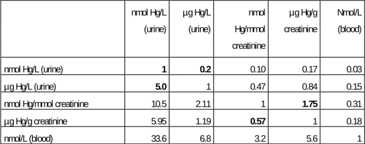
moniroting index. At steady state the daily urinary excretion is about 1% of the body burden. Measurement of the urinary excretion requires full sampling of all the urine voided during 24 hours and in field conditions sensitive to incomplete sampling. Therefore, as a proximate the urinary excretion divided by the creatinine excretion is often used. This value can be measured in a spot sample and is expressed in μg Hg/g creatinine equal to 0.57 μmol Hg/mol creatinine. The excretion of creatinine is roughly proportional to the muscle mass, in average mmol /kg body weight per day.
The raw concentration of mercury in urine in μg/l equal to 5 nmol/L is very often used. The problem is varying dilution of the urine why correction for density, osmolarity, or more often creatinene has been used. This has in various studies reported here been done by expressing the amount of mercury by mass of creatinine (μg Hg/g creatinine = 0.57 nmol/ mmol creatinine). An empiric conversion factor assuming a concentration of 1 g creatinine/l gives a conversion factor as shown in table 1-1. This may give some imprecisions, which, however, in the these group means will be diminished.
In steady state some empiric conversion factors between blood and urinary mercury (7). This article showed high correlations between the three values, average
concentration in breathing zone, end shift blood values, and next morning urinary concentration in urine. The conversion factor between blood and urine could be calculated to 2.80-3.48 (3.2) nmol/mmol creatinine in urine per nmol/l in blood. Mercury in other matrices as hair (head or pubic) or nails have been used because the values theoretically reflect an exposure over a longer time than blood or urinary values. However as no accumulation is seen in these matrices and there is a
considerable risk of contamination these measures are of generally lower value even though some studies have used them.
In order to facilitate the comparison of the exposure in the different articles a conversion table is used (see table 1-2). Some of the factors are empirical in which case an average was used even though this may imply some imprecision in converting the indices of personal exposure or body burden.
Table 1-2 Conversion factors between the different units of measurements for inorganic mercury used in the reviewed articles. The factors show the factors used when converting from the values in the left column to the values in the top row. Bold figures are exact factors, while the other are empiric.
nmol Hg/L (urine) µg Hg/L (urine) nmol Hg/mmol creatinine µg Hg/g creatinine Nmol/L (blood) nmol Hg/L (urine) 1 0.2 0.10 0.17 0.03 µg Hg/L (urine) 5.0 1 0.47 0.84 0.15 nmol Hg/mmol creatinine 10.5 2.11 1 1.75 0.31
µg Hg/g creatinine 5.95 1.19 0.57 1 0.18
nmol/L (blood) 33.6 6.8 3.2 5.6 1
The ACGIH (The American Conference of Governmental Industrial Hygienists) uses the ratio of 1 μg Hg/m3 in air to 1.22 μg Hg/g creatinine (1 nmol Hg/l equals
to 0.14 nmol Hg/mmol creatinine in urine to convert air concentrations to urinary excretion rates for long-term exposures. (6) In the present report, however air concentrations and biological concentrations will be handled separately. 1.4 Dental amalgam
Amalgam consists of about 50% metallic mercury in an amalgam with silver or cupper, with small amounts of other metals such as zinc. It was introduced when Auguste Taveau in Paris in 1826 used a “silver paste” made from five French franc pieces mixed with mercury. The silver coins also contained tin and a small amount of cupper, which gave the mixture more plasticity and a quicker setting time. (8)
1.4.1 Procedures for the use of amalgam in dentistry.
Today most dental amalgam is sold as an encapsulated preparation. The powdered metals and elemental mercury are packed into separate compartments, and the physical divider is broken just prior to use.
Silver-mercury fillings contain 50% mercury and a metal alloy consisting of silver (approx. 70%), tin (approx. 25%), cupper (1-6%) and zinc (0-2%). After mixing there may be an excess of mercury in the amalgam, which is removed just prior placement of the amalgam filling.
1.4.2
1.4.3
1.4.4
Mixing of amalgam
Another previously frequently used method to make amalgam was mixing of mercury and metal alloy in a mortar. Excess mercury was squeezed through a cloth, and the amalgam was then often moulded in the palm of the hand to keep it soft. Some dental clinics had their own production of capsules filled with mercury and metal alloy and subsequent shakened in a mixing device. Excess mercury was also here squeezed through a cloth.
Another method frequently used in the 1970s and 1980s in Norway was mixing of amalgam in a Dentomat or a similar mixing device. Mercury and metal alloy were added in separate chambers in the device and dosed in a semi-closed system. The device dosed the mixture itself, but it was possible to set it on dry or soft amalgam. The soft amalgam was also here in excess of mercury that had to be squeezed out. Filling of the Dentomat or a similar mixing device involved risk of spills.
Prefabricated closed capsules containing mercury and metal alloy were introduced in the 1980s. The mixture was so precise that it was not necessary to squeeze out any excess mercury. (9).
Cupper amalgam, previously used as filling material in decidual teeth, contained approx. 70% mercury and 30% cupper. The cupper amalgam was easier to use than traditional amalgam because it was more flexible to work with and it was considered to have a better anti-bacterial effect that prevented further attack of caries. When cupper amalgam was prepared from tablets, the tablets was heated using a burner to about 225 °C in a little pan until the mixture was fluent. The heated mixture was transferred to a mortar for further processing before the excess mercury was squeezed through a cloth. Most dental clinics stopped using cupper amalgam in the early 1980s.
Condensation of amalgam
Several methods have been used when condensating amalgam either manually or by ultrasound, vibrator (pneumatic, Cavitron, or electro-mallet).
Polishing and removal of amalgam
Cutting, placing, and polishing of amalgam restorations has been done by various methods. Drilling may be air cooled or water cooled. During this various kinds of
suction devices can be used; High-volume evacuator with an evacuating capacity of 150 L/min., Saliva-extractor (SE) with an evacuating capacity of less than 20 L/min. in air, or Mirror-evacuator (ME) which is a combination of a dental mirror and suction device with an evacuating capacity of 40 L/min (10).
1.4.5 Waste removal
During time waste amalgam has been removed and either storaged under water or different kinds of sealed containers.
1.5 Exposure to mercury by amalgam in own teeth
The amalgam fillings liberate mercury which is evaporated into the oral cavity. When mouth-breathing mercury vapour is carried to the lung where it is absorbed and distributed to the tissues. The rate of mercury vapour release is
stimulated/determined by; hot liquids and excessive chewing. Excessive chewing may lead to urinary mercury levels in excess of 120 nmol/L.
The number of amalgam surfaces, especially occlusal surfaces determines the release as 10 amalgam surfaces will on the average cause an increase in urine levels of 1 μg Hg/L urine. Urinary concentrations in people with amalgams are typically about 2-4
μg Hg/L. The rate of release in people with amalgam restoration is 2-17 μg Hg/day. The removal of amalgam fillings first causes a rise in blood plasma levels followed by an exponential decline lasting about 1 year. In the immediate postremoval phase, the peak plasma mercury level may rise to three to four times higher than the
pre-treatment level.
An allergic response can occur in the person having amalgam fillings, but it is so rare that a practicing dentist may not see one case in his or her professional lifetime. The allergic response to the placement of amalgam takes the form of ulceration and inflammation of oral tissues in contact with the amalgam. Allergic skin reaction to metallic mercury is very rarely seen.
For a review of the toxicity of dental amalgam in own teeth see Bates 2006 (11). 1.6 Clinical toxicology of inorganic mercury
Acute poisoning with high doses of inorganic mercury appears to occur in three phases: An initial phase with flu-like symptoms lasting 1-3 days. An intermediate phase with signs and symptoms of severe pulmonary toxicity, and a final phase with
gingivo-stomatitis, tremor and erethism (memory loss, emotional lability, depression, insomnia, and shyness) (6;12;13).
The more chronic toxicity is characterized by tremor and psychological disturbances (excessive timidity, diffidence, increasing shyness, loss of self-confidence, anxiety, and a desire to remain unobserved and unobtrusive) as the main features.
Tremor is characteristic, intentional during guided movements (finger-to-nose test) in milder cases, postural in more severe cases (tremor in the extended arm).
Gingivitis, stomatitis, and excessive salivation are seen.
Severe kidney damage sometimes associated with the nephrotic syndrome (rare), characterized by albuminuria and edema.
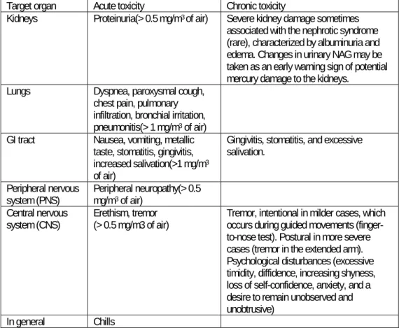
Table 1-2 shows the different signs in relation to the air concentrations above which they have been documented.
Target organ Acute toxicity Chronic toxicity
Kidneys Proteinuria(> 0.5 mg/m3 of air) Severe kidney damage sometimes associated with the nephrotic syndrome (rare), characterized by albuminuria and edema. Changes in urinary NAG may be taken as an early warning sign of potential mercury damage to the kidneys.
Lungs Dyspnea, paroxysmal cough,
chest pain, pulmonary infiltration, bronchial irritation, pneumonitis(> 1 mg/m3 of air) GI tract Nausea, vomiting, metallic
taste, stomatitis, gingivitis, increased salivation(>1 mg/m3 of air)
Gingivitis, stomatitis, and excessive salivation.
Peripheral nervous
system (PNS) Peripheral neuropathy(> 0.5 mg/m3 of air) Central nervous
system (CNS) Erethism, tremor (> 0.5 mg/m3 of air) Tremor, intentional in milder cases, which occurs during guided movements (finger-to-nose test). Postural in more severe cases (tremor in the extended arm). Psychological disturbances (excessive timidity, diffidence, increasing shyness, loss of self-confidence, anxiety, and a desire to remain unobserved and unobtrusive)
In general Chills
1.7 Permissible exposure levels
During time the occupational exposure levels set by the national board of labour or board of health have decreased. Mainly the Time Weighted Average concentration for 8 hours work has been the standard. The Danish value is 0.025 mg/m3 and in
most European countries the level varies between 0.025 and 0.1 mg/m3. The
European Union has recommended a value of 0.02 mg/m3.
2
Exposure in dentistry
The exposure assessments in Danish dental clinics and dental personnel, however, are scarce and unsystematic. Two older published studies have been found (14;15) and only a few and not well documented analyses have been made by the Danish National Institute of Occupational Health (16;17).
The tables 2-1, 2-2, and 2-3 describe shortly the results of the various studies showing measurements in air and urine, Detailed information of the measured values in air, urine, blood and other body compartments are shown in appendix 3.
Urinary excretion of mercury has both in dentristry and other exposures been most frequently used due to the easy sampling and because the measurement reflect a relevant time of exposure due to a half time of about 60 days. Some studies have used blood levels of mercury reflecting a shorter exposure period while some studies were based on hair and nail concentrations of mercury theoretically reflecting a long term exposure.
2.1 Mercury in air in relation to the type of clinic and procedures
The most relevant information about air measurement in relation to different circumstances in dental clinics is shown in table 2-1.
2.1.1 Older studies, observations before 1990
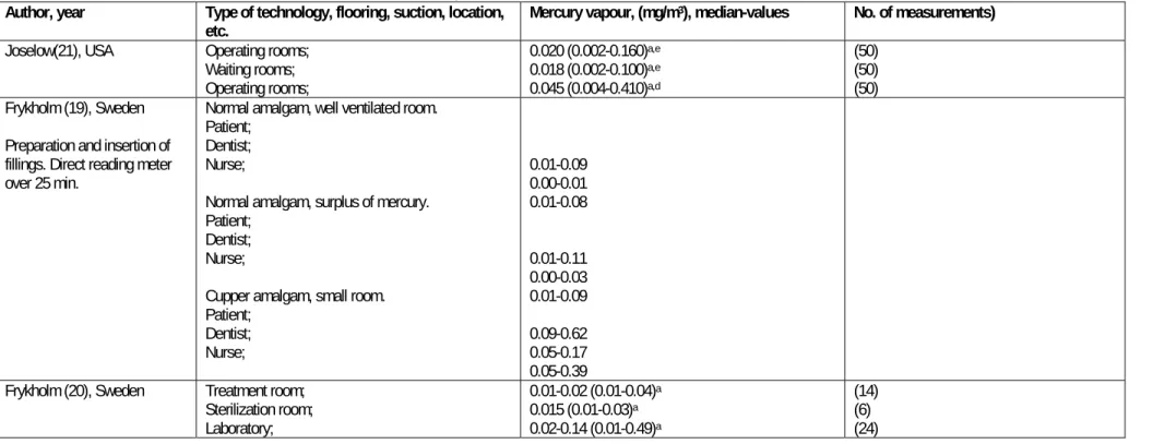
Frykholm has in his thesis from 1957 reported measurement of the concentration of mercury in the breathing zone of patient, dentist and dentist´s nurse during preparation and application of amalgam including preparation and insertion of cupper amalgam (18).
Figure 2-1 Illustration from Frykholm 1957. The air concentrations of mercury in inhaled air of the patient, dentist, and nurse during mixing (M), insertion (I), and completion (C) of silver amalgam (two figures at the left) and cupper amalgam (right) (18).
Concentrations were measured using a directly reading detector(19). The exposure were characterized by peaks in the exposures, up to 0.1 mg/m3 for traditional
amalgam and 0.39 mg/m3 for cupper amalgam but with very fast decline in
concentrations within 5 min after end of the procedures.
Measurements at four different departments at the Royal Swedish School of Dentistry from 1946 to1957 is reported by Frykholm. More than half of the 110 measurements were below 0.01 mg/m3 while a few ranged up to 0.13 mg/m3, the
laboratory department showing slightly higher values than the operative departments.
A later study by Frykholminvestigated Swedish dental personnel (10 dentists, 10 dental nurses, 10 dental technicians) from five workplaces (20). Analysis of air, blood and urine were made. A recommended upper limit of 0.05 mg Hg/m3 air was not
exceeded in the work-places used by dentists and dental nurses. However, during occasional brief periods these limits were reached or exceeded in several dental technical laboratories, e.g., during preparing and polishing amalgam models. The total mercury and vapour concentrations and the urinary excretion of mercury in 50 dentists were measured in a group of dental offices in New York (21). The means and ranges for vapour mercury concentrations in the operating rooms were 0.020 mg/m3 (0.002-0.160) and for the waiting rooms 0.018 mg/m3 (0.002-0.100).
The mean total mercury concentration (vapour and particulate) in the operating rooms at a height approximating the breathing zone of the dentist was 0.045 mg/m3
(0.004-0.410). Fourteen percent of the operating rooms exceeded the actual TLV for mercury of 0.1 mg/m3. The mean urinary mercury contents of the dentists were 200
(0-775) nmol/L.
The work environment and procedures of 22 Norwegian dentists and their 33 assistants were evaluated by Norseth (22). Determinations were made of the mercury vapour concentrations in the offices, the urinary excretion of mercury and the mercury concentration in blood. The mean mercury vapour concentration of all 24 offices were 0.043 mg/m3 ranging from 0.00 to 0.400 mg/m3. In three of the 24
offices surveyed, mercury vapour concentration exceeded the threshold limit value of 0.05 mg/m3. Especially heating of cupper amalgam, used in children, raised the
concentration of mercury vapour in all offices (e.g. from 0.001 to 0.005 mg/m3 and
from 0.01 to 0.08 mg/m3).
In Denmark Lundgaard 1981 measured the Hg in air at various places (14). The mercury vapour level was measured for 6 days, 3 days with open windows and 3 days with closed windows. By a Dentomat the values were between 0.01 and 0.065 mg/m3 irrespective of the use of windows. The concentrations were slightly higher
when the Dentomat was in use and further increased by a factor 1.5-2 when the lid was not fastened. Residues of amalgam on the work table gave rise to higher
concentrations.Comparing the levels of the ventilated and not ventilated rooms, the mercury vapour level was somewhat lower in the ventilated rooms (0.002->0.1 mg/m3 vs. 0.008->0.1 mg/m3).
Buchwald surveyed the work environment and procedures associated with the preparation of mercury amalgam fillings of 23 US dentists and their assistants (23). Amalgam dust was measured which cannot be compared with the vapours
concentrations. Measuring dust during removal of old fillings the use of suction devices decreased dust values by a factor 14. Water coolant caused lower levels of dust compared with air cooling.
2.1.2 Studies later than 1990
Pohl and Bergman conducted a study in Sweden with the aim to evaluate directly the dentist’s exposure to mercury vapour during the cutting, and filling of 50 amalgam restorations and during the polishing of 80 restorations, while using various dental suction devices (10). During the cutting, filling, and polishing operations using a high-volume evacuator, the mean mercury vapour levels in the breathing zone of the dentist were in the range of 0.001-0.002 mg/m3. However, when only a saliva
extractor was used, the cutting of amalgam fillings caused at least a factor 10 higher
and highly fluctuating mercury vapour levels. During condensing and polishing lower values were seen.
During a clinical simulation of insertion and removal of dental amalgams Powell recorded mercury vapour levels (24). The levels increased slightly, but never exceeded the TLV of 0.05 mg/m3.
The Scottish study by (25)measured in relation to the study of health effects the environmental measurements of mercury in 180 dental clinics (25). One hundred and twenty two (67.8%) of the 180 surgeries visited had environmental mercury measurements in one or more areas above the Occupational Exposure Standard (OES) set by the Health and Safety Executive (0.025 mg/m3). In the majority of these
surgeries the high levels of mercury were found at the skirting and around the base of the dental chair. In 45 surgeries (25%) the personal dosimetry measurement (i.e. in the breathing zone of dental staff) was above the OES.
The use of an amalgam mixer showed higher values than when using prefabricated capsules. The concentration varied considerably between measurements within each procedure while the differences between procedures were smaller. However the highest values were around the chair and these did reflect the dosimeter values of breathing air zones concentrations as well as the biological measurements. The air concentrations at the various sites were correlated with the reported number of amalgams placed.
2.2 Urinary mercury in dental personnel
See table 2-2
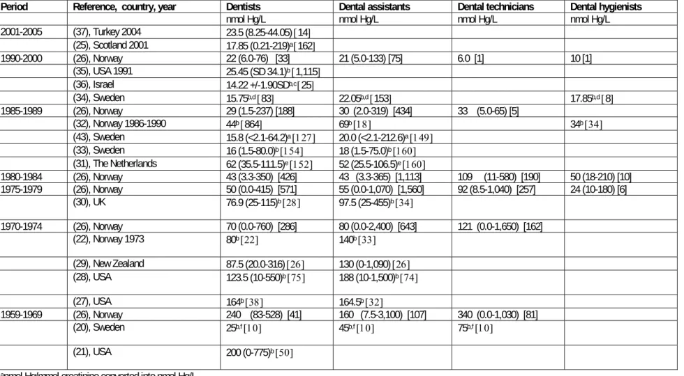
In Norway a comprehensive monitoring program has been carried out for urinary mercury in dental personnel. Lenvik et al.(26) have conducted a statistical analysis of data also shown in a recent report (STAMI). Data consists of 6283 samples from 3112 participants for the period 1959-2000. The number of measurements before 1970, however were small compared with the next decades. Main results are shown in table 2-2.
The results show a regular decline in urinary mercury levels in dental personnel from the 1960s and on to 2000. As the table above shows, dental technicians had the highest mean concentration with a median of 340 nmol/l urine in the 1960s and 33 nmol/l urine at the end of the 1980s.
Dentists had a median of 240 nmol/l in the 1960s decreasing to 22 nmol/l in the
1990s. Corresponding values for dental assistants were 160 and 21 nmol/l. Dental hygienists are represented with 17 samples for the period 1975-2000 with a median of 26 nmol/l, which is on the same level as in the general population. These median values cover a considerable variation. For dentists the highest value in the early period was 528 nmol/L and the 10 % highest according the figure was dimished from 500 nmol/L in the 60s to below 100 nmol/l in the 90s.
Up to 1970 64% and 63% of the samples from dental technicians and dentists, respectively, exceeded 200 nmol/l, which in EU has been suggested as biological threshold value for mercury in urine. Corresponding values in dental assistants were 41%. After 1990 no measurements above 200 nmol/L have been registered in any of the groups.
The representativity of the material is, however, not documented in the available literature. In the monitoring program persons with high values have a higher probability for being reexaminated, thereby giving an overrepresentation of the high values. This effect is very often seen in surveys of exposure. A further analysis of this very large material in case better background information could be provided, an analysis of the within person variation would probably give a better description of risk groups.
Battistone (27) determined mercury levels in the urine of 38 dentists and 32 assistants in six different clinics in the US army. Twenty non-dental personnel were used as controls. Mean urinary mercury values, ranging from 15-85 nmol/L and 25-620 nmol/L, were obtained for the controls and dental personnel, respectively. The six clinics included were of two basic designs. Two clinics consisted of large,
rectangular rooms with a number of dental units on both sides of the length of each room. The mean urinary mercury values for these two clinics were 280 and 180 nmol/L, respectively. The rest of the clinics consisted of rows of adjoining operatories, a central corridor, and rooms opposite the operatories devoted to supply, administration, etc. The mean urinary mercury values for these clinics were 110, 105, 130 and 65 nmol/L, respectively. This shows that with regard to mercury hygiene, the single-large-room design is less desirable than individual dental units located in separate but adjoining rooms.
Schneider (28) conducted an environmental health survey of 19 US dental offices with 284 dental workers to determine sources of uncontrolled exposure to mercury vapour. An additional objective was to assess the degree of risk of the various dental groups, in the handling of mercury. Dental assistants who handle mercury and
amalgam had higher mean urinary mercury levels than dental assistants whose duties did not include the handling of amalgam (188 vs. 84 nmol/L, respectively),
significant p< 0.01. The corresponding concentration of the dentists was 123.5 nmol/L for those handling mercury versus 96.5 nmol/L for dentists not handling mercury, not significant. No significant differences were found between dentists who handle mercury and dental assistants who also handle amalgams.
Brooks (29) compared 25 dentists’ surgeries in New Zealand with a control group of 20 persons with regards to urinary mercury levels and mercury in hair. Median-values for mercury in the urine samples were for the dentists 87.5 nmol/L (20-316), for the chairside assistants 130 nmol/L (0-1090) and for the control group 17.5 nmol/L (0-178.5).
In the older Norwegian study urinary mercury contents of one dentist and four dental assistants exceeded 250 nmol/L. The mean urinary mercury concentrations in all 22 dentists were 80 nmol/L and in the 33 dental assistants 140 nmol/L (22) Kelman (30) reports the results of a survey of urinary mercury excretion in 62 Area Health Authority (AHA) and 49 National Health Service (NHS) dentists and dental surgery assistants (DSAs) in Leicestershire, UK. AHA personnel comprised 28 dentists and 34 DSAs; NHS personnel consisted of 21 dentists and 28 DSAs. In both groups of workers the DSAs had a higher mean and greater range of urinary mercury concentrations than the dentists; the NHS workers had similarly, in both dentists and DSAs, a higher mean and greater range of concentrations. Urinary mercury concentrations in AHA; 76.9 (25-115) and 97.5 (25-455) nmol/L for dentists and DSAs, respectively and in NHS; 110.7 (25-500) and 191.3 (25-940) nmol/L for dentists and DSAs, respectively.
In The Netherlands Herber conducted a study of 162 dentists’ (all men) and their assistants’ (all women) mercury levels in hair and urine (31)30). A threshold level of 0.5 g creatinine/L allowed the determination of 160 urines from the dentists and 152 from the assistants. The mercury concentrations in urine (Hg-U) were somewhat higher in the dentists than the assistants (nmol/L, respectively, vs. 52 nmol/L.)). The method of condensation of amalgam, the number of amalgam fillings per year and hours in own practice were positively related to Hg-U.
A Norwegian study conducted by Jokstad assessed the relationship between mercury exposure and the urinary mercury excretion (32). Morning urine samples and questionnaires were collected from 672 participants in 1986 and 273 participants in
1987. The mean values of the urinary mercury excretion were 39 nmol/L (SD=29) in 1986, and 43 nmol/L (SD=36) in 1987. The mean mercury value was lower for the female (40 nmol/L) than for the male participants (44 nmol/L), (p< 0.05). Elevated mercury values correlated with the number of placed, polished, and replaced amalgam restorations/week. Furthermore, participants working in clinics with wooden floors had significantly higher mean mercury values than other dental personnel.
Skare (33) studied mercury exposure among dental personnel in Stockholm, with the use of urinary mercury excretion rates and questionnaires. The study covered 314 dentists (n= 154; 76 men, 78 women) and dental nurses (n= 160, all women). Male dentists had a higher mean concentration, 18 (3.5-80) nmol/L, than their female colleagues, 14 55) nmol/L. Dental nurses had a mean concentration of 18 (1.5-75) nmol/L compared to dentists with a mean concentration of 16 (1.5-80) nmol/L. Dental nurses and dentists employed in private practice had a higher mean
concentration than their colleagues in public clinics. The reported mean of the mercury concentration in the urine samples of the dentists was 19 nmol/L for private practices versus 11.5 nmol/L for public clinics, and the corresponding concentration of the dental nurses was 20.5 nmol/L for private practices versus 15 nmol/L for public clinics.
In Sweden Akesson (34) investigated Hg-levels in whole blood (B-Hg), plasma (P-Hg), and urine (U-Hg), in various groups of dental personnel (dentists, nurses, dental hygienists) and 81 matched referents, with no known occupational exposure to Hg. The study group consisted of 244 dental personnel in the public dental service, 83 dentists, 153 nurses, and 8 dental hygienists. U-Hg and P-Hg levels were higher in the dental personnel than in the matched referents (U-Hg 18.9 and 11.55 nmol/L urine, respectively; p< .001; P-Hg: 6.7 and 6.2 nmol/L, respectively; p= .03). Higher U-Hg levels were found in nurses than in dentists (22.05 and 15.75 nmol/L urine, respectively; p= .02). No statistically significant differences were observed for P-Hg and B-Hg. Levels of P-Hg and B-Hg were significantly higher in individuals who worked in large clinics than in individuals who were employed at smaller facilities. The oral amalgam status of the dental personnel was determined and statistically significant associations were found between the amount of amalgam in own teeth and U-Hg, P-Hg, and B-Hg (p< .0001 for all), with the highest correlation seen between the total amalgam surface area and U-Hg.
Martin (35) examined personal (diet, age and non-occupational mercury exposures), professional (number of amalgams placed per week, whether the dentist uses squeeze
cloths and how amalgam scrap was stored) and office (prior accidential mercury spills, flooring material and number of operatories) characteristics of American dentists to determine which factors contribute most to exposure. Complete data sets were obtained from 1277 dentists, 92.3% male. General dentists constituted 92.4 percent of the study sample. The mean mercury concentration for all subjects was 24.7 (SD=33.05) nmol/L. General dentists had a greater mean concentration, 25.45 (SD=34.1) nmol/L than specialists, 15.8 (SD=11) nmol/L. Men had a higher mean concentration, 24.95 (SD=33.45) nmol/L, than women, 20.6 (SD=24.2) nmol/L. This could be explained by the women having been in their current offices less time, having been in practice less time and having reported fewer accidental spills.
Mercury concentration increased with age, from a mean of 19.4 (SD=21.25) nmol/L for those 20 to 30 years of age, to 51.9 (SD=94.25) nmol/L for those older than 70 years of age. Mercury concentration increased with length of practice, from a mean of 24.15 (SD=32.8) nmol/L for the group that had practiced less than five years to 39.7 (SD=70.2) nmol/L for those who had practiced more than 40 years. Urinary mercury concentration increased with numbers of amalgams in the dentist’s own mouth, from a mean of 20.55 nmol/L for dentists with no amalgams to 33 for dentists with more than 12 amalgams.
In Israel Steinberg (36) compared urinary mercury levels of 25 dental personnel with 22 controls, not exposed to mercury in their daily occupations, and explored
possible correlations between environmental factors in the dental office and the urinary level of the personnel. The results indicated that the urinary mercury levels of the tested dental professionals were significantly higher than those of the control group (14.22 +/- 1.9 vs. 5.35 +/- 2.023 nmol mercury/L urine). Of the 25 dental
personnel and 22 control subjects tested, 18 of the dental personnel (72%) had detectable mercury levels in their urine compared to only 6 (27%) of the control subjects. None of the participants had mercury levels exceeding 150 nmol Hg/L urine. No significant correlation was found between the amount of mercury and years in the profession, weekly working hours, amalgam restorations performed per week, age of the dental office or size of the room. A weak correlation of r = 0.263 was found between the amount of urinary mercury levels and number of amalgam restorations in the mouths of the dental personnel.
Ritchie (25) conducted a study of 180 dentists in the West of Scotland to determine their exposure to mercury during the course of their work and the effects on their health and cognitive function and compared it to 180 controls. Data were obtained from questionnaires distributed to dentists and measurements of environmental mercury in surgeries. Furthermore the dentists were asked to give samples of urine,
hair and nails for mercury analysis. Dentists were found to have, on average, urinary mercury levels over 4 times that of control subjects (27.09 and 7.04 nmol Hg/L urine, respectively) although all but one dentist had urinary mercury below the Health and Safety Executive health guidance value of 20 μmol mmol-1 creatinine (~
200 nmol/L).
In a study by Karahalil (37) the urinary Hg excretion levels of 20 Turkish dentists and nine control subjects, matched for age. The levels of Hg in the urine samples of the dentists was about three times higher than the control subjects (31 +/- 17.5 and
9.85 +/- 4.5 nmol/L, respectively). Some 90% of the dentists wore both gloves and
masks. Only one subject wore neither mask nor gloves. Coincidentally, his urinary Hg level was the second highest among exposed subjects. The length of the work experience of the dentists did influence the urinary Hg excretion. Dentists who had been working less than 10 years had a mean urinary Hg level of 25.0 nmo.l/L (SD 12.5 )compared to dentists with 10 or more years of work experience 44.5 nmol/L (SD21.5).
2.3 Hg in Blood
In Denmark Moller-Madsen (15)analyzed blood samples from a group of 130 dentists and a control group of 40 blood-donors to evaluate the extent of mercury exposure. The median blood concentration of mercury was 20.0 (range: 6.0-96) nmol/L for dentists and 10.0 (5.5-23) nmol/L for controls. These values are according to Table 1-1 roughly equivalent with urinary concentrations of 100 nmol/L and 50 nmol/L for dentist and controls, respectively.
No statistically significant differences were observed between dentists with different practice characteristics (address of office, private practice/school dentist, number of amalgam restorations performed per week, days since last filling, method of
trituration). A statistically significant 47% increase in blood mercury was seen among the examined dentist concerning fish consumption.
Chang (38)collected blood samples from 205 dentists participating in the 1985 Health Assessment Program of the American Dental Association Annual Session. Control blood samples were obtained from the non-dental employees of the Association. The total, inorganic, and organic mercury contents of blood were determined. The results indicated that the total and inorganic mercury levels in blood were significantly different between dentists (30.5 and 10.5 nmol/L, respectively) and nondental controls (20.0 and 3.5 nmol/L, respectively). The organomercurial levels were, however, insignificant. This implies therefore, that
significant enzymatic conversion of inorganic to organic mercury compounds does not occur in vivo.
Atesagaoglu (39)compared blood mercury levels of 10 Turkish dentists with 10 amalgam-free controls. The mean mercury concentration in the blood was 178.5 nmol/L for dentists and 260.5 nmol/L for the controls and showed non-significant differences. The values of this study are a factor 5 higher than in the two above mentioned studies.
2.4 Hg in hair and nails
A number of studies have used hair or nail mercury as biomarkers of exposure (40-42)
Ritchie et al 2004 included mercury in hair (scalp and pubic) and nails in addition to the urinary measurements (1;25). The concentrations were elevated in dentists in comparison with control but the hair and nail measurements did not give additional information about the exposure.
2.5 Gender difference in the biological measurements
Table 2-3 shows the biological exposure indices for males and females in the studies, where this information could be retrieved. Generally the values were 10-20% lower for females although urinary concentrations in two of the Swedish studies show the opposite trend (33;43).
In the Norwegian survey female dentists in all periods, except for the few
measurements before 1970 had substantially lower urinary mercury concentrations than their male colleagues, while no systematic difference was seen in dental technicians (44). The female dental assistants had in the first periods higher values than the other groups, probably because of differences in exposure and not gender specific toxicokinetics.
2.6 Discussion and conclusion
2.6.1 Mercury in workroom air.
From the studies it is seen that the exposure in the older studies were higher and more variable up to 1980. After this time the general concentrations in the
workroom air were considerably lower although still at least in Brittish dental clinics contaminated with air concentrations exceeding the occupational limits in several places (25). Studies from Scandinavia, other European countries, or from US