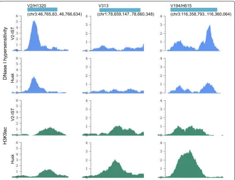
Genome wide mapping of transcriptional enhancer candidates using DNA and chromatin features in maize
Full text
Figure
Related documents
The multimode Radio cellular mobile system has improved performance on the basis of BER and constellation size for different modulation algorithms.. Thus, the mode may improve
To identify the physiological targets of PieE during infection, we established a new purification method for which we created an A549 cell line stably expressing the Escherichia
(A) Type I IFN production by SV40T MEFs from matched wild-type and Sting -deficient mice stimulated with 0.1 M CPT for 48 h, measured by bioassay on LL171 cells (data shown as
It was decided that with the presence of such significant red flag signs that she should undergo advanced imaging, in this case an MRI, that revealed an underlying malignancy, which
So, the aim of the present was to screen this endemic plant extracts has been earlier to have antimicrobial activity against the pathogens causing
21 Department of Neurosurgery, Tangdu Hospital, The Second Affiliated hospital of the Fourth Military Medical University, 1 Xinsi Road, Xian, Shanxi Province 710038, People ’ s
The maximum adsorption capacity for Mn(II) removal from an aqueous solution was 41.67 mg/g at pH 5 and their adsorption state was completed within 30 min.. The adsorption kinetics was
We first assessed the expression of LSH in a panel of lung cells using a Western blot analysis (Figure 1A) and found an increased expression of LSH in the lung cancer cell
