Evaluation of a TaqMan Array Card for
Detection of Central Nervous System
Infections
Clayton O. Onyango,
aVladimir Loparev,
bShirley Lidechi,
cVinod Bhullar,
bD. Scott Schmid,
dKay Radford,
dMichael K. Lo,
bPaul Rota,
dBarbara W. Johnson,
bJorge Munoz,
bMartina Oneko,
cDeron Burton,
eCarolyn M. Black,
bJohn Neatherlin,
a,fJoel M. Montgomery,
a,fBarry Fields
a,fDivision of Global Health Protection, Centers for Disease Control and Prevention, Nairobi, Kenyaa; National Center for Emerging and Zoonotic Infectious Diseases, Centers for Disease Control and Prevention, Atlanta, Georgia, USAb; Kenya Medical Research Institute, Nairobi, Kenyac; National Center for Immunization and Respiratory Diseases, Centers for Disease Control and Prevention, Atlanta, Georgia, USAd; National Center for HIV/AIDS, Viral Hepatitis, STD, and TB Prevention, Centers for Disease Control and Prevention, Atlanta, Georgia, USAe; Division of Global Health Protection, Center for Global Health, Centers for Disease Control and Prevention, Atlanta, Georgia, USAf
ABSTRACT
Infections of the central nervous system (CNS) are often acute, with
sig-nificant morbidity and mortality. Routine diagnosis of such infections is limited in
developing countries and requires modern equipment in advanced laboratories that
may be unavailable to a number of patients in sub-Saharan Africa. We developed a
TaqMan array card (TAC) that detects multiple pathogens simultaneously from
cere-brospinal fluid. The 21-pathogen CNS multiple-pathogen TAC (CNS-TAC) assay includes
two parasites (
Balamuthia mandrillaris
and
Acanthamoeba
), six bacterial pathogens
(
Streptococcus pneumonia
e,
Haemophilus influenzae
,
Neisseria meningitidis
,
Myco-plasma pneumoniae
,
Mycobacterium tuberculosis
, and
Bartonella
), and 13 viruses
(parechovirus, dengue virus, Nipah virus, varicella-zoster virus, mumps virus, measles
virus, lyssavirus, herpes simplex viruses 1 and 2, Epstein-Barr virus, enterovirus,
cytomeg-alovirus, and chikungunya virus). The card also includes human RNase P as a nucleic
acid extraction control and an internal manufacturer control, GAPDH
(glyceraldehyde-3-phosphate dehydrogenase). This CNS-TAC assay can test up to eight samples for all
21 agents within 2.5 h following nucleic acid extraction. The assay was validated for
linearity, limit of detection, sensitivity, and specificity by using either live viruses
(dengue, mumps, and measles viruses) or nucleic acid material (Nipah and
chikungu-nya viruses). Of 120 samples tested by individual real-time PCR, 35 were positive for
eight different targets, whereas the CNS-TAC assay detected 37 positive samples
across nine different targets. The CNS-TAC assays showed 85.6% sensitivity and 96.7%
specificity. Therefore, the CNS-TAC assay may be useful for outbreak investigation
and surveillance of suspected neurological disease.
KEYWORDS
central nervous system, TaqMan PCR, meningitis, encephalitis
I
nfections of the central nervous system (CNS) such as meningitis, encephalitis, or
meningoencephalitis may present as an acute illness with significant mortality and
extended sequelae (1). Because these infections are often difficult to diagnose in the
laboratory, clinical diagnoses often rely upon modern noninvasive techniques,
includ-ing computerized tomography scans and in some cases magnetic resonance imaginclud-ing.
Access to these advanced clinical diagnostic techniques is limited to large metropolitan
hospitals rarely found in developing countries. Even with neuroimaging, the
determi-nation of etiologic agents causing meningitis and encephalitis remains complicated,
requiring confirmation using other laboratory tests (2, 3). Laboratory tests can identify
Received15 December 2016Returned for modification12 January 2017 Accepted6 April 2017
Accepted manuscript posted online12 April 2017
CitationOnyango CO, Loparev V, Lidechi S, Bhullar V, Schmid DS, Radford K, Lo MK, Rota P, Johnson BW, Munoz J, Oneko M, Burton D, Black CM, Neatherlin J, Montgomery JM, Fields B. 2017. Evaluation of a TaqMan array card for detection of central nervous system infections.
J Clin Microbiol 55:2035–2044.https://doi.org/
10.1128/JCM.02469-16.
EditorKaren C. Carroll, Johns Hopkins University School of Medicine
Copyright© 2017 American Society for
Microbiology.All Rights Reserved.
Address correspondence to Clayton O. Onyango, xwl4@cdc.gov.
crossm
on May 16, 2020 by guest
http://jcm.asm.org/
a number of etiological agents responsible for bacterial and viral meningitis. Timely
identification of these agents continues to be challenging in developing countries,
where physicians frequently resort to empirical treatment with little or no benefit.
Advances in molecular diagnostic technology have fostered the development of
multiple pathogen detection systems based on PCR. The advent of real-time PCR and
multiplexing technologies has facilitated the detection of multiple targets from a single
clinical sample (4–7). Although multiplex PCR is susceptible to reduced efficiency and
sensitivity due to competition for PCR reagents by the different targets, specificity may
also be affected if closely related targets are not selected and validated carefully. These
problems can be overcome by using the TaqMan array card (TAC), which utilizes
microfluidic technology and single-plex PCRs configured in a 384-well array format. The
TAC has previously been used to detect pathogens responsible for respiratory (8, 9),
enteric (10), and neonatal (11) infections as well as other acute febrile illnesses (12).
Even though a number of multiplex pathogen detection assays, e.g., BioFire, SeeGene,
and Fast Track diagnostics, are commercially available, there are numerous advantages
to the TAC, including ease of use, low risk of contamination attributable to the sealed
format, the ability to modify or replace individual targets without additional
optimiza-tion, and a small sample volume requirement compared to using multiple single agent
real-time PCR assays (13, 14).
In this study, we evaluated a CNS multiple-pathogen TAC (CNS-TAC) assay for 21
etiologies and validated the CNS-TAC assay results alongside individual real-time PCR
(IRTP) assays for nine pathogens (four viruses, four bacteria, and one parasite). The
purpose of this evaluation was to determine the sensitivity and specificity of the
CNS-TAC assay compared to IRTP assays in detecting multiple pathogens from clinical
samples. We likewise propose the use of this tool in outbreak settings, providing
reduced turnaround times resulting in timely and agent appropriate interventions. In
addition, the method will be used to improve our understanding of the epidemiology
of the CNS.
RESULTS
Analytical performance (plasmid controls).
All assays exhibited a linear
relation-ship between threshold cycle (
CT
) values and the concentrations of nucleic acids.
CNS-TAC assays demonstrated linearity with
R
2values ranging between 0.987 and
0.998, except for the measles assay which had a
R
2value of 0.920. The PCR efficiency
for detection of all targets in the plasmid ranged from 98.7 to 99.7%, whereas measles
virus had an efficiency of 92.0% (Table 1).
The lower limit of detection (LOD) for all targets was 1.6
⫻
10
⫺7ng/well, an
equivalent of 54 copies per well (Fig. 1). At a concentration of 1.6
⫻
10
⫺2ng/well, the
assay sensitivity ranged from 80 to 100% for the plasmid targets and was 55% for the
measles virus nucleic material. The specificity for all of the positive controls was 100%.
The assays showed an accuracy range of 96.7 to 100% at a concentration of 1.6
⫻
10
⫺2ng/well. The variation in reproducibility of the
CT
values for the 21 targets ranged
from 0.9 to 2.2% for the high-concentration control, and it was 8.6% for the measles
assay. The variation in the reproducibility of the low-concentration control material
ranged from 1.0 to 7.5% for the plasmid targets, and it was 6.7% for the measles
assay (Table 1).
Clinical performance.
We tested by CNS-TAC assay 120 specimens, 35 of which
were initially positive upon IRTP analysis to validate the assays. The specimens were
positive for nine targets: 4% (5/120) cytomegalovirus (CMV), 4% (5/120)
Neisseria
meningitidis
, 8% (10/120) Epstein-Barr virus (EBV), 3% (3/120) varicella-zoster virus (VZV),
5% (6/120)
Streptococcus pneumoniae
, 2% (2/120) mumps virus, 6% (7/120)
Mycobac-terium tuberculosis
, 2% (2/120)
Acanthamoeba
, and 2% (2/120)
Haemophilus influenzae
.
The specificity for all nine targets across 120 samples ranged from 87.5 to 100%.
Streptococcus pneumoniae
had the lowest specificity (87.5%), whereas CMV, VZV,
mumps virus, and
Haemophilus influenzae
all had specificities of 100%. Compared to
IRTP, the overall sensitivity of the CNS-TAC assay ranged from 33.3% for VZV to 100%
on May 16, 2020 by guest
http://jcm.asm.org/
for
Neisseria meningitidis
(Table 2). Low sensitivities (
⬍
50%) were observed in cases
where there were small sample sizes of positive targets as seen with the mumps virus
and VZV assays (Table 2). Although
Acanthamoeba
was not detected by IRTP in any of
the samples, the CNS-TAC assay detected this target in two samples. Further attempts
to detect this by IRTP failed to yield positive results.
DISCUSSION
[image:3.585.42.374.84.335.2]We describe our evaluation of an in-house-developed CNS-TAC assay that can be
used to test cerebrospinal fluid (CSF) for infections associated with meningitis and
encephalitis. Infections of the CNS comprise a number of serious and often fatal
infections, and yet such infections often pose challenges in diagnosis (15). Many of the
pathogens associated with CNS infections are detected by culture, microscopy, or
TABLE 1Analytical performance of the CNS-TAC assaya
Target
Linearity
Accuracy (sensitivity %)
Reproducibility (CV%)
Linearity (R2)
Efficiency (%)
High concn
Low concn
High concn
Low concn
Bartonella 0.994 99.4 100 80.0 1.6 2.3
Chikungunya 1 virus 0.991 99.1 100 100 1.3 5.7 Chikungunya 2 virus 0.994 99.4 100 100 1.8 5.5
CMV 0.994 99.4 100 100 1.5 4.1
EBV 0.987 98.7 100 100 1.5 6.8
HSV 1 0.987 98.7 96.7 100 2.3 4
HSV 2 0.993 99.3 100 100 0.9 4
Measles virus 0.92 92.0 100 55.0 8.6 6.7
Mumps virus 0.997 99.7 100 100 1.1 2
Mycobacterium tuberculosis 0.993 99.3 96.7 100 1.0 5.1
VZV 0.993 99.3 100 95.0 2.4 2.7
Mycoplasma pneumoniae 0.993 99.3 100 100 1.0 2.9
Nipah virus 0.980 98.0 100 100 1.4 1
Neisseria meningitidis 0.994 99.4 100 100 1.8 4
Pan-dengue virus 0.993 99.3 100 100 2.1 1.7
Haemophilus influenza 0.993 99.3 100 100 1.2 5.2
Parechovirus 0.995 99.5 96.6 85.0 1.9 5.6
Streptococcus pneumoniae 0.992 99.2 100 95.0 1.2 7.4
RNase P 0.993 99.3 100 95.0 1.8 1.6
Acanthamoeba 0.990 99.0 100 100 1.8 4.1
Balamuthia mandrillaris 0.990 99.0 100 100 1.7 5.4
aHigh concentration⫽1.6⫻10⫺2ng/well or 5.3⫻106copies per well; low concentration⫽1.6⫻10⫺7
ng/well or 54 copies per well. CV, coefficient of variance.
FIG 1Test of linearity for all targets in the TAC. Dilutions from 1.6⫻10⫺2to 1.6⫻10⫺7are shown as bars on thexaxis.
on May 16, 2020 by guest
http://jcm.asm.org/
[image:3.585.45.476.541.728.2]antigen detection techniques. PCR is generally more reliable at detecting pathogens in
the CSF, with substantially higher sensitivity than other diagnostic methods such as
culture and enzyme-linked immunosorbent assay if the samples are collected at the
appropriate time during the infection (16–18). PCR-based approaches for detecting
multiple pathogens in a single array not only increase the number of pathogens that
can be detected but also reduce the overall amount of time needed to rule out multiple
pathogens. Therefore, we utilized previously published real-time PCR assays
incorpo-rated into a CNS-TAC assay. With improvements in sensitivity and ease of use, such
multipathogen TAC assays have been used in the detection of both human respiratory
and enteric pathogens (8–10). All CNS-TAC assays had LODs similar to what has been
described elsewhere for respiratory and enteric pathogens (8, 10). Although this
CNS-TAC method was designed for East Africa, many of the pathogens evaluated here
cause CNS infections worldwide, and therefore the card is suitable for broader use. In
this study, we designed and evaluated a CNS-TAC method that was able to detect 13
viruses, 6 bacteria, and 2 parasites. Clinical evaluation was against 120 patient samples;
TABLE 2Sensitivity of CNS-TAC assays compared to IRTP assays using aCTof 40 as a cutoffa
Target and parameter IRTP assay TAC assay % Sensitivity (95% CI) CMV
No. positive 7 5 100 (59.0–100)
MeanCT⫾SD 35.5⫾2.8 33.0⫾2.3 MedianCT(range) 36 (30.7–38.9) 33.2 (29.9–36)
Neisseria meningitidis
No. positive 5 5 100 (47.8–100)
MeanCT⫾SD 24.2⫾1.9 23.16⫾1.6 MedianCT(range) 24.4 (21.5–26.7) 23 (21.1–24.9)
EBV
No. positive 8 10 80.0 (44.4–97.5)
MeanCT⫾SD 34.9⫾2.0 32.2⫾1.1 MedianCT(range) 35.5 (31.5–37.0) 32.1 (29.8–34.0)
VZV
No. positive 1 3 33.3 (0.8–90.6)
MeanCT⫾SD 30.6 28.3⫾3.5 MedianCT(range) 29.0 (24.4–31.3)
Streptococcus pneumoniae
No. positive 5 6 83.3 (35.9–99.6)
MeanCT⫾SD 23.0⫾3.2 22.4⫾4.0 MedianCT(range) 22.8 (18.8–27.5) 22.3 (17.6–27.6)
Mumps virus
No. positive 1 2 50.0 (1.3–98.7)
MeanCT⫾SD 31.8 27.3⫾6.2 MedianCT(range) 27.3 (22.9–31.8)
Mycobacterium tuberculosis
No. positive 6 7 85.7 (42.1–99.6)
MeanCT⫾SD 33.4⫾2.5 32.2⫾4.0 MedianCT(range) 33.1 (29.9–37.7) 34.0 (24.8–36.8)
Haemophilus influenzae
No. positive 2 2 100 (47.8–100)
MeanCT⫾SD 22.1⫾6.6 23.2⫾5.6 MedianCT(range) 22.1 (17.4–26.8) 23.2 (19.2–27.2)
Acanthamoeba
No. positive 0 2
MeanCT⫾SD 34.9⫾3.2
MedianCT(range) 34.1 (33.9–38.4)
aIRTP, individual real-time PCR; TAC, TaqMan array card; CI, confidence interval.
on May 16, 2020 by guest
http://jcm.asm.org/
35 of these specimens were found to be positive for eight pathogens using IRTP. The
patient samples used in clinical validation were collected from subjects presenting at
either Mbagathi District Hospital or Siaya District Hospital for patient care.
Analysis of the clinical validation demonstrated an average sensitivity of 79% across
the TAC. This excluded the two
Acanthamoeba
positive specimens that failed to amplify
by IRTP, suggesting that these were false-positive reactions. However, this average
sensitivity was skewed by the VZV assay, which had a suboptimal sensitivity of 33.3%;
eliminating these results from the calculation yields an average sensitivity of 85.6%. The
low sensitivity for VZV and mumps virus may be attributed to the low numbers of
positive samples tested. A parasite with global distribution,
Balamuthia mandrillaris
,
was not detected by the assay in this card. This could have been due to the small
sample size tested or to the absence of this pathogen in patients from the two
geographical regions sampled in Kenya. Additional positive samples for these targets
are needed to accurately determine the sensitivity of these assays. Alternatively, we
advise the exclusion of these targets with low sensitivities from cards designed for
future studies.
Acanthamoeba
spp. and
Balamuthia mandrillaris
are free-living amoebas
that can potentially cause infections in humans and have been implicated in CNS
infections worldwide (19). However, the prevalence of these pathogens is not well
documented in sub-Saharan Africa. On the other hand,
Plasmodium falciparum
infec-tion can present in a severe form of cerebral malaria, with a mortality rate of 10 to 25%,
and is most common in sub-Saharan Africa (20, 21). Similarly, several studies have
implicated cryptococcal meningitis as the major cause of meningitis among
HIV-infected individuals in sub-Saharan Africa (22–24). Therefore, we recommend
replace-ment of the
Acanthamoeba
and
Balamuthia mandrillaris
targets with
Plasmodium
falciparum
and cryptococcal meningitis, which are more prevalent in sub-Saharan
Africa, in future versions of these cards. The average specificity for CNS-TAC assay for
the eight targets was 96.7%. A subset of samples failed to amplify RNase P in CNS-TAC
(13%) and IRTP (6%) analyses. Usually, this would suggest inappropriate specimen
collection, sample degradation, or the complete absence of human DNA in some CSF
samples. However, the concentration of RNase P should reflect the concentration of
white blood cells in the specimen, since CSF is usually free of human DNA. This suggests
that a different marker should be used in the future as a control for specimen integrity
for CSF.
The CNS-TAC assay detected seven targets that were not detected by the IRTP
assays. These additional detections failed to yield positive results using IRTP despite
numerous attempts, and as such, are possibly false positives that would negatively
impact the specificity of the TAC assays since the IRTP method was considered the gold
standard for these comparisons. These discrepancies could possibly be explained by
additional freeze-thaw cycles negatively impacting nucleic acid integrity for the IRTP
assay. However, the observed mean
CT
of 30.8
⫾
4.2 indicates significant amplification
and would argue against this possibility.
In our protocol, two of eight lanes were occupied on the first TAC: one of the eight
lanes of the card was designated a no-template negative control, and another was
designated the combined positive control. Subsequent cards tested would hold a
negative control and seven specimens on a card. Up to three cards were tested per day
in one ViiA-7 machine, which allowed for 20 specimens to be tested for 21 pathogens
each per day. This greatly reduces the turnaround time for specimen testing compared
to IRTP assays. Despite the discrepancies observed between TAC and IRTP, we think
there is added value in the use of CNS-TAC as a screening assay in outbreak settings.
Indeed, samples with positive IRTP or TAC results require further investigations,
includ-ing gene sequencinclud-ing, among other confirmatory tests. Our future plans are therefore to
confirm all the TAC positives by sequencing, as well as to further validate the CNS-TAC
assay using a larger sample size from patients presenting with CNS infections from
other geographical sites within Kenya. This will help us better understand the utility of
CNS-TAC in outbreak investigations.
on May 16, 2020 by guest
http://jcm.asm.org/
MATERIALS AND METHODS
CNS-TAC design. The CNS card includes assays for the detection of six bacterial pathogens,
Streptococcus pneumoniae,Haemophilus influenzae,Neisseria meningitidis,Mycoplasma pneumoniae, My-cobacterium tuberculosis, andBartonella(genus specific); 13 viruses, parechovirus, pan-dengue virus (detects all four serotypes), Nipah virus, VZV, mumps virus, measles virus, pan-Lyssa virus, HSV-1 and -2, EBV, enterovirus, CMV, and chikungunya virus (detection is based upon two different gene targets for all three genotypes); and two parasites,Balamuthia mandrillarisandAcanthamoeba(Fig. 2). All primers and probes were adapted from previously published assays, except for theM. tuberculosisassay, which is described for the first time here. Primers and probes for the targets were titrated individually by real-time PCR using genomic DNA, plasmid DNA, or RNA on a Bio-Rad CFX 96 platform and AgPath-ID One-Step RT-PCR master mix (Life Technologies, United Kingdom). Once titrated, the primers and probe for each assay were preloaded and dried by a ViiA7 Applied Biosystems (Foster City, CA) instrument in duplicate wells on the TAC, which included three intrinsic controls: (i) an extraction control, RNase P; (ii) measles virus RNA acting as both a target control and a RNA control; and (iii) an internal manufacturer control, glyceraldehyde-3-phosphate (GAPDH) (Table 3). All of the assays in this card apart from lyssavirus and pan-dengue virus assays were developed at the Centers for Disease Control and Prevention (CDC) and are used routinely for clinical diagnosis in the United States. The other two assays were developed at the University of Pretoria, South Africa, and the Bernhard Nocht Institute, Germany, respectively.
Design of combined positive control.Customized combined positive controls were designed and synthesized in two different plasmids. The design and orientation of the positive-control plasmid maps are similar to that described by Kodani and Winchell (25). The forward primer sequence was placed downstream of the plasmid pUC57 T7 sequence, followed by the probe sequence and finally the reverse primer sequence. The plasmid comprised these concatenated sequences for all targets. The two plasmids were designated A and B. Plasmid A contained sequences forBartonella, CMV, EBV, HSV-1 and -2, mumps virus, Mycobacterium tuberculosis, VZV, Mycoplasma pneumoniae, Nipah virus, Neisseria meningitidis, pan-dengue virus,Haemophilus influenzae, parechovirus,Streptococcus pneumoniae, and RNase P. Plas-mid B contained sequences for chikungunya virus targets 1 and 2, Acanthamoeba, and Balamuthia mandrillaris. Measles virus RNA was spiked into plasmid B preparation as an exogenous control for the virus, as well as an RNA control. Positive-control extracts were titrated following a 10-fold dilution to determine the LOD.
Analytical validation.The LOD, linearity, repeatability, and reproducibility were determined using 10-fold dilutions of the control material using infection-free CSF as the diluent. The positive-control materials were derived from nucleic acid materials from the respective targets. Nucleic acid material from RNA viruses was transcribed into cDNA and prepared for gene cloning. Repeatability was tested using eight repeats on a single card, whereas reproducibility was tested with 10 serial dilutions of each plasmid and assayed over 5 days. The lower LOD was defined as the lowest concentration at which the target could be detected in all of the diluted samples. Analytical validation of these assays was performed at the National Center for Emerging and Zoonotic Infectious Diseases at the CDC.
Testing of CSF.CSF samples were obtained from patients in either Mbagathi District Hospital in Nairobi or Siaya District Hospital in rural Western Kenya. Children older than 6 weeks and adults of all ages were eligible for lumbar puncture if they presented with two or more signs and symptoms of CNS infection, such as fever (ⱖ38°C) and/or history of reported fever in the last 3 days, neck stiffness and/or
FIG 2CNS-TAC layout with 22 encephalitis targets, as well two human DNA/RNA controls, GAPDH and RNase P. PCRs for all the targets, including intrinsic controls, were customized for testing in duplicates.
on May 16, 2020 by guest
http://jcm.asm.org/
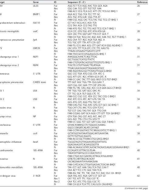
[image:6.585.86.326.69.266.2]TABLE 3Oligonucleotide sequences for CNS-TAC assaysa
Target Gene ID Sequence (5=–3=) Reference
CMV UL55 For AGG TCT TCA AGG AAC TCA GCA AGA 26
Rev CGG CAA TCG GTT TGT TGT AAA
Pr FAM-ACC CCG TCA GCC ATT CTC TCG GC-BHQ 1
EBV BNRF1 For GGA ACC TGG TCA TCC TTT GC 27
Rev ACG TGC ATG GAC CGG TTA AT
Pr FAM-CGC AGG CAC TCG TAC TGC TCG CT-BHQ 1
Mycobacterium tuberculosis IS6110 For CCT ACT ACG ACC ACA TCA —b
Rev CCG TAA ACA CCG TAG TTG
Pr FAM-ATG TGC TCC TTG AGT TCG CCA T-BHQ 1
Neisseria meningitidis sodC For CCA CCC GTG TGG ATC ATA ATA GA 28
Rev GCA CAC TTA GGT GAT TTA CCT GCA T
Pr FAM-CA TGA TGG CAC AGC AAC AAA TCC TGT TT-BHQ 1
Streptococcus pneumoniae lytA For ACG CAA TCT AGC AGA TGA AGC A 29
Rev TCG TGC GTT TTA ATT CCA GCT
Pr FAM-TG CCG AAA ACG CTT GAT ACA GGG AG-BHQ 1
VZV ORF29 For CAC GTA TTT TCA GTC CTC TTC AAG TG 30
Rev TTAGACGTGGAGTTGACATCGTTT Pr FAM-TACCGCCCGTGGAGCGCG-BHQ 1
Chikungunya virus 1 NSP1 For AAAGGGCAAACTCAGCTTCAC 31
Rev GCCTGGGCTCATCGTTATTC
Pr FAM-CTGTGATACAGTGGTTTCGTGTG-BHQ 1
Chikungunya virus 2 NSP4 For TCACTCCCTGTTGGACTTGATAGA 31
Rev TTGACGAACAGAGTTAGGAACATACC Pr FAM-AGGTACGCGCTTCAAGTTCGGCG-BHQ1
Enterovirus 5=UTR For GGC CCC TGA ATG CGG CTA ATC C 32
Rev GCG ATT GTC ACC ATWA GCA GYC A
Pr FAM-CC GAC TAC TTT GGG WGT CCG TGT-BHQ1
Mycoplasma pneumoniae CARDS toxin For TTT GGT AGC TGG TTA CGG GAA T 33
Rev GGT CGG CAC GAA TTT CAT ATA AG
Pr FAM-TG TAC CAG AGC ACC CCA GAA GGG CT-BHQ1
HSV 1 US4 For TAT TGG TGC GAT GGC GAC AC 34
Rev CTT TCC GCA TGT GGG CTC TC
Pr FAM-CCC CGC CCC ATA CCC TAC CCG C-BHQ1
HSV 2 US6 For AGC ATC CCG ATC ACT GTG TAC TA 34
Rev GCG ATG GTC AGG TTG TAC GT
Pr FAM-CAG TGC TGG AAC GTG CCT GCC GC-BHQ 1
Measles virus N For TGG CAT CTG AAC TCG GTA TCA C 35
Rev TGT CCT CAG TAG TAT GCA TTG CAA
Pr FAM-CCGAG GAT GCA AGG CTT GTT TCA GA-BHQ1
Mumps virus NP For GTA TGA CAG CGT ACG ACC AAC CT 36
Rev GCG ACC TTG CTG CTG GTA TT
Pr FAM-CC GGG TCT GCT GAT CGG CGA T-BHQ 1
Parechovirus 5=UTR For GTAACASWWGCCTCTGGGSCCAAAAG 37
Rev GGCCCCWGRTCAGATCCAYAGT
Pr FAM-CCTRYGGGTACCTYCWGGGCATCCTT-BHQ 1
Bartonella ssrA For GCTATGGTAATAAATGGACAATGAAATAA 38
Rev GCTTCTGTTGCCAGGTG
Pr FAM-ACCCCGCTTAAACCTGCGACG-BHQ1
Haemophilus influenzae bexA For TGCGGTAGTGTTAGAAAATGGTATTATG 39
Rev GGACAAACATCACAAGCGGTTA
Pr FAM-ACAAAGCGTATCAATACTACAACGAGACGCAAAAA-BHQ1
Acanthamoeba 18S rRNA For CCCAGATCGTTTACCGTGAA 40
Rev TAAATATTAATGCCCCCAACTATCC
Pr FAM-CTGCCACCGAATACATTAGCATGG-BHQ1
Lyssavirus N For GTRCTCCARTTAGCRCACAT 41
Rev CACMGSNAAYTAYAARACNAA
Pr FAM-CATCACACCTTGATGACAACTCACAA-BHQ1
Balamunthia mandrillaris 18S rRNA For TAA CCT GCT AAA TAG TCA TGC CAA T 40
Rev CAA ACT TCC CTC GGC TAA TCA
Pr FAM-AG TAC TTC TAC CAA TCC AAC CGC CA- BHQ1
Pan-dengue virus 3=NCR For GGA TAG ACC AGA GAT CCT GCT GT 42 Rev 1 CAT TCC ATT TTC TGG CGT TC
Rev 2 CAA TCC ATC TTG CGG CGC TC
Pr FAM CA GCA TCA TTC CAG GCA CAG-BHQ1
(Continued on next page)
on May 16, 2020 by guest
http://jcm.asm.org/
bulging fontanel, headache, reduced level of consciousness, or new-onset seizures. A total of 120 samples, including 35 that were positive for any of the CDC in-house IRTP assays, were also tested using the CNS-TAC method. CDC in-house assays were designed to include targets for CMV, EBV, mumps virus,
Mycobacterium tuberculosis, VZV, Neisseria meningitidis, Haemophilus influenzae, Acanthamoeba, and
Streptococcus pneumoniae. In addition, 85 randomly selected samples, determined to be negative in IRTP assays, were tested by the CNS-TAC assay to determine specificity.
Nucleic acids were extracted from CSF specimens using the KingFisher ML extraction platform (Thermo Scientific, Waltham, MA) and MagMax nucleic isolation kit (Life Technologies, Carlsbad, CA). Portions (100l) of CSF specimens were mixed with 260l of lysis binding solution and added to the columns. The column was washed once with 600l of wash solution 1 and then twice with 450l of wash solution 2 according to the manufacturer’s recommendations. After the wash steps, the nucleic acids were eluted with 60l of elution buffer. An additional 166l of previously PCR-positive samples was reextracted for IRTP testing using the same platform and kit. We used 433l of lysis binding solution to adjust for the increased sample volume. The samples were then eluted in 100l of elution buffer. An increased extraction volume was required for the eight IRTP assays, and this increased volume did not alter the sensitivity of the assays. The CNS-TAC assays were compared to the cognate IRTP assays on 96-well plates under the same thermocycling conditions using the same PCR master mix and 5l of nucleic acids as the template. Samples with aCTofⱖ40 were interpreted as negative, and those with a CTof 35 to 40 were classified as indeterminate and retested. If theseCTvalues remained within the range
of 35 to 40, they were ultimately classified as weak positives.
The CNS-TAC assays were run on a ViiA-7 real-time PCR system using an AgPath-ID One-Step real-time PCR kit (Applied Biosystems, Foster City, CA). The PCR master mix for each card included 1⫻ RT-PCR buffer, RT-PCR enzyme in a final 100-l reaction volume. A 46-l portion of nucleic acid extract was added to the master mix. Each run consisted of a negative control and a positive control for the first card of the day to be tested. A minimum of three cards were tested per day, with thermal cycling conditions as follows: 45°C for 10 min, 94°C for 10 min, and then 45 cycles of 94°C for 30 s and 60°C for 1 min. These clinical analyses were performed at the Centre for Global Health Research of the Kenya Medical Research Institute (KEMRI) in western Kenya.
Data analysis.Receiver operating characteristic analysis was used to deriveCTcutoffs. TheCTvalues
for CNS-TAC and IRTP assays were compared using attest, whereas dichotomous measures of the presence or absence of extrinsic controls were compared using a Fisher exact test. Linearity was tested by fitting linear regression models ofCTvalues against the concentrations of nucleic acids and
inter-preting theR2. The sensitivity of the CNS-TAC assay was also calculated against the gold standard: IRTP
for nine targets. All analyses were performed using STATA v13 (StataCorp).
Ethical approval.This study was covered under an investigational protocol reviewed by human subject review experts from the institutional review boards at the CDC (protocol 6092) and KEMRI (SSC protocol 1948). Informed written consent for survey participation, and CSF collection was obtained from all adult participants 18 years of age and older and from mature minors 13 to 17 years old. Verbal assent from minors (children 13 to 17 years old) and written consent from parents or guardians of those minors were obtained, and written consent from parents was obtained for childrenⱕ13 years old. If a patient of any age was unable to provide consent or assent because of altered mental status, consent was obtained from the patient’s responsible family member or guardian.
ACKNOWLEDGMENTS
We thank Jan Pohl, Maya Kodani, and Dean Erdman for assistance in the analytical
validation. We also acknowledge the KEMRI staff who helped collect specimens used in
clinical validation and laboratory testing. Work in Kenya was made possible through
assistance by a number of personnel, including Janet Awando, Bryan Nyawanda,
Weldon Korir, Newton Wamola, Patrick Emojoong, Jeremiah Nyaundi, Fredrick Ade,
Victoria Mwende, and Jim Katieno.
This study was partly funded by the 2012 U.S. Department of Defense, Defense
Threat Reduction Agency/Cooperative Biologic Engagement Program (DTRA/CBEP)
funding (KE 06; Deployment of Diagnostic Tests, Including Rapid Diagnostic Tests for
TABLE 3(Continued)
Target Gene ID Sequence (5=–3=) Reference
Nipah virus N For CTG GTC TCT GCA GTT ATC ACC ATC GA 43
Rev ACG TAC TTA GCC CAT CTT CTA GTT TCA Pr FAM-CAG CTC CCG ACA CTG CCG AGG AT-BHQ
RNase P RPP30 For AGA TTT GGA CCT GCG AGC G 44
Rev GAG CGG CTG TCT CCA CAA GT
Pr FAM-TTC TGA CCT GAA GGC TCT GCG CG-BHQ
aID, oligonucleotide identity; For, forward; Rev, reverse; Pr, probe; CMV, cytomegalovirus; EBV, Epstein-Barr virus; VZV, varicella-zoster virus; HSV, herpes simplex virus;
UTR, untranslated region; NCR, noncoding region; ORF, open reading frame.
b—, J. Posey, unpublished data.
on May 16, 2020 by guest
http://jcm.asm.org/
Diagnosis of Human and Animal Infections to District Laboratories in Kenya). Additional
funding was from internal CDC-Kenya funds.
The findings and conclusions in this report are those of the authors and do not
necessarily represent the official position of the U.S. Centers for Disease Control and
Prevention.
REFERENCES
1. Mace SE. 2010. Central nervous system infections as a cause of an altered mental status? What is the pathogen growing in your central nervous system? Emerg Med Clin North Am 28:535–570.https://doi.org/10.1016/ j.emc.2010.03.002.
2. Venkatesan A, Tunkel AR, Bloch KC, Lauring AS, Sejvar J, Bitnun A, Stahl JP, Mailles A, Drebot M, Rupprecht CE, Yoder J, Cope JR, Wilson MR, Whitley RJ, Sullivan J, Granerod J, Jones C, Eastwood K, Ward KN, Durrheim DN, Solbrig MV, Guo-Dong L, Glaser CA. 2013. Case definitions, diagnostic algorithms, and priorities in encephalitis: consensus state-ment of the International Encephalitis Consortium. Clin Infect Dis 57: 1114 –1128.https://doi.org/10.1093/cid/cit458.
3. Whitley RJ. 1990. Viral encephalitis. N Engl J Med 323:242–250.https:// doi.org/10.1056/NEJM199007263230406.
4. Briese T, Palacios G, Kokoris M, Jabado O, Liu Z, Renwick N, Kapoor V, Casas I, Pozo F, Limberger R, Perez-Brena P, Ju J, Lipkin WI. 2005. Diagnostic system for rapid and sensitive differential detection of pathogens. Emerg Infect Dis 11:310 –313. https://doi.org/10.3201/ eid1102.040492.
5. Dominguez SR, Briese T, Palacios G, Hui J, Villari J, Kapoor V, Tokarz R, Glode MP, Anderson MS, Robinson CC, Holmes KV, Lipkin WI. 2008. Multiplex MassTag-PCR for respiratory pathogens in pediatric nasopha-ryngeal washes negative by conventional diagnostic testing shows a high prevalence of viruses belonging to a newly recognized rhinovirus clade. J Clin Virol 43:219 –222.https://doi.org/10.1016/j.jcv.2008.06.007. 6. Pabbaraju K, Tokaryk KL, Wong S, Fox JD. 2008. Comparison of the Luminex xTAG respiratory viral panel with in-house nucleic acid ampli-fication tests for diagnosis of respiratory virus infections. J Clin Microbiol 46:3056 –3062.https://doi.org/10.1128/JCM.00878-08.
7. Reijans M, Dingemans G, Klaassen CH, Meis JF, Keijdener J, Mulders B, Eadie K, van Leeuwen W, van Belkum A, Horrevorts AM, Simons G. 2008. RespiFinder: a new multiparameter test to differentially identify fifteen respiratory viruses. J Clin Microbiol 46:1232–1240.https://doi.org/10 .1128/JCM.02294-07.
8. Kodani M, Yang G, Conklin LM, Travis TC, Whitney CG, Anderson LJ, Schrag SJ, Taylor TH, Jr, Beall BW, Breiman RF, Feikin DR, Njenga MK, Mayer LW, Oberste MS, Tondella ML, Winchell JM, Lindstrom SL, Erdman DD, Fields BS. 2011. Application of TaqMan low-density arrays for simul-taneous detection of multiple respiratory pathogens. J Clin Microbiol 49:2175–2182.https://doi.org/10.1128/JCM.02270-10.
9. Weinberg GA, Schnabel KC, Erdman DD, Prill MM, Iwane MK, Shelley LM, Whitaker BL, Szilagyi PG, Hall CB. 2013. Field evaluation of TaqMan Array Card (TAC) for the simultaneous detection of multiple respiratory viruses in children with acute respiratory infection. J Clin Virol 57:254 –260. https://doi.org/10.1016/j.jcv.2013.03.016.
10. Platts-Mills JA, Gratz J, Mduma E, Svensen E, Amour C, Liu J, Maro A, Saidi Q, Swai N, Kumburu H, McCormick BJ, Kibiki G, Houpt ER. 2014. Associ-ation between stool enteropathogen quantity and disease in Tanzanian children using TaqMan array cards: a nested case-control study. Am J Trop Med Hyg 90:133–138.https://doi.org/10.4269/ajtmh.13-0439. 11. Diaz MH, Waller JL, Napoliello RA, Islam MS, Wolff BJ, Burken DJ, Holden
RL, Srinivasan V, Arvay M, McGee L, Oberste MS, Whitney CG, Schrag SJ, Winchell JM, Saha SK. 2013. Optimization of multiple pathogen detec-tion using the TaqMan array card: applicadetec-tion for a populadetec-tion-based study of neonatal infection. PLoS One 8:e66183.https://doi.org/10.1371/ journal.pone.0066183.
12. Liu J, Ochieng C, Wiersma S, Stroher U, Towner JS, Whitmer S, Nichol ST, Moore CC, Kersh GJ, Kato C, Sexton C, Petersen J, Massung R, Hercik C, Crump JA, Kibiki G, Maro A, Mujaga B, Gratz J, Jacob ST, Banura P, Scheld WM, Juma B, Onyango CO, Montgomery JM, Houpt E, Fields B. 2016. Development of a TaqMan array card for acute-febrile-illness outbreak investigation and surveillance of emerging pathogens, including Ebola virus. J Clin Microbiol 54:49 –58.https://doi.org/10.1128/JCM.02257-15. 13. Driscoll AJ, Karron RA, Bhat N, Thumar B, Kodani M, Fields BS, Whitney
CG, Levine OS, O’Brien KL, Murdoch DR. 2014. Evaluation of fast-track diagnostics and TaqMan array card real-time PCR assays for the detec-tion of respiratory pathogens. J Microbiol Methods 107:222–226.https:// doi.org/10.1016/j.mimet.2014.10.009.
14. Liu J, Kabir F, Manneh J, Lertsethtakarn P, Begum S, Gratz J, Becker SM, Operario DJ, Taniuchi M, Janaki L, Platts-Mills JA, Haverstick DM, Kabir M, Sobuz SU, Nakjarung K, Sakpaisal P, Silapong S, Bodhidatta L, Qureshi S, Kalam A, Saidi Q, Swai N, Mujaga B, Maro A, Kwambana B, Dione M, Antonio M, Kibiki G, Mason CJ, Haque R, Iqbal N, Zaidi AK, Houpt ER. 2014. Development and assessment of molecular diagnos-tic tests for 15 enteropathogens causing childhood diarrhoea: a multicentre study. Lancet Infect Dis 14:716 –724.https://doi.org/10 .1016/S1473-3099(14)70808-4.
15. Weber MW, Herman J, Jaffar S, Usen S, Oparaugo A, Omosigho C, Adegbola RA, Greenwood BM, Mulholland EK. 2002. Clinical predictors of bacterial meningitis in infants and young children in The Gambia. Trop Med Int Health 7:722–731. https://doi.org/10.1046/j.1365-3156.2002 .00926.x.
16. Meyer T, Franke G, Polywka SK, Lutgehetmann M, Gbadamosi J, Magnus T, Aepfelbacher M. 2014. Improved detection of bacterial central ner-vous system infections by use of a broad-range PCR assay. J Clin Microbiol 52:1751–1753.https://doi.org/10.1128/JCM.00469-14. 17. Welinder-Olsson C, Dotevall L, Hogevik H, Jungnelius R, Trollfors B, Wahl
M, Larsson P. 2007. Comparison of broad-range bacterial PCR and culture of cerebrospinal fluid for diagnosis of community-acquired bac-terial meningitis. Clin Microbiol Infect 13:879 – 886.https://doi.org/10 .1111/j.1469-0691.2007.01756.x.
18. Wu HM, Cordeiro SM, Harcourt BH, Carvalho M, Azevedo J, Oliveira TQ, Leite MC, Salgado K, Reis MG, Plikaytis BD, Clark TA, Mayer LW, Ko AI, Martin SW, Reis JN. 2013. Accuracy of real-time PCR, Gram stain, and culture forStreptococcus pneumoniae,Neisseria meningitidis, and Hae-mophilus influenzaemeningitis diagnosis. BMC Infect Dis 13:26.https:// doi.org/10.1186/1471-2334-13-26.
19. Visvesvara GS, Moura H, Schuster FL. 2007. Pathogenic and opportunistic free-living amoebae: Acanthamoeba spp., Balamuthia mandrillaris,
Naegleria fowleri, andSappinia diploidea. FEMS Immunol Med Microbiol 50:1–26.https://doi.org/10.1111/j.1574-695X.2007.00232.x.
20. Mishra SK, Newton CR. 2009. Diagnosis and management of the neuro-logical complications of falciparum malaria. Nat Rev Neurol 5:189 –198. https://doi.org/10.1038/nrneurol.2009.23.
21. Taylor TE, Fu WJ, Carr RA, Whitten RO, Mueller JS, Fosiko NG, Lewallen S, Liomba NG, Molyneux ME. 2004. Differentiating the pathologies of cerebral malaria by postmortem parasite counts. Nat Med 10:143–145. https://doi.org/10.1038/nm986.
22. Britz E, Perovic O, von Mollendorf C, von Gottberg A, Iyaloo S, Quan V, Chetty V, Sriruttan C, Ismail NA, Nanoo A, Musekiwa A, Reddy C, Viljoen K, Cohen C, Govender NP. 2016. The epidemiology of men-ingitis among adults in a South African province with a high HIV prevalence, 2009-2012. PLoS One 11:e0163036. https://doi.org/10 .1371/journal.pone.0163036.
23. Jarvis JN, Meintjes G, Williams A, Brown Y, Crede T, Harrison TS. 2010. Adult meningitis in a setting of high HIV and TB prevalence: findings from 4961 suspected cases. BMC Infect Dis 10:67. https://doi.org/10 .1186/1471-2334-10-67.
24. Rajasingham R, Rhein J, Klammer K, Musubire A, Nabeta H, Akampurira A, Mossel EC, Williams DA, Boxrud DJ, Crabtree MB, Miller BR, Rolfes MA, Tengsupakul S, Andama AO, Meya DB, Boulware DR. 2014. Epidemiology of meningitis in an HIV-infected Ugandan cohort. Am J Trop Med Hyg 92:274 –279.https://doi.org/10.4269/ajtmh.14-0452.
25. Kodani M, Winchell JM. 2012. Engineered combined-positive-control template for real-time reverse transcription-PCR in multiple-pathogen-detection assays. J Clin Microbiol 50:1057–1060.https://doi.org/10.1128/ JCM.05987-11.
on May 16, 2020 by guest
http://jcm.asm.org/
26. Boppana SB, Fowler KB, Pass RF, Rivera LB, Bradford RD, Lakeman FD, Britt WJ. 2005. Congenital cytomegalovirus infection: association be-tween virus burden in infancy and hearing loss. J Pediatr 146:817– 823. https://doi.org/10.1016/j.jpeds.2005.01.059.
27. Ramamurthy M, Alexander M, Aaron S, Kannangai R, Ravi V, Sridharan G, Abraham AM. 2011. Comparison of a conventional polymerase chain reaction with real-time polymerase chain reaction for the detection of neurotropic viruses in cerebrospinal fluid samples. Indian J Med Micro-biol 29:102–109.https://doi.org/10.4103/0255-0857.81777.
28. Dolan Thomas J, Hatcher CP, Satterfield DA, Theodore MJ, Bach MC, Linscott KB, Zhao X, Wang X, Mair R, Schmink S, Arnold KE, Stephens DS, Harrison LH, Hollick RA, Andrade AL, Lamaro-Cardoso J, de Lemos AP, Gritzfeld J, Gordon S, Soysal A, Bakir M, Sharma D, Jain S, Satola SW, Messonnier NE, Mayer LW. 2011.sodC-based real-time PCR for detection of Neisseria meningitidis. PLoS One 6:e19361.https://doi.org/10.1371/ journal.pone.0019361.
29. Carvalho MDGS, Tondella ML, McCaustland K, Weidlich L, McGee L, Mayer LW, Steigerwalt A, Whaley M, Facklam RR, Fields B, Carlone G, Ades EW, Dagan R, Sampson JS. 2007. Evaluation and improvement of real-time PCR assays targetinglytA,ply, andpsaAgenes for detection of pneumococcal DNA. J Clin Microbiol 45:2460 –2466.https://doi.org/10 .1128/JCM.02498-06.
30. Quinlivan ML, Ayres K, Ran H, McElwaine S, Leedham-Green M, Scott FT, Johnson RW, Breuer J. 2007. Effect of viral load on the outcome of herpes zoster. J Clin Microbiol 45:3909 –3914.https://doi.org/10.1128/ JCM.00874-07.
31. Lanciotti RS, Kosoy OL, Laven JJ, Panella AJ, Velez JO, Lambert AJ, Campbell GL. 2007. Chikungunya virus in US travelers returning from India, 2006. Emerg Infect Dis 13:764 –767. https://doi.org/10.3201/ eid1305.070015.
32. Kilpatrick DR, Yang CF, Ching K, Vincent A, Iber J, Campagnoli R, Man-delbaum M, De L, Yang SJ, Nix A, Kew OM. 2009. Rapid group-, serotype-, and vaccine strain-specific identification of poliovirus isolates by real-time reverse transcription-PCR using degenerate primers and probes containing deoxyinosine residues. J Clin Microbiol 47:1939 –1941. https://doi.org/10.1128/JCM.00702-09.
33. Winchell JM, Thurman KA, Mitchell SL, Thacker WL, Fields BS. 2008. Evaluation of three real-time PCR assays for detection ofMycoplasma pneumoniaein an outbreak investigation. J Clin Microbiol 46:3116 –3118. https://doi.org/10.1128/JCM.00440-08.
34. Miari VF, Wall GR, Clark DA. 2015. Evaluation of non-extracted genital swabs for real-time HSV PCR. J Med Virol 87:125–129.https://doi.org/10 .1002/jmv.23967.
35. Hummel KB, Lowe L, Bellini WJ, Rota PA. 2006. Development of
quan-titative gene-specific real-time RT-PCR assays for the detection of mea-sles virus in clinical specimens. J Virol Methods 132:166 –173.https://doi .org/10.1016/j.jviromet.2005.10.006.
36. Rota JS, Rosen JB, Doll MK, McNall RJ, McGrew M, Williams N, Lopareva EN, Barskey AE, Punsalang A, Jr, Rota PA, Oleszko WR, Hickman CJ, Zimmerman CM, Bellini WJ. 2013. Comparison of the sensitivity of laboratory diagnostic methods from a well-characterized outbreak of mumps in New York city in 2009. Clin Vaccine Immunol 20:391–396. https://doi.org/10.1128/CVI.00660-12.
37. Nix WA, Maher K, Johansson ES, Niklasson B, Lindberg AM, Pallansch MA, Oberste MS. 2008. Detection of all known parechoviruses by real-time PCR. J Clin Microbiol 46:2519 –2524. https://doi.org/10 .1128/JCM.00277-08.
38. Diaz MH, Bai Y, Malania L, Winchell JM, Kosoy MY. 2012. Development of a novel genus-specific real-time PCR assay for detection and differenti-ation of Bartonella species and genotypes. J Clin Microbiol 50: 1645–1649.https://doi.org/10.1128/JCM.06621-11.
39. Wang X, Mair R, Hatcher C, Theodore MJ, Edmond K, Wu HM, Harcourt BH, Carvalho MDGS, Pimenta F, Nymadawa P, Altantsetseg D, Kirsch M, Satola SW, Cohn A, Messonnier NE, Mayer LW. 2011. Detection of bacterial pathogens in Mongolia meningitis surveillance with a new real-time PCR assay to detectHaemophilus influenzae. Int J Med Micro-biol 301:303–309.https://doi.org/10.1016/j.ijmm.2010.11.004. 40. Qvarnstrom Y, Visvesvara GS, Sriram R, da Silva AJ. 2006. Multiplex
real-time PCR assay for simultaneous detection ofAcanthamoebaspp.,
Balamuthia mandrillaris, and Naegleria fowleri. J Clin Microbiol 44: 3589 –3595.https://doi.org/10.1128/JCM.00875-06.
41. Coertse J, Weyer J, Nel LH, Markotter W. 2010. Improved PCR methods for detection of African rabies and rabies-related lyssaviruses. J Clin Microbiol 48:3949 –3955.https://doi.org/10.1128/JCM.01256-10. 42. Drosten C, Gottig S, Schilling S, Asper M, Panning M, Schmitz H, Gunther
S. 2002. Rapid detection and quantification of RNA of Ebola and Marburg viruses, Lassa virus, Crimean-Congo hemorrhagic fever virus, Rift Valley fever virus, dengue virus, and yellow fever virus by real-time reverse transcription-PCR. J Clin Microbiol 40:2323–2330. https://doi.org/10 .1128/JCM.40.7.2323-2330.2002.
43. Lo MK, Lowe L, Hummel KB, Sazzad HM, Gurley ES, Hossain MJ, Luby SP, Miller DM, Comer JA, Rollin PE, Bellini WJ, Rota PA. 2012. Characteriza-tion of Nipah virus from outbreaks in Bangladesh, 2008-2010. Emerg Infect Dis 18:248 –255.https://doi.org/10.3201/eid1802.111492. 44. Luo W, Yang H, Rathbun K, Pau CP, Ou CY. 2005. Detection of human
immunodeficiency virus type 1 DNA in dried blood spots by a duplex real-time PCR assay. J Clin Microbiol 43:1851–1857.https://doi.org/10 .1128/JCM.43.4.1851-1857.2005.