BIROn - Birkbeck Institutional Research Online
Kerai, L.V. and Hilton, S. and Maugueret Minerve, Tafie and Kazi, Bilkis Banu
and Nicklin, Jane and Bhakta, Sanjib and Murdan, S. (2016) UV-curable
gels as topical nail medicines: in vivo residence, anti-fungal efficacy and
influence of gel components on their properties. International Journal of
Pharmaceutics 514 (1), pp. 244-254. ISSN 0378-5173.
Downloaded from:
Usage Guidelines:
Please refer to usage guidelines at or alternatively
1 UV-curable gels as topical nail medicines: in vivo residence, anti-fungal efficacy and
1
influence of gel components on their properties 2
3
L V Kerai1, S Hilton1, M Maugueret2, B B Kazi2, J Faull2, S Bhakta2, S Murdan1*
4
5
6
1UCL School of Pharmacy, 29-39 Brunswick Square, London, WC1N 1AX, UK. 7
2Department of Biological Sciences, Institute of Structural and Molecular Biology, Birkbeck, 8
University of London, Malet Street, London, WC1E 7HX, UK.
9
10
*Corresponding author
11
T +44-207 753 5810
12
F +44-207-753-5942
13
14
15
16
17
Keywords: UV gel, acrylate, Ungual, Release, Permeation, Anti-fungal efficacy, In vivo, 18
Residence, Onychomycosis, amorolfine, terbinafine, TOWL 19
*Manuscript
2 Abstract
20
UV-curable gels, used as nail cosmetics for their in vivo durability, were reported to be
21
promising as topical nail medicines. Our firstaim was thus to investigate whether such
22
durability applies to drug-loaded formulations. This was found to be true. However, ethanol
23
inclusion in the pharmaceutical formulation (to enable drug loading) reduced the in vivo
24
residence. The second aim was therefore to determine any other effects of ethanol, and if
25
ethanol could be avoided by the choice of monomers. Thus, three methacrylate monomers,
26
ethyl methacrylate, isobornyl methacrylate and 2 -hydroxyethyl methacrylate (HEMA) were
27
selected, and their influence on the formulation properties were determined. Results:
28
Ethanol and the methacrylate monomer influenced some (but not all) of the formulation
29
properties. The most significant was that HEMA could dissolve drug and enable the
30
preparation of ethanol-free, drug-loaded formulations, which would benefit in vivo
31
residence. The absence of ethanol reduced drug loading, release and ungual flux, but had
32
no negative impact on the in vitro anti-fungal efficacy. Conclusion: judicious selection of gel
33
components enables the exclusion of ethanol. The long in vivo residence, little residual
34
monomers, sufficient ungual permeation and in vitro anti-fungal activity of the gels indicate
35
their potential as anti-onychomycotic topical medicines.
36
3 1. Introduction
38
Onychomycosis which is defined as fungal infection of the nail plate and/or nail bed, is a
39
significant medical burden, affecting 10% of the general population, 20% of those over 60
40
years old, up to 50% of those over 70 years old and up to one-third of diabetics (Thomas et
41
al., 2010) . The disease is often of long duration, is ‘more than a cosmetic problem’ (Scher,
42
1994), significantly affects patients’ quality of life (Lubeck et al., 1993) and is recalcitrant to
43
treatment (Arrese and Pierard, 2003; Daniel and Jellinek, 2010). Oral therapy with
anti-44
fungal agents, such as terbinafine and itraconazole, is the treatment mainstay. However,
45
drug efficacy is far from ideal, with pooled mycological cure rates in randomised controlled
46
trials being 76 ±3% for terbinafine and 63% ±7% for itraconazole (Gupta et al., 2004) . The
47
disadvantages of oral therapy, such as the potential for adverse systemic effects and drug
48
interactions means that topical therapy is highly attractive, and a number of topical
49
formulations have been tested, patented and a few have been marketed, compiled in, for
50
example (Saner et al., 2014; Shivakumar et al., 2012) . Unfortunately, the success rates of
51
topical therapy are even lower, as indicated in the medicines’ patient information leaflets,
52
and compiled in (Murdan, 2016) . A number of possible reasons for the low clinical cure of
53
therapy have been proposed, such as inaccurate diagnosis, incorrect antifungal or delivery
54
modality, presence of dormant fungal conidia (which can germinate and give rise to further
55
infection), resistant fungal species, dermatophytoma (areas of the nail filled with debris of
56
keratin and a large amount of fungi material which is more impermeable to drug), low
57
patient compliance, and insufficient drug permeation into and through the nail plate for
58
anti-fungal kill (Baran et al., 2005; Goodfield and Evans, 2000).
59
60
Insufficient drug in the nail could be due to the fact that the commercially available topical
61
formulations do not have a continuous residence on the nail plate following application, and
62
thus do not maximise the duration of ungual drug permeation. For example, the most
63
recently marketed formulations Jublia®, Kerydin® and Onytec® are easily washed off, and
64
the patient information leaflets recommend application prior to bedtime to enable ungual
65
drug permeation to occur for at least the duration of sleep (AnacorPharmaceuticals, 2014;
66
KakenPharmaceutical, 2014; Onytec). Loceryl® is meant for weekly application (Galderma,
4
2015). However, in vivo experiments have shown that the Loceryl film does not stay intact
68
on the nail for the duration of a whole week, at least on fingernails (Murdan et al., 2015).
69
Non-continuous residence of drug formulations on the nail could be compounded by poor
70
patient compliance. While the latter is not known, the extensive literature on (fairly low)
71
patient compliance in general, suggests that compliance to topical nail medicines is likely to
72
be lower than required.
73
74
Patient compliance could be improved by longer-lasting topical formulations and we have
75
previously proposed UV-curable gels as topical formulations which are likely to have long in
76
vivo residence on the nail (Kerai et al., 2015). In the work discussed in this paper, the first 77
aim was thus to investigate the in vivo residence of these formulations. During this study, it
78
was found that, while the UV-cured films have a fairly long in vivo residence, inclusion of
79
ethanol in the formulation (which enabled sufficient drug loading) reduced the in vivo
80
residence. The second aim was therefore to determine what other effects ethanol had, if
81
these effects were independent of the other gel components, and whether the inclusion of
82
ethanol could be avoided.
83
84
Cosmetic UV curable gel formulations typically consist of three components: i) a urethane
85
methacrylate, ii) a (meth)acrylate based monomer and iii) a polymerisation photoinitiator.
86
The pharmaceutical UV curable gel formulation reported previously consisted of these,
87
specifically, diurethane dimethacrylate (DUDMA), ethyl methacrylate (as the (meth)acrylate
88
based monomer) and 2-hydroxy-2-methylpropiophenone as the photoinitiator, as well as a
89
drug (amorolfine HCl or terbinafine HCl) and a solvent (ethanol). In order identify more
90
appropriate gel components (to avoid the use of ethanol), one could screen alternative
91
urethane-based methacrylates such as Di-HEMA trimethylhexyl dicarbomate,
92
photoinitiators such as 1-hydroxycyclohexyl phenyl ketone and different (meth)acrylate
93
based monomers. In this work, we decided to keep DUDMA as the backbone of the
94
formulation as it produces a strong film with exceptional abrasion resistance and durability,
95
and 2-hydroxy-2-methylpropiophenone as the photoinitiator, and to screen different
96
methacrylate based monomers. Thus, 2-hydroxyethyl methacrylate (HEMA) and isobornyl
5
methacrylate (IBOMA), in addition to ethyl methacrylate (EMA) were selected as the
98
methacrylate based monomers. The influence of these monomers and of ethanol on the
99
gel and film properties were determined, and correlations among the different properties of
100
the films formed by photo-polymerisation were explored.
101
102
2. Materials and Methods 103
2.1 Materials 104
Amorolfine HCl was purchased from Ranbaxy Research Laboratories (Haryana, India) and
105
terbinafine HCl from AK Scientific (CA, USA). Diurethane dimethacrylate, ethyl methacrylate,
106
isobornyl methacrylate, 2-hydroxyethyl methacrylate, 2-hydroxy-2-methylpropiophenone,
107
absolute ethanol, methanol, propan-2-ol, sodium chloride, triethylamine, phosphoric acid
108
85% wt solution in water and trifluoroacetic acid and a dialysis tubing cellulose membrane
109
(MW 10281) were purchased from Sigma–Aldrich (Dorset, UK). Acetonitrile HPLC gradient
110
grade was purchased from Fisher Scientific (Hertfordshire, UK). A 36 Watt Cuccio
111
Professional UVA nail lamp was purchased from Amazon UK. Nail & Beauty Emporium
lint-112
free wipes (4 ply) were purchased from Just Beauty UK, an online retailer specialising in
113
professional beauty, hair and skin products. Sabouraud dextrose agar was purchased from
114
Oxoid Ltd (Basingstoke, UK). One dermatophyte strain, Trichophyton rubrum (CBS 118892),
115
was used. Curanail® nail lacquer was used as a control in some experiments and was
116
purchased from pharmacies in the UK. Human nail clippings (fingernails) were obtained
117
from healthy volunteers aged between 18 and 65 years (following ethics approval).
118
119
2.2 Determination of drug (amorolfine HCl and terbinafine HCl) solubility in methacrylate 120
monomers 121
The saturation solubility of the two drugs in EMA, HEMA and IBOMA was determined as
122
described in (Kerai et al., 2015) .
123
6 2.3 Preparation of UV gels and films
125
Drug-free gel formulations were prepared by mixing DUDMA with a methacrylate monomer
126
(EMA or IBOMA or HEMA) at 85:15 or 75:25 (for HEMA only) v/v ratios, the photoinitiator at
127
3% v/v of the methacrylate mixture, with or without ethanol at 25% v/v of the methacrylate
128
mixture, and leaving the mixture to stir overnight, which produced a clear homogenous
129
solution. Drug-loaded formulations were prepared by first dissolving the drug in ethanol,
130
and then adding the methacrylates and photoinitiator and leaving the mixture to stir
131
overnight, which also produced a clear homogenous solution. To prepare the films, the gel
132
mixture was applied on a microscope glass slide using a pipette tip (30 µl to an area of 15
133
mm x 15 mm as a single layer), which was then placed under the UVA lamp for two minutes.
134
This caused curing of the formulation and formation of a film. The surface of the film was
135
wiped with propan-2-ol using a super absorbent 4 ply lint-free nail wipe to remove the
136
oxygen inhibition layer (an unreacted monomer layer). This revealed a glossy polymer film,
137
which was then removed from the glass slide using a scalpel and characterised.
138
139
A curing time of 2 minutes was selected after experimenting with 0.5-5 minutes of UVA
140
exposure, and measuring the gel blend to polymer film mass yield, the alkene-to-alkane
141
degree of conversion (the route of polymerisation), and the levels of residual monomers in
142
the polymer film. The measurements (Supplementary Data 1a) showed that a cure time of
143
two minutes was sufficient for maximal mass yield and degree of conversion and for
144
minimal residual monomers.
145
146
2.4 In vitro characterisation of the gels and UV-cured films 147
The in vitro properties of the gels and of the films, such as gel viscosity, maximum drug
148
loading, gel-to-film mass yield, degree of conversion and the films’ appearance,
149
microstructure, crystallinity, level of residual monomers, drug-polymer interactions, glass
150
transition temperature, adhesion, water sensitivity, drug release and ungual permeation
151
were determined as described in (Kerai et al., 2015) . The films’ mass change with time was
152
monitored over 28 days under the following testing conditions: 30°C ± 2°C and 50% RH ± 5%
7
RH, by placing the films in a desiccator containing a saturated solution of magnesium
154
nitrate and placing this in a 30⁰C oven. The film’s mass was recorded on days 0, 1, 2, 3, 7, 14,
155
21 and 28. The film thickness was measured using a Sealey AK9635D 0–25 mm Digital
156
External Micrometer (PVR Direct, Bristol, UK). Each film was measured at three separate
157
points and an average was taken, and each test was carried out in triplicate. Precautions
158
were taken to measure the thickness without compressing the polymer film. The uniformity
159
of thickness within each film and between films was calculated using the following formula:
160
161
2.5 In vivo fingernail residence of UV-cured polymer films 162
Following approval by the UCL School of Pharmacy's ethics committee (Project 5337/002),
163
12 volunteers (aged between 18 – 65 years) with healthy fingernails were recruited. For
164
each volunteer, a single layer of the UV-curable gel formulation was applied on the five
165
fingernail plates of one hand using a pipette tip, leaving a formulation-free margin
166
(approximately 1-2 mm) near the nail plate perimeter, ensuring that skin contact was
167
avoided. This hand was then placed under a UVA nail lamp for two minutes. Subsequently,
168
the surface of the cured gel on each nail plate was wiped with propan-2-ol using a super
169
absorbent nail wipe to remove the oxygen inhibition layer, leaving behind a thin, smooth,
170
transparent film. The procedure was repeated on the other hand. The nails were visually
171
observed daily to estimate the percentage of UV-cured film remaining on the fingernails.
172
Estimation was facilitated by visually dividing the nail plate into quadrants which were then
173
further divided into sub-quadrants. Daily observation was continued until the UV-cured
174
films had completely dislodged from the nail plate or for up to 28 days, whichever was
175
sooner. After 28 days, the remaining film was removed using a wooden stick 10 minutes
176
following the application of a cotton pad soaked with acetone over the film. The experiment
177
was then repeated with a different formulation. A total of four UV-curable gel formulations
178
(which contained the monomer EMA, ± solvent and drug) were tested, and each
179
formulation was tested on a minimum of six volunteers.
180
8 2.6 In vivo occlusivity of UV-cured polymer films
182
The occlusivity of UV-cured films was assessed by measuring trans-onycheal water loss
183
(TOWL) from the healthy fingernail plates of volunteers before and after the application of
184
the gel formulations (which were applied as described in Section 2.5). Curanail® lacquer film
185
was used as control, and was applied as instructed in the packaging (i.e. after filing and
186
cleansing). Prior to TOWL measurements, the volunteers had rested for at least 60 minutes
187
in the laboratory, and avoided contact with water. TOWL measurements were obtained
188
using a Biox Aquaflux Model AF200 equipped with an in vivo nail cap (Biox Systems Ltd,
189
London, UK). The finger was placed on a flat surface for support and the Aquaflux
190
measurement head was placed on the centre of the nail plate. No pressure was applied as
191
the weight of the measurement head itself ensured a good seal, and measurements were
192
collected using AquaFlux version 4.8. Water vapour flux density versus time curves were
193
recorded until a steady-state TOWL reading was obtained (between 90 - 120 seconds, with a
194
mean of 10 points), and five repeats were obtained per formulation. All measurements were
195
conducted in the same laboratory, where the ambient temperature and air humidity
196
fluctuated between 22 - 26⁰C and 38 - 48% respectively. The % reduction in TOWL was
197
calculated as follows:
198
199
A greater reduction in TOWL indicates higher occlusivity of the film.
200
201
2.7 In vitro antifungal efficacy of UV gels against T. rubrum
202
The dermatophyte T. rubrum was chosen as it is the most common causative organism of
203
onychomycosis. The ability of the formulations to inhibit fungal growth was used as a
204
measure to compare formulations. The classical disc diffusion method was adapted, where
205
the ‘disc’ was replaced by a circular piece of nail clipping onto which a gel formulation had
206
been cured, taking care to leave a formulation-free margin at the nail edge, such that the
207
drug would have to be permeate into and through the nailplate to reach the agar gel. This
9
method was used as there is currently no standard in vitro method to assess the anti-fungal
209
efficacy of anti-onychomycotic formulations.
210
211
Preparation of SDA plates: A Sabouraud Dextrose Agar (SDA) solution (65 g SDA/L of distilled
212
water) was prepared, sterilised (by autoclaving at 1210C for 15 minutes) and poured into
213
90mm petri dishes (25 ml of agar solution/dish) and allowed to solidify overnight.
214
Preparation of test plates: A cork borer (8mm-internal diameter, sterilised by dipping in
215
ethanol then flamed) was punched into the centre of a SDA plate (prepared above) to
216
remove a plug of agar, which was discarded. The cork borer was sterilised once more, and
217
used to punch out a T. rubrum plug from a fresh culture plate of T. rubrum. The fungal plug
218
was inserted into the slot created in the first SDA plate, and the latter was then incubated at
219
32⁰C for three days to ensure that the fungal culture grew sufficiently and to identify (and
220
remove) defective plates, if any. During this time, the T. rubrum culturegrew to 10 mm in
221
diameter.
222
Preparation of nail clippings with drug formulations: Human fingernail clippings were
223
washed with water, cut to size (circular, with a diameter of 3 mm) and autoclaved at 1200C
224
for 20 minutes in order to sterilise them (autoclaving did not affect the nail properties
-225
indicated by the lack of change in solvent uptake). Two µl of a formulation was applied on
226
the nail surface, ensuring a small formulation-free perimeter at the edge of the nail surface.
227
For the control Curanail, the lacquer was allowed to air-dry. For the UV gels, the nail was
228
placed under an UVA lamp for 2 minutes, after which the surface of the film produced was
229
wiped with propan-2-ol using a nail wipe.
230
Details of the formulations tested are shown in Table 1. Drug-free counterparts of these
231
formulations were also tested to assess the influence, in any, of the UV-cured film itself
232
against T. rubrum. In addition, Curanail® nail lacquer (containing 5% w/v amorolfine HCl)
233
was tested as a positive control.
234
Evaluation of anti-fungal efficacy: Three days following the insertion of the fungal plug into
235
a prepared SDA plate, the latter was marked into four quadrants (which enabled the testing
236
of one test formulation on three nail clippings, and one drug-free formulation on a fourth
237
nail clipping per plate). Using a sterile tweezer, the prepared nail clippings were carefully
10
placed on the agar gel surface at a distance of 20 – 25 mm from the centre of the plate. This
239
distance was chosen to allow sufficient time for the drug to penetrate into and through the
240
nail into the agar gel, based on the ungual drug permeation results and T. rubrum growth
241
rates from preliminary studies. Thus, each plate contained a triplicate set of the same
242
formulation in addition to a drug-free control.
243
In addition to the test plates, two controls were set up: i) a formulation-free, nail-free
244
negative control was set up to monitor the growth of T. rubrum, and ii) a control plate
245
containing 3 equally-spaced nail clippings with drug-free UV-cured films on their surface,
246
with the clippings being placed at a distance of 20 – 25 mm from the centre of the plate.
247
The SDA plates were incubated at 32⁰C for as long as it took the T. rubrum culture to grow
248
over the entire SDA plate’s surface (or when no further growth was detected). During this
249
period, the plates were observed daily to monitor the growth of T. rubrum and to monitor
250
the formation of a zone of inhibition, if any, around the nail clippings.
251
252
2.8 Statistical analysis 253
All the experiments described above were repeated three times (except for the in vitro drug
254
permeation study which was repeated six times, in vivo residence studies where n=60 (10
255
fingernails x 6 volunteers) and in vivo TOWL measurements where n=5). Statistical
256
calculations were conducted using SPSS 22. Repeated measures ANOVA and post hoc Tukey
257
tests were performed on the drug release, ungual permeation and in vivo residence profiles
258
over time. When n ≥5, Students t-tests were performed to compare two data sets while
259
ANOVA, with post hoc Tukey were conducted to compare more than two data sets.
Non-260
parametric tests were used when n=3; specifically, Mann-Whitney to compare two groups,
261
and Kruskal-Wallis, followed by Nemenyi’s tests to compare more than two groups.
262
263
3. Results and Discussion 264
11
The in vivo residence of UV-cured films, and of Curanail nail lacquer (used as a commercially
266
available control) is shown in Figure 1. It can be seen that the UV-cured films have a much
267
longer residence on the nail compared to Curanail (p<0.05). Four weeks after gel
268
application and film formation, a considerable proportion of the film remained on the nails.
269
This shows that the UV-cured films have promise as long-term topical nail medicines,
270
continuously allowing drug to permeate into the nail, and thereby improve the success of
271
topical nail medicines. A longer residence on the nail could also improve patient compliance
272
as less frequent drug application will be needed.
273
274
The longer residence of the free UV-cured formulation compared to
ethanol-275
containing ones (p<0.05) indicates that ethanol-free pharmaceutical formulations would be
276
advantageous for nail medicines. The presence of ethanol had enabled drug loading in the
277
formulations and had influenced some, but not all of the formulation properties
278
investigated (Kerai et al., 2015) . In order to further explore the influence of ethanol on the
279
formulation properties and determine whether such influence was independent of the
280
nature of the other gel components, and evaluate the possibility of developing ethanol-free
281
formulations, two additional methacrylate-based monomers (in addition to ethyl
282
methacrylate) were selected and their formulations were characterised as discussed below.
283
284
3.2 Selection of the nature and proportion of the methacrylate monomers in the gel 285
In contrast to cosmetic UV gels examined, which contain a mixture of acrylate- and
286
methacrylate-based monomers, only methacrylate-based monomers were included in our
287
pharmaceutical formulations due to their lower toxicity and lower sensitising capacity
288
(Clemmensen, 1984; Rietschel et al., 2008; Yoshii, 1997), which render them more
289
favourable for nail products (Schoon, 2010). Of the possible methacrylate-based monomers,
290
ethyl methacrylate (EMA), hydroxyethyl methacrylate (HEMA) and isobornyl methacrylate
291
(IBOMA) were selected due to their unique structures (Figure 2), which could differently
292
impact the resulting films’ properties, and which would, in turn, enable the exploration of
293
correlations among the formulation properties. EMA was expected to enhance film
12
flexibility, IBOMA – with its cyclic group - could affect the resulting film’s mechanical
295
strength and glass transition temperature while HEMA – with its hydroxyl group – could
296
affect the hydrophilic/hydrophobic character of the resulting film. The three monomers
297
also have different solvencies for the two drugs being tested (Table 2), and the higher
298
solvency of HEMA towards the drugs enables the development of ethanol-free gels.
299
300
The ratio of diurethane dimethacrylate (DUDMA) to methacrylate monomer (EMA or HEMA
301
or IBOMA) in the gel formulations were generally kept at 85:15 % v/v as in (Kerai et al.,
302
2015) , except for the ethanol-free HEMA-based formulations. As HEMA could dissolve the
303
drugs to some extent (Table 2), it was possible to produce drug-loaded gels without ethanol.
304
In this case, the proportion of HEMA was increased to enable sufficient drug loading. Thus
305
HEMA-containing formulations were also produced at a DUDMA:HEMA ratio of 75:25 %v/v.
306
307
The photoinitiator (2-hydroxy-2-methylpropiophenone) was kept at 3% v/v of the
308
methacrylate mixture as this was found to be sufficient for maximal gel-to-polymer degree
309
of conversion and mass yield and minimal levels of residual monomers (supplementary data
310
1b). Ethanol was included at a concentration of 25% v/v as this concentration increased the
311
formulations’ drug loading and did not compromise the films’ water-resistance (Kerai et al.,
312
2015). Ethanol-free formulations of HEMA were also produced.
313
314
3.3 Influence of gel components on its properties 315
316
3.3.1 Gel viscosity: The viscosities of the gels (± ethanol and ± drug) are shown in the
317
Supplementary Data 2. In ethanol-free gels, the nature and proportion of the methacrylate
318
monomer had a direct influence on viscosity (p<0.05), which was between 162 mPas and
319
672 mPas. Inclusion of ethanol (whose viscosity is 1 mPas) considerably reduced the gel and
320
all ethanol-containing gels (with/without drug) had similar viscosities of 15-25 mPas
321
(p>0.05). Inclusion of the very low viscosity ethanol overrides any influence of the
13
methacrylate monomer on gel viscosity. Gel viscosity is an important parameter: too high
323
viscosity will cause difficulty during gel application, while insufficient viscosity will result in
324
the gel spreading from the nail plate into the nail folds. Excessive gel spreading is
325
undesirable to avoid contact between the gel (containing potentially allergenic and
326
sensitising monomers) and the skin. The viscosities of these gels are deemed to be
327
appropriate as the gel could be applied to the nail in volunteers, without any problems
328
(Section 2.5).
329
330
3.3.2 Drug loading and stability in gels: Drug loading in a gel (Table 3) depended on the
331
drug’s solubility in ethanol and in the methacrylate-based monomer, the drugs being
332
insoluble in DUDMA (Kerai et al., 2015). Thus, gels containing HEMA and ethanol dissolved
333
the most drug due to the greater drug solvency of HEMA. Terbinafine loading was higher
334
than that of amorolfine, due to its greater solubility in ethanol (Kerai et al., 2015) . Over a
335
period of six months, all the drug-loaded gels (stored under accelerated stability conditions)
336
showed no change in colour, no visible signs of drug precipitation, and no change in drug
337
concentration. The UV-curable gel formulations were therefore considered stable and
338
suitable for further development.
339
340
3.3.3 Polymerisation, degree of conversion and levels of residual monomers: Exposure of
341
the gels to UVA initiates polymerisation between DUDMA and the methacrylate-based
342
monomer (EMA or IBOMA or HEMA), where the original alkene bonds in the acrylate
343
moieties are converted to alkane ones. The degree of alkene-to-alkane conversion (DC) was
344
between 56% and 68% (Supplementary data 3a). Ethanol inclusion in the gels led to
345
significantly higher (p<0.05) degrees of conversion, possibly due to the lower gel viscosities,
346
which could have enabled increased reaction among the more mobile reactants. In contrast,
347
the nature of the methacrylate-based monomer (EMA, IBOMA, HEMA) and the presence of
348
drug did not influence DC (p>0.05).
349
14
Very low levels of residual monomers (≤1% DUDMA, ≤ 0.005% EMA, ≤0.004% IBOMA and
351
≤0.005% HEMA) were found in drug-containing films (Supplementary data 3b), which
352
indicates that the much less than 100% alkene-to-alkane degrees of conversion seen are
353
due to the presence of unreacted alkene groups within the polymer, rather than unreacted 354
monomers. It appears that most of the methacrylate monomers in the UV gel are involved
355
in the polymerisation process, while a large number of the alkene groups are not. The very
356
low levels of residual monomers make the films attractive drug carriers, as the potential for
357
allergic reactions (which is linked to monomers) is reduced.
358
359
360
3.4 Influence of gel components on the properties of the UV-cured films 361
362
3.4.1 Film macro-and micro- structure, amorphicity, FTIR: All the UV-cured films
363
(irrespective of monomer nature, and presence or absence of ethanol and of drug) were
364
visually smooth, transparent, had uniform thickness (≥95%), were amorphous, and were
365
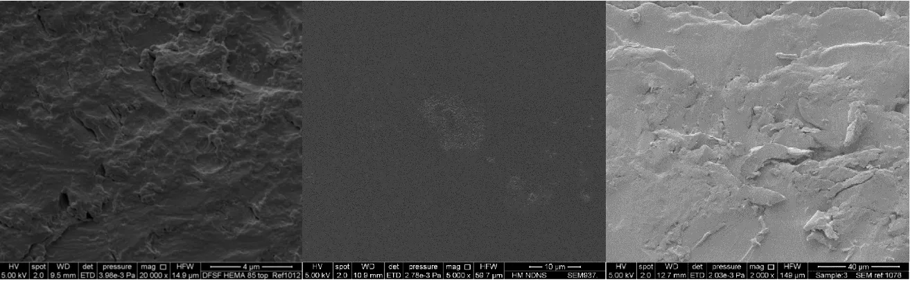
aesthetically acceptable as topical drug carriers. Scanning electron microscopy revealed a
366
generally rough film surface on the side that was exposed to UV light, a much smoother
367
surface where the film had been formed in contact with the support and a fairly dense film
368
cross-section (Figure 3). Film thickness was about 200 µm, with no influence of drug or of
369
nature of the monomer (p>0.05). Inclusion of ethanol significantly (p<0.05) reduced film
370
thickness to about 165 µm, due to the formulations’ lower methacrylate content. FT-IR of
371
the films showed no drug-polymer interactions.
372
373
3.4.2 Drug loading and stability: Drug in the film was in the dissolved state, and its
374
concentration in the film was similar to that in the gel shown in Table 3 (p>0.05). Upon
375
storage of the films for 28 days under accelerated stability conditions, the drug levels
376
remained the same, and the drug remained dissolved for at least two weeks, with a few
377
drug crystals appearing in week 3. There were no differences among films with/without
15
ethanol, with/without drug and containing the different methacrylate-based monomers
379
(Supplementary data 4).
380
381
3.4.3 Glass transition temperatures of films. The films produced from ethanol-free
382
formulations showed two transition temperatures (Table 4). It is thought that the lower Tg
383
value is the polymer’s Tg caused by UV curing of the gel mixture, and that the second Tg is
384
related to a thermal cure which occurred during the DMA measurement. The UV-cure
385
related Tg is influenced by the nature of the methacrylate-based monomer, with EMA-
386
based gel having higher Tg than HEMA-based one, which in turn had higher Tg than
IBOMA-387
based one (p<0.05). Such a difference in gel Tgs could be due to their different viscosities,
388
which were 367 mPas, 598 mPas and 672mPas for EMA- , HEMA- and IBOMA- based gels
389
respectively. The negative correlation between gel viscosity and polymer Tg can be
390
explained by the fact that, in a reaction mixture with lower viscosity, reactants are likely to
391
be more mobile, which would increase the likelihood of polymerisation and of cross-linking
392
and which would in turn increase the resulting polymer’s Tg.
393
Inclusion of ethanol in the formulation had a major influence on the films’ glass transition
394
temperatures, resulting in films with only one Tg, and increasing the UV-cure Tg (p<0.05).
395
The higher Tgs reflect the greater extent of polymerisation (shown by higher degrees of
396
conversion and lower levels of residual monomers in Supplementary Data 3b) in
ethanol-397
containing formulations and could also be a result of these gels’ much lower viscosities.
398
399
3.4.4 In vitro adhesion and water sensitivity of films: In contrast to the in vivo situation
400
where the presence of ethanol reduced the in vivo residence of the films (Figure 1), the
401
three in vitro tests used (Instron pull-off, the cross-cut and water sensitivity) showed no
402
influence of ethanol, or of the nature and concentration of the methacrylate monomer on
403
film adhesion and water sensitivity (p>0.05, Supplementary data 5). It seems that DUDMA
404
(present at a much higher content than the methacrylate monomer in the film) is the main
405
factor responsible for the adhesion and water-resistance properties of the films in vitro.
406
16
3.4.5 In vitro drug release: Drug release profiles (Figures 4-5) of all the UV-cured films
408
(containing the maximum amount of drug that could be loaded in the dissolved state in each
409
film) showed a burst release over the first 24 hours, followed by a slower phase, followed by
410
a plateau. Examination of the films at the end of the release study showed negligible
411
change in film mass over the study duration and no drug precipitation. Fitting of the release
412
data using zero order, first order and Higuchi models showed the best fit to be the Higuchi
413
model (r2 ≥ 0.8), indicating drug release to be diffusion-controlled through the film matrix
414
following the burst release phase.
415
416
For both drugs, release was incomplete over the 30-day experiment as shown when % drug
417
release was plotted against time (Supplementary data 6). The amorolfine-containing films
418
released about 46% of their drug load, while the terbinafine-containing films released about
419
26% of their drug load during the 30-day study. The reason(s) for the greater amorolfine
420
release is as yet unclear. The respective amorolfine- and terbinafine- loaded gels have
421
similar reactants-to-film mass conversion, alkene-to-alkane degree of conversion, film
422
thickness, glass transition temperatures, water sensitivity and dense internal structure. It is
423
possible that terbinafine binds to a greater extent to the polymer film compared to
424
amorolfine, which would hinder its release.
425
426
In addition to the nature of the drug, the nature of the methacrylate-based monomer also
427
has an impact on the extent of drug release, via its influence on drug solvency, and
428
consequently, on drug loading in the film. Films produced using EMA and IBOMA (both
non-429
solvents for the drugs) contained the same drug loadings (3% amorolfine or 4% terbinafine),
430
which was solely due to the presence of the ethanol. In contrast, films produced using
431
HEMA (a solvent for the drugs) could have a certain drug loading in the absence of ethanol
432
(2% of amorolfine or of terbinafine), and the drug loading was considerably increased in the
433
presence of ethanol (to 4% amorolfine or 6% terbinafine). As the release studies were
434
conducted using films containing the maximum drug loading, the drug concentrations in the
435
films varied from 2% to 4% for amorolfine, and from 2% to 6% for terbinafine. For both
436
drugs, the highest release was obtained from the films containing the highest drug
17
concentration, i.e. the film containing HEMA and ethanol (p<0.05, Figures 4-5). Drug
438
concentration in the film is thus an important parameter governing the extent of drug
439
release. Both the nature of the methacrylate-based monomer and the inclusion of ethanol
440
influenced drug release, via their influence on drug concentration in the film.
441
442
When drug release from the control Curanail lacquer film was examined , it was found that
443
like the UV-cured films, the Curanail lacquer film containing amorolfine also showed a burst
444
of drug release (Supplementary data 6a). In contrast to the UV-cured films however, the
445
Curanail polymer film released most of its drug load, possibly due to the fact that the film
446
disintegrated during the 30-day study.
447
448
3.4.6 In vitro ungual drug permeation: The ungual drug permeation profiles and the
449
permeation parameters are shown in Figures 6-7 and Table 5. Mass balance calculations are
450
shown in Supplementary Data 7. The fairly lengthy lag times in Table 5 are typical of ungual
451
permeation (McAuley et al., 2016) and are due to the poor permeability of the nailplate. The
452
latter is also evidenced by the very small percentage (maximum of 6%) of applied drug
453
which permeated into and through the nail (Supplementary Data 7).
454
455
Examination of the ungual permeation profiles shows significant influence of the drug
456
nature and concentration, with amorolfine and the higher drug loadings resulting in greater
457
ungual permeation (p<0.05). Thus, amorolfine-containing formulations showed significantly
458
higher steady-state flux and permeability coefficient (p<0.05) compared to the respective
459
terbinafine-containing ones. This reflects the greater amorolfine released (discussed in
460
section 3.4.5), and thereby its greater availability for ungual permeation. An additional
461
cause for the higher amorolfine permeation could be its lower affinity to nail keratin,
462
compared to that of terbinafine (Tatsumi et al., 2002). Drug-keratin affinity could also
463
explain the similar drug-in-nail concentrations at the end of the permeation experiments
464
(p>0.05; Table 5) despite the lower terbinafine release from the films.
18 466
For both drugs, drug loading in the film was the maximum possible (while keeping the drug
467
dissolved) and was related to the nature of the methacrylate-based monomer, and the
468
presence of ethanol as discussed in section 3.4.5. From Figures 6-7, it can be seen that as
469
drug concentration in the film increased, drug permeation increased. This reflects the
470
greater amounts of drug released at the highest drug loadings (section 3.4.5) and
471
consequently, greater drug availability for ungual permeation. Thus, the choice of the
472
monomer and addition of ethanol had important consequences on ungual drug permeation,
473
with the addition of ethanol to HEMA-based formulations significantly increasing drug
474
loading and flux (p<0.05).
475
476
The ungual permeation of amorolfine from the control Curanail was similar to that from the
477
‘best’ UV-cured film (p>0.05 for the permeation curve, permeability coefficients, diffusion
478
coefficients and drug-in-nail at day 30). Curanail® nail lacquer however showed a slightly
479
greater steady-state flux (p<0.05) probably due to its higher drug-load (32% w/w in the film
480
vs. 4% w/w) and much greater drug release (shown in Supplementary Data 6a). However,
481
the UV-cured film displayed a shorter lag time possibly due to its greater occlusivity
482
compared to the lacquer film (Fig. 9), which could increase nail hydration, which is known to
483
enhance permeation (Gunt and Kasting, 2007; Gunt et al., 2007).
484
485
3.4.7 In vitro anti-fungal efficacy: The in vitro anti-fungal efficacy of the formulations was
486
measured using an anti-fungal whole cell phenotypic assay, that we adapted from the
487
classical disc diffusion method, in our laboratories. In the negative controls (i.e. where a
488
drug-free formulation was cured onto a nail clipping), the T. rubrum culture grew over the
489
entire plate’s surface (Table 6). In contrast, no T. rubrum growth was observed when
drug-490
loaded formulations had been cured on the nail clippings. This shows that sufficient
anti-491
fungal drug was released from the UV-cured film, permeated into the nailplate, and
492
subsequently permeated out of the nail plate and into the agar gel to inhibit fungal growth.
493
The UV-cured gels are thus promising topical carriers for anti-onychomycotic drugs.
19
All the gels tested showed complete inhibition of fungal growth (example shown in Table 6),
495
and no difference could be discerned among the formulations (in Supplementary data 7).
496
This could be related to the fact that the amounts of drug present in the nail and that had
497
permeated through the nail plate were fairly similar for all the formulations (as shown in
498
Figures 6-7). In these anti-fungal assays, the ethanol-free gels with the lowest drug load was
499
as good as the ethanol-containing gels with the highest drug load. It seems that the
500
increased drug loading afforded by the inclusion of ethanol in the gels, which results in
501
increased flux was not necessary for anti-fungal kill in this in vitro set-up. Optimisation of
502
the in vitro anti-fungal methodology is needed and it might enable discrimination among
503
the formulations or confirm anti-fungal equivalence despite different drug loadings, release
504
and ungual permeation.
505
506
3.4.8 In vivo occlusivity of the UV-cured films: The UV-cured films reduced trans-onycheal
507
water loss (TOWL) by about 40% (Figure 8), and were more occlusive than the control
508
Curanail lacquer film. Occlusion is desirable, as a reduction in TOWL has been hypothesised
509
to hyper-hydrate the nail plate (Marty, 1995; Spruit, 1971), which is in turn beneficial for
510
ungual drug permeation (Gunt and Kasting, 2007; Gunt et al., 2007). A second benefit of
511
increased nail plate hydration in the treatment of onychomycosis is that hydration could
512
induce the germination of drug-resistant fungal spores into drug-susceptible hyphae, which
513
would enable fungi eradication (Flagothier et al., 2005). From Figure 8, it can be seen that
514
ethanol or drug or the nature of the methacrylate monomer in the UV-cured films did not
515
have any influence on the films’ occlusivity (p>0.05). This indicates that DUDMA (present in
516
the film at a much higher content than the methacrylate monomer) is the main factor
517
responsible for the occlusivity of the films.
518
519
4. Conclusions 520
Drug-loaded diurethane dimethacrylate-based UV gels, with or without ethanol, and
521
containing one of three different monomers (EMA, IBOMA or HEMA) were produced and
522
characterised to determine the influence of the monomers, ethanol and drugs on the gel
20
and resulting films’ properties, and to explore correlations among the films’ characteristics.
524
The drugs – amorolfine HCl and terbinafine HCl – had negligible influence on the films’
525
properties, except for the extent of their release and ungual flux, with amorolfine release
526
from the film and ungual flux being much higher compared to that of terbinafine. The major
527
influence of ethanol was its ability to increase drug loading, reduce gel viscosity, increase
528
polymerisation and alter the films’ Tg. The greater drug loading enabled by ethanol resulted
529
in higher drug release and ungual flux. However, ethanol inclusion had a negative impact on
530
the film’s in vivo residence, which shows the importance of screening the other
531
methacrylate monomers in order to identify suitable ones. The major difference among the
532
monomers was the fact that HEMA dissolves the drugs to some extent and enabled
533
sufficient drug loading, such that ethanol-free formulations could be developed. All the
534
formulations inhibited anti-fungal growth to the same extent i.e. fully, and differences
535
among the formulations regarding anti-fungal efficacy could not be discerned in the tests
536
used i.e there was no influence of methacrylate monomer nature, ethanol inclusion and
537
drug concentration on anti-fungal activity. The UV gels’ anti-fungal activities and long in
538
vivo residence further show the promise of these formulations as nail medicines.
539
540
Acknowledgments 541
The authors thank the UCL School of Pharmacy and Birkbeck, University of London for
542
funding this work. We are also grateful for the volunteers for donating their nail clippings
543
and for participating in the in vivo studies.
544
References 545
AnacorPharmaceuticals, 2014. Kerydin Highlights of Prescribing Information, US. 546
Arrese, J.E., Pierard, G.E., 2003. Treatment failures and relapses in onychomycosis: A stubborn 547
clinical problem. Dermatology 207, 255-260. 548
Baran, R., Gupta, A.K., Pierard, G.E., 2005. Pharmacotherapy of onychomycosis. Expert Opinion on 549
Pharmacotherapy 6, 609-624. 550
Clemmensen, S., 1984. Cross-Reaction Patterns in Guinea Pigs Sensitised to Acrylic Monomers. 7, 551
527-540. 552
Daniel, C.R., Jellinek, N.J., 2010. Commentary: The illusory tinea unguium cure. J. Am. Acad. 553
Dermatol. 62, 415-417. 554
Flagothier, C., Pierard-Franchimont, C., Pierard, G.E., 2005. New insights into the effect of amorolfine 555
nail lacquer. Mycoses 48, 91-94. 556
21 Goodfield, M.J.D., Evans, E.G.V., 2000. Combined treatment with surgery and short duration oral 558
antifungal therapy in patients with limited dermatophyte toenail infection. Journal of Dermatological 559
Treatment 11, 259-262. 560
Gunt, H.B., Kasting, G.B., 2007. Effect of Hydration on the Permeation of Ketoconazole Through 561
Human Nail Plate In Vitro. European Journal of Pharmaceutical Sciences 32, 254-260. 562
Gunt, H.B., Miller, M.A., Kasting, G.B., 2007. Water Diffusivity in Human Nail Plate. Journal of 563
Pharmaceutical Sciences 96, 3352-3362. 564
Gupta, A.K., Ryder, J.E., Johnson, A.M., 2004. Cumulative meta-analysis of systemic antifungal agents 565
for the treatment of onychomycosis. Br. J. Dermatol. 150, 537-544. 566
KakenPharmaceutical, 2014. Jublia Highlights of Prescribing Information, US. 567
Kerai, L.V., Hilton, S., Murdan, S., 2015. UV-curable gel formulations: Potential drug carriers for the 568
topical treatment of nail diseases. Int. J. Pharm. 492, 177-190. 569
Lubeck, D.P., Patrick, D.L., McNulty, P., Fifer, S.K., Birnbaum, J., 1993. Quality-of-life of persons with 570
onychomycosis. Qual. Life Res. 2, 341-348. 571
Marty, J.-P.L., 1995. Amorolfine Nail Lacquer: A Novel Formulation. Journal of the European 572
Academy of Dermatology and Venereology 4, S17-S21. 573
McAuley, W.J., Jones, S.A., Traynor, M.J., Guesne, S., Murdan, S., Brown, M.B., 2016. An 574
Investigation of how Fungal Infection influences Drug Penetration through Onychomycosis Patient's 575
Nail Plates. European Journal of Pharmaceutics and Biopharmaceutics 102, 178-184. 576
Murdan, S., 2016. Nail disorders in older people, and aspects of their pharmaceutical treatment. Int. 577
J. Pharm., in press. 578
Murdan, S., Kerai, L., Hossin, B., 2015. To what extent do in vitro tests correctly predict the in vivo 579
residence of nail lacquers on the nail plate? Journal of Drug Delivery Science and Technology 25, 23-580
28. 581
Onytec, Package leaflet: Information for the user Onytec 80 mg/g medicated nail lacquer, Ireland, p. 582
Patient Information Leaflet. 583
Rietschel, R.L., Fowler, J.F., Fisher, A.A., 2008. Medical Devices, Implants, and Equipment, in: 584
Rietschel, R.L., Fowler, J.F., Fisher, A.A. (Eds.), Fisher's Contact Dermatitis 6. BC Decker Inc., Ontario, 585
pp. 370-392. 586
Saner, M.V., Kulkarni, A.D., Pardeshi, C.V., 2014. Insights into drug delivery across the nail plate 587
barrier. J. Drug Target. 22, 769-789. 588
Scher, R.K., 1994. ONYCHOMYCOSIS IS MORE THAN A COSMETIC PROBLEM. Br. J. Dermatol. 130, 15-589
15. 590
Schoon, D., 2010. Cosmetic Prostheses as Artificial Nail Enhancements. Blackwell Publishing Ltd., 591
Oxford. 592
Shivakumar, H.N., Juluri, A., Desai, B.G., Murthy, S.N., 2012. Ungual and Transungual drug delivery. 593
Drug Dev. Ind. Pharm. 38, 901-911. 594
Spruit, D., 1971. Measurement of Water Vapor Loss Through Human Nail In Vivo. Journal of 595
Investigative Dermatology 56, 359-361. 596
Tatsumi, Y., Yokoo, M., Senda, H., Kakehi, K., 2002. Therapeutic Efficacy of Topically Applied KP-103 597
against Experimental Tinea Unguium in Guinea Pigs in Comparison with Amorolfine and Terbinafine. 598
Antimicrobial Agents and Chemotherapy 46, 3797-3801. 599
Thomas, J., Jacobson, G.A., Narkowicz, C.K., Peterson, G.M., Burnet, H., Sharpe, C., 2010. Toenail 600
onychomycosis: an important global disease burden. J. Clin. Pharm. Ther. 35, 497-519. 601
Yoshii, E., 1997. Cytotoxic Effects of Acrylates and Methacrylates: Relationships of Monomer 602
Structures and Cytotoxicity. Journal of Biomedical Materials Research 37, 517-524. 603
604
22 Figure legends
606
Figure 1 In vivo residence of UV-cured films (± ethanol and ± drug) on the ten fingernails in
607
six volunteers. Curanail lacquer (containing amorolfine HCl) was used as a commercially
608
available control. Abbreviations: ETOH – ethanol; AH – amorolfine HCl; TH – terbinafine HCl.
609
Means and standard deviations are shown, n=60.
610
Figure 2: Chemical structures and viscosities of the different methacrylate-based monomers
611
Figure 3: Scanning electron micrographs of the top surface (i.e. exposed to UV light), under
612
surface (i.e. in contact with the support), and cross-sectional surface of a drug-free,
ethanol-613
free UV-cured film (DUDMA:HEMA 85:15). Films with the other methacrylate monomers,
614
and ± ethanol and ± drug (amorolfine HCl or terbinafine HCl) had similar micrographs.
615
Figure 4: Cumulative amount of drug release from the amorolfine HCl loaded UV-cured films.
616
Means and standard deviations are shown, n=3. Abbreviations: AH, amorolfine HCl; ETOH,
617
ethanol.
618
Figure 5: Cumulative amount of drug release from the terbinafine HCl loaded UV-cured.
619
Means and standard deviations are shown, n=3. Abbreviations: TH, terbinafine HCl; ETOH,
620
ethanol.
621
Figure 6: Cumulative amount of amorolfine HCl permeated across the nail with time from the UV-cured and Curanail® films, and the % of drug permeated across the nail and remaining in the nail at day 30. Means and standard deviations are shown, n=6. Abbreviations: AH, amorolfine HCl; ETOH, ethanol.
Figure 7: Cumulative amount of terbinafine HCl permeated across the nail with time from the UV-cured films, and the % of drug permeated across the nail and remaining in the nail at day 30. Means and standard deviations are shown, n=6. Abbreviations: TH, terbinafine HCl; ETOH, ethanol.
Table 1: Table shows details of test formulations used for anti-fungal efficacy assessment. In addition, drug-free counterparts of these and Curanail were used as controls.
Nature of monomer Ethanol concentration Drug nature and concentration
EMA 25% Amorolfine HCl; 3%
IBOMA 25% Amorolfine HCl; 3%
HEMA 25% Amorolfine HCl; 4%
HEMA None Amorolfine HCl; 2%
HEMA 25% Terbinafine HCl; 6%
Table 2 Solubilities of amorolfine HCl and terbinafine HCl in the three methacrylate monomers; means and standard deviations are shown, n=3.
Solubility (mg/ml)
EMA IBOMA HEMA
[image:25.842.200.645.116.171.2]Table 3 Theoretical and actual drug-load in UV-curable gel formulations. AH- amorolfine HCl, TH- terbinafine hydrochloride. N=3. Sd=0 for all. The theoretical (based on drug solubility in the gel components) and actual (determined by the absence/presence of drug crystals in the formulation) drug loadings were fairly similar for all the gels except for ethanol-free HEMA-containing gels, where the actual drug-load was much lower than anticipated, due to drug precipitation upon mixing with DUDMA and the photoinitiator.
Solvent Methacrylate-based
monomer
Drug Expected Drug load (% w/v) Actual drug load (% w/v)
Ethanol (25% v/v) EMA AH 3.0 3
TH 3.5 4
IBOMA AH 3.0 3
TH 3.5 4
HEMA AH 5.2 4
TH 6.0 6
None HEMA AH 5.1 2
Table 4: Transition temperatures (Tg) of UV-cured films. Means ± standard deviations are shown, n=3.
Tg values of UV-cured films produced from formulations containing
Excipients DUDMA Solvent
Ethanol
Drug EMA IBOMA HEMA
Formulations
DUDMA 85 % v/v : diluent monomer 15 % v/v
None None 85.7 ± 1.2 and
146.0 ± 2.5
67.2 ± 2.4 and 164.0 ± 0.2
74.4 ± 2.5 and 147.2 ± 0.3
25% v/v
None 112.5 ± 1.8 139.2 ± 9.0 98.1 ± 2.3
AH 109.2 ± 1.1 137.2 ± 0.02 99.2 ± 1.8
TH 109.6 ± 0.8 133.4 ± 3.6 94.7 ± 4.8
None
None 95.1 ± 1.5 NF NF
AH 94.8 ± 2.0 NF NF
TH 95.0 ± 1.8 NF NF
DUDMA 75 % v/v : diluent monomer 25 v/v
None
None NF NF 81.8 ± 0.5 and
142.6 ± 0.4
AH NF NF 92.0 ± 0.9 and
138.9 ± 0.5
TH NF NF 92.4 ± 0.4 and
133.2 ± 0.2 Tg of homopolymers
(⁰C)
Table 5 Ungual permeation parameters. Lag time, steady-state flux, permeability coefficient, diffusion coefficient and amount of drug in nail clippings. Means and standard deviations are shown, n=6. Abbreviations: AH, amorolfine HCl; TH, terbinafine HCl; ETOH, ethanol.
Amorolfine HCl (AH) Terbinafine HCl (TH)
% in
formula-tion (% w/v)
Lag time (day)
Steady-state flux
(µg/cm2/day)
Permeability coefficient
x 10-5
(cm/day)
Diffusion coefficient
x 10-5
(cm2/day)
Drug in nail clipping
(mg/cm3)
% in
formula-tion (% w/v)
Lag time (day)
Steady-state flux
(µg/cm2/day)
Permeability coefficient
x 10-5
(cm/day)
Diffusion coefficient
x 10-5
(cm2/day)
Drug in nail clipping
(mg/cm3)
Curanail® nail lacquer 5 10.4 ± 1.3 2.9 ± 0.2 5.8 ± 0.3 2.5 ± 0.5 0.6 ± 0.2 NA NA NA NA NA NA
DUDMA & EMA gel containing ETOH & AH or TH
3 10.4 ± 0.9 2.4 ± 0.2 7.9 ± 0.7 2.7 ± 0.8 0.3 ± 0.2 4 9.5 ± 1.2 1.5 ± 0.1 3.7 ± 0.3 1.6 ± 0.4 0.9 ± 0.2
DUDMA & IBOMA gel containing ETOH & AH or TH
3 10.5 ± 0.6 2.3 ± 0.3 7.8 ± 1.0 2.2 ± 0.4 0.4 ± 0.1 4 8.3 ± 0.8 1.4 ± 0.3 3.4 ± 0.8 3.1 ± 0.9 0.4 ± 0.1
DUDMA & HEMA gel containing ETOH & AH or TH
4 7.1 ± 1.7 2.2 ± 0.3 5.5 ± 0.7 3.5 ± 1.4 0.6 ± 0.1 6 7.1 ± 1.0 1.6 ± 0.2 2.6 ± 0.4 3.0 ± 0.8 0.7 ± 0.2
DUDMA & HEMA gel containing
AH or TH
Table 6 Photographic images (at day 30) of T. rubrum inoculated SDA plates containing nail clippings with drug-free and drug-loaded formulations cured on the surface. An example, using the ethanol-free HEMA containing formulation is shown. All the other formulations showed similar anti-fungal activity and the images are shown in the Supplementary file.
Formulation applied on nail clipping
surface
T. rubrum inoculated SDA plates containing nail clippings with drug-loaded formulations cured on the surface (n=3) and the corresponding drug-free formulation cured on the surface (C)
T. rubrum inoculated SDA plates containing nail clippings with drug-free formulations cured on
the surface (n=3)
Top surface Under surface Top surface Under surface
None
(nail clipping-free & formulation-free
control)
DUDMA & HEMA
0 2 4 6 8 10 12 14 16 18 20 22 24 26 28 0
20 40 60 80 100
% of
fil
m r
emain
in
g
on
fi
ng
erna
il
in
vi
vo
Time (days)
EMA gel (drug-free, solvent-free)
EMA gel containing ETOH
EMA gel containing ETOH & AH
EMA gel containing ETOH & TH
Curanail nail lacquer
Figure 1 In vivo residence of UV-cured films (± ethanol and ± drug) on the ten fingernails in six volunteers; n=60, means and standard deviations are shown. Curanail lacquer (containing amorolfine HCl) was used as a commercially available control. Abbreviations: ETOH – ethanol; AH – amorolfine HCl; TH – terbinafine HCl.
Ethyl methacrylate, MW 114, Viscosity 0.62 mPas
Isobornyl methacrylate, MW 222, Viscosity 7.40 mPas
Hydroxyethyl methacrylate, MW 130, Viscosity 6.79 mPas
Top surface Under surface Cross-section
0 5 10 15 20 25 30 0
20 40 60 80 100 120 140 160 180 200 220
DUDMA & HEMA gel containing ETOH & 4% w/v AH DUDMA & IBOMA gel containing ETOH & 3% w/v AH DUDMA & HEMA gel 2% w/v AH
DUDMA & EMA gel containing ETOH & 3% w/v AH
Cumula
tiv
e a
moun
t o
f
amorolf
in
e HCl (
AH) relea
se
d
(
g/
cm
3 )
[image:33.842.249.611.75.320.2]Time (days)
0 5 10 15 20 25 30 0
20 40 60 80 100 120 140 160
DUDMA & HEMA gel containing ETOH & 6% w/v TH DUDMA & IBOMA gel containing ETOH & 4% w/v TH DUDMA & HEMA gel containing 2% w/v TH DUDMA & EMA gel containing ETOH & 4% w/v TH
Cumula
tiv
e a
moun
t o
f
te
rbin
af
in
e HCl (
TH)
relea
se
d
(
g/
cm
3 )
[image:34.842.246.619.70.331.2]Time (days)
0 5 10 15 20 25 30 0
5 10 15 20 25 30 35 40 45 50
0 1 2 3 4
% of AH permeated and in nail
% permeated % in nail
Cum
ulat
ive amo
un
t o
f am
orolf
ine HCl (
AH)
permeat
ed across t
he n
ail
(
g/cm
2 )
Time (days)
Curanail naill lacquer (containing 5% w/v AH)
DUDMA & HEMA gel
containing ETOH & 4% w/v AH
DUDMA & IBOMA gel containing ETOH & 3% w/v AH
DUDMA & EMA gel containing ETOH & 3% w/v AH
[image:35.842.146.705.85.323.2]DUDMA & HEMA gel containing 2% w/v AH
Figure 6 Cumulative amount of amorolfine HCl permeated across the nail with time from the UV-cured and Curanail® films, and the % of amorolfine HCl permeated across the nail and remaining in the nail at day 30. Means and standard deviations are shown, n=6. Abbreviations: AH, amorolfine HCl; ETOH, ethanol.
The permeation profiles for the gel containing the highest drug load (4% amorolfine HCl) was significantly different (p<0.05) to those
0 5 10 15 20 25 30 0
5 10 15 20 25 30 35
Cum
ulat
ive amo
un
t o
f t
erbinaf
ine HCl (
TH)
permeat
ed across t
he n
ail
(
g/cm
2 )
Time (days)
DUDMA & HEMA gel
containing ETOH & 6% w/v TH
DUDMA & IBOMA gel containing ETOH & 4% w/v TH
DUDMA & EMA gel containg ETOH & 4% w/v TH
DUDMA & HEMA gel containing 2% w/v TH
0.0 0.5 1.0 1.5 2.0 2.5 3.0 3.5 4.0
% permeated % in nail
[image:36.842.138.706.86.334.2]% of TH permeated and in nail
Figure 7 Cumulative amount of terbinafine HCl permeated across the nail with time from the UV-cured films, and the % of terbinafine HCl permeated across the nail and remaining in the nail at day 30. Means and standard deviations are shown, n=6. Abbreviations: TH, terbinafine HCl; ETOH, ethanol.
The permeation profiles for the gel containing the highest drug load (6% terbinafine HCl) was significantly different (p<0.05) to those
0 10 20 30 40 50 60
Reduction in TOWL %
Curanail nail lacquer
k
DUDMA & EMA gel containing ETOH & 3% w/v AH
k
DUDMA & EMA gel containing ETOH & 4% w/v TH
k
DUDMA & EMA gel containing ETOH (drug-free)
k
DUDMA & EMA gel (drug-free, solvent-free)
k
DUDMA & IBOMA gel (drug-free, solvent-free)
k
DUDMA & HEMA gel (drug-free, solvent-free)
Fo
rm
ul
at
io
[image:37.842.235.592.77.246.2]n
Figure 8: Reduction of TOWL by the different UV-cured films and the Curanail lacquer film control. Means and standard deviations are shown. n=5
Supplementary Material