Copyright © 2001, American Society for Microbiology. All Rights Reserved.
Nosocomial Fungemia Due to
Exophiala jeanselmei
var.
jeanselmei
and
a
Rhinocladiella
Species: Newly Described Causes
of Bloodstream Infection
MARCIO NUCCI,1* TIYOMI AKITI,1GLORIA BARREIROS,1FERNANDA SILVEIRA,1
SANJAY G. REVANKAR,2† DEANNA A. SUTTON,3ANDTHOMAS F. PATTERSON2,4
Mycology Laboratory, Hospital Universita´rio Clementino Fraga Filho, Universidade Federal do Rio de Janeiro, Brazil,1
and Departments of Medicine2and Pathology,3The University of Texas Health Science Center at San Antonio,
and Audie L. Murphy Division, South Texas Veterans Health Care System,4San Antonio, Texas
Received 25 August 2000/Returned for modification 10 October 2000/Accepted 16 November 2000
Fungi have become increasingly important causes of nosocomial bloodstream infections. The major cause of nosocomial fungemia has beenCandida spp, but increasingly molds and other yeasts have caused disease.
Exophiala jeanselmeiand members of the genus Rhinocladiella are dematiaceous moulds, which have been
infrequently associated with systemic infection and have not been described as causes of fungemia. In this paper, the occurrence of 23 cases of fungemia due to these organisms over a 10-month period is reported and the clinical characteristics of patients and outcomes are described. The majority of patients were immuno-suppressed; 21 of 23 (91%) had received blood products and 78% had a central venous catheter. All patients had at least one manifestation of fever, but only one patient had signs or symptoms suggesting deep-seated infection. Antifungal therapy was given to 19 of the 23 patients; of those who did not receive therapy, 3 died prior to the culture result and 1 had been discharged without therapy. Antifungal susceptibility of the organ-isms showed activity of amphotericin B, itraconazole, and the new triazole antifungals voriconazole and posa-conazole. E. jeanselmei and Rhinocladiella species are potential causes of nosocomial fungemia and may be associated with systemic infection.
Systemic fungal infections are increasingly frequent in hos-pitalized patients (4). WhereasCandidaspecies account for the majority of fungal infections, the spectrum of fungi that may cause infection is growing (2).Exophiala jeanselmei and
Rhi-nocladiellaspecies are dematiaceous fungi widely distributed in
the environment, especially in soil, wood, polluted water, and sewage (7, 17). The clinical spectrum of infection caused by these organisms include mycetomas, chromoblastomycosis, and pheohyphomycosis, either superficial, cutaneous, subcuta-neous, or systemic (10, 25). Deep-seated or systemic infections due to E. jeanselmeiorRhinocladiellaare rare, with case re-ports of infection in the lungs (14, 26), brain (9, 30), perito-neum (1, 12, 22), and esophagus (6, 27). In addition, there is a single case of possible hematogenous dissemination ofE.
jean-selmei in a patient who developed endocarditis and arthritis
(24). However, there have been no reports of fungemia due to these fungi. In this paper we report 23 cases of fungemia due
to E. jeanselmei alone, E jeanselmei in combination with a
Rhinocladiellaspecies, or aRhinocladiellaspecies alone.
MATERIALS AND METHODS
The University Hospital of the Universidade Federal do Rio de Janeiro is a tertiary-care hospital with 540 beds, including a 6-bed bone marrow transplant unit, a 20-bed intensive care unit, and a 6-bed semi-intensive postoperative unit. Laboratory records were reviewed to identify patients with positive blood
cul-tures from December 1996 through October 1997. In December 1996,E. jean-selmeiwas isolated from blood cultures of two patients. During 1997, 21 other patients had positive blood cultures for eitherE. jeanselmeior aRhinocladiella
species.
We reviewed the medical records of these 23 patients to determine the clinical characteristics and the outcome of this infection. Fungemia due toE. jeanselmei
or aRhinocladiellaspecies was defined as the isolation of these fungi from at least one blood culture taken from a peripheral vein or a central venous catheter. Blood specimens were inoculated in bottles containing brain-heart infusion medium. The bottles were incubated at 37°C and examined daily for the first week and once a week until discharge. Blind subcultures were performed on the second day of incubation.E. jeanselmeiwas first identified as the growth of black colonies of yeasts from the subculture plate. The colonies were then isolated, plated onto Sabouraud dextrose agar, and incubated at room temperature. Spe-cies identification ofE. jeanselmeiwas based on macroscopic, microscopic, and physiologic characteristics. All 23 isolates were initially identified asE. jeanselmei
and sent to a reference laboratory for confirmation. Identification of all isolates was confirmed at the Fungus Testing Laboratory at the University of Texas Health Science Center at San Antonio, Tex. Isolates for identification were subcultured onto potato flakes agar (PFA) slants, a PFA plate, and a PFA slide cultures (prepared in-house) (23). Colonies on PFA at 25°C were black and initially moist to mucoid with a yeast-like appearance. Microscopically, these young colonies consisted predominantly of the annellated black yeast synana-morph characteristic of severalExophialaspecies. After 2 weeks of incubation, the colonies were greater than 10 mm in diameter and were olivaceous black and velvety. The microscopic morphology examined by slide culture revealed medi-um-length annellophores, as well as annellides that were both terminary and intercalary (borne on short conidiogenous loci between septa). Annelloconidia accumulated in balls near the apex of the annellides and measured 2 to 3 by 4 to 8m. Temperature studies revealed no growth at 40°C, and nitrate was assim-ilated (20). On the basis of the above characteristics, most isolates were con-firmed to beE. jeanselmeivar.jeanselmei(29).E. jeanselmeivar.lecanii-corniis differentiated fromE. jeanselmeivar.jeanselmeiby having conidia being formed predominantly from intercalary conidiogenous loci and by forming a distinct cluster in an ITS1 phylogenetic tree (30). The other isolates identified as Rhi-nocladiellaspecies were similar to those ofE. jeanselmeivar.jeanselmei, both macroscopically and physiologically, but they differed microscopically. The Col-onies were initially black, mucoid, and yeast-like, displayed a black yeast synana-* Corresponding author. Mailing address: Hospital Universita´rio
Clementino Fraga Filho, Universidade Federal do Rio de Janeiro, Av. Brigadeiro Trompovsky s/n 21941-590, Rio de Janeiro, Brazil. Phone and Fax: 5521-5622460. E-mail: mnucci@hucff.ufrj.br.
† Present address: The University of Texas Southwestern Medical School, Dallas, Tex.
514
on May 15, 2020 by guest
http://jcm.asm.org/
morph, assimilated nitrate, and failed to grow at 40°C. Significant differences, however, were noted in the microscopic morphology of the filamentous forms for these two species. While both species contain intercalary conidiogenous loci (conidia formed from very short openings on the hyphae), the genera differ by the formation of balls of conidia at the apices of annellides inExophialaand conidia borne on closely packed denticles inRhinocladiella. All isolates identified asRhinocladiellaproduced their conidia on crowded denticles, a feature not seen inExophialaspecies.Exophiala (Wangiella) dermatitidis, another species display-ing a black yeast synanamorph, is differentiated from the above by faildisplay-ing to assimilate nitrate and by having the ability to grow at 40°C.
Additionally, broth macrodilution MICs and MLCs of amphotericin B, itra-conazole, voriitra-conazole, and posaconazole (SCH59562) were obtained for nine of the clinical isolates ofE. jeanselmeivar.jeanselmeifollowing National Committee for Clinical Laboratory Standards (NCCLS) procedures (16). Testing was per-formed by the Fungus Testing Laboratory, University of Texas Health Science Center at San Antonio.
RESULTS
Epidemiology.Between December 1996 and October 1997, 23 cases of fungemia due toE. jeanselmeior aRhinocladiella
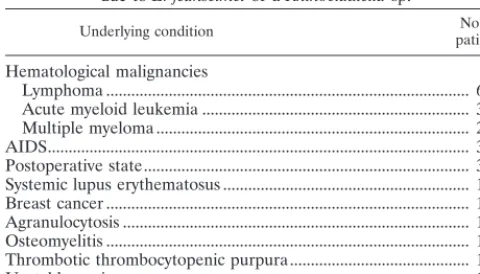
species were diagnosed. The median age of the patients was 50 years, with a range between 8 and 76 years. There were 11 males and 12 females. Table 1 shows the underlying conditions of the patients. Cancer was the underlying disease in 12 pa-tients (52%) and included 11 hematological malignancies and 1 case of breast cancer. An immunodeficiency was present in the other six patients: AIDS in three, agranulocytosis in one, and systemic lupus erythematosus and thrombotic thrombocy-topenic purpura (both in patients receiving corticosteroids) in one each. Three patients were in the postoperative period (cardiac revascularization, Fournier syndrome, and gastric sur-gery), one had osteomyelitis, and one had unstable angina.
As shown in Table 2, 21 of the 23 patients (91%) had re-ceived a blood transfusion, either red blood cells (11 patients), red blood cells plus platelets (7 patients), red blood cells plus plasma (2 patients), or red blood cells plus platelets plus plasma (1 patient). In 78% of the patients a central venous catheter was in place, and 61% had received broad-spectrum antibiotics. Neutropenia was present in 13 (56%) of the pa-tients, and 8 patients had undergone an autologous bone mar-row transplantation. The positive blood cultures had been taken from peripheral blood in 14 patients, peripheral blood plus the central catheter in 3 patients, and the central catheter in only 6 patients. In addition, in one patient with positive blood cultures taken from a peripheral vein,E. jeanselmeigrew
from the bag of peripheral blood stem cells collected for trans-plantation. The median number of positive blood cultures was 1 (range 1 to 6; mean, 1.7).
Clinical manifestations.All patients presented with at least one manifestation of infection at the time a positive culture was drawn. Fever was the most frequent clinical manifestation of the fungemia, occurring in all but one patient, who pre-sented with hypotension. This manifestation also occurred in five other cases. Only one patient presented signs suggestive of a deep-seated infection. The patient had the first positive blood culture forE. jeanselmeiduring a period of neutropenia due to the administration of chemotherapy for the treatment of a relapsing large-cell non-Hodgkin’s lymphoma. She had a Hickman catheter in place, and since the only positive blood cultures had been collected from the catheter and the patient had no complaints, the device was removed and no antifungal treatment was given. Two weeks later she was admitted for an autologous peripheral blood stem cell transplantation. There was no sign of infection, and the chemotherapy was started. After 3 days of neutropenia, she developed fever and empirical antibiotic therapy was started. The blood cultures taken from a new Hickman catheter, as well as from peripheral blood, grew
E. jeanselmei. The patient had positive blood cultures for 22
days, despite catheter removal and the use of amphotericin B (1 mg/kg daily). She subsequently developed thoracic pain, dry cough, and dyspnea. A chest radiograph showed nodular lung opacities. The patient developed respiratory failure and died after having received 975 mg of amphotericin B. Autopsy was not performed.
Species identification.The isolates were identified asE.
jean-selmeivar.jeanselmeiin 19 patients,E. jeanselmeivar.
jeansel-mei plus a Rhinocladiellaspecies in 1 patient, and
Rhinocla-diellaspecies in 3 patients.
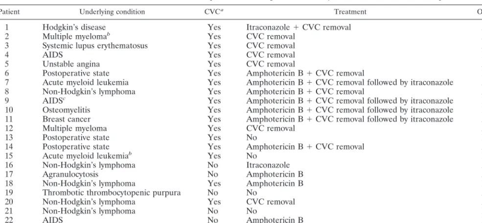
[image:2.612.53.293.90.227.2]Therapy and outcome. Table 3 shows the treatment and outcome of the 23 patients. Four patients did not receive any treatment: three patients died before the blood culture become positive, and one patient was discharged before the blood culture become positive. This patient was admitted for the treatment of thrombotic thrombocytopenic purpura. She had no central venous catheter in place, and the blood culture had been taken because of one spike of fever. Since no new fever developed and the patient was well, she was discharged. Fol-low-up evaluation up to 6 months after discharge did not show any abnormality. Among the 18 patients with a central venous catheter in place, the device was removed in 15. This was the sole treatment in seven patients. Amphotericin B was given to
TABLE 2. Coexisting exposures of 23 patients with fungemia due toE. jeanselmeior aRhinocladiellasp.
Coexisting exposure No. (%) ofpatientsa
Blood transfusion... 21 (91) Central venous catheter ... 18 (78) Antibiotic use ... 14 (61) Neutropenia ... 13 (56) Chemotherapy ... 12 (52) Bone marrow transplantation... 8 (35) Foley catheter... 5 (22) Total parenteral nutrition ... 4 (17)
[image:2.612.314.551.617.717.2]aA total of 23 patients were involved.
TABLE 1. Underlying conditions in 23 patients with fungemia due toE. jeanselmeior aRhinocladiellasp.
Underlying condition patientsNo. of
Hematological malignancies
Lymphoma ... 6
Acute myeloid leukemia ... 3
Multiple myeloma ... 2
AIDS... 3
Postoperative state... 3
Systemic lupus erythematosus ... 1
Breast cancer ... 1
Agranulocytosis ... 1
Osteomyelitis ... 1
Thrombotic thrombocytopenic purpura... 1
Unstable angina ... 1
on May 15, 2020 by guest
http://jcm.asm.org/
10 patients, with a median duration of 3 days (range, 3 to 15 days). The daily dose of amphotericin B varied between 0.5 and 1 mg/kg. Itraconazole was given to six patients; in four of these the azole was given after some days of amphotericin B use, and in two it was given alone.
Of the 23 patients, 9 (39%) died. The underlying diseases were AIDS (3 patients), postoperative period (2 patients), non-Hodgkin’s lymphoma (2 patients), acute myeloid leuke-mia (1 patient), and osteomyelitis (1 patient). The median time between the positive blood culture and death was 25 days (range, 2 to 95 days). In eight of the nine patients who died, the death was attributed to the underlying condition or other com-plications rather than the fungemia. The patient whose death was attributed to the fungemia was the one who developed nodular opacities in the lungs (see above). No autopsy was performed.
Antifungal susceptibility. Antifungal susceptibility results for nine of the clinical isolates ofE. jeanselmeiare shown in Table 4. The MICs of each agent tested for all isolates were very similar to each other, and the MLCs were not increased. Significant antifungal activity was demonstrated against all strains tested with amphotericin B as well as itraconazole and the newer triazole antifungals voriconazole and posaconazole.
DISCUSSION
Fungi have increased in importance as nosocomial patho-gens in the last decade, and the most dramatic increases have occurred in the rates of fungemia (3). WhileCandidaspecies account for the vast majority of cases, fungemia due to other fungi has been increasingly reported. This study extends the list of dematiaceous genera known to incite fungemia. To our knowledge, fungemia due to E. jeanselmei or Rhinocladiella
species has not been previously reported.
[image:3.612.58.551.83.311.2]Infections due to dematiaceous fungi are usually restricted to the skin and soft tissues, but dissemination may occur (19). Catheter-associated fungemia due toE. (W.) dermatitidishas, however, been occasionally reported (11, 15, 28). In the present series, fungemia occurred in association with a wide range of underlying conditions, the majority of them in pa-tients with some degree of immunodeficiency either due to the underlying disease itself such as cancer, agranulocytosis, and AIDS or due to the treatment (use of steroids). In addition, many of the coexisting exposures usually associated with nos-ocomial fungemia were present: postoperative state, central venous catheter, use of broad-spectrum antibiotics, and neu-tropenia. Infections were also associated with blood product transfusion in all but one of the patients, suggesting a potential role of contaminated transfusions in acquisition of infection (21). This outbreak appeared to be related to contaminated TABLE 3. Treatment and outcome of 23 patients with fungemia due toE. jeanselmeior aRhinocladiellasp.
Patient Underlying condition CVCa Treatment Outcome
1 Hodgkin’s disease Yes Itraconazole⫹CVC removal Alive
2 Multiple myelomab Yes CVC removal Alive
3 Systemic lupus erythematosus Yes CVC removal Alive
4 AIDS Yes CVC removal Dead
5 Unstable angina Yes CVC removal Alive
6 Postoperative state Yes Amphotericin B⫹CVC removal Dead
7 Acute myeloid leukemia Yes Amphotericin B⫹CVC removal followed by itraconazole Alive
8 Non-Hodgkin’s lymphoma Yes Amphotericin B⫹CVC removal Alive
9 AIDSc Yes Amphotericin B⫹CVC removal followed by itraconazole Dead
10 Osteomyelitis Yes Amphotericin B⫹CVC removal followed by itraconazole Dead 11 Breast cancer Yes Amphotericin B⫹CVC removal followed by itraconazole Alive
12 Multiple myeloma Yes CVC removal Alive
13 Postoperative state Yes No Dead
14 Postoperative state Yes Amphotericin B⫹CVC removal Alive
15 Acute myeloid leukemiab Yes No Dead
16 Non-Hodgkin’s lymphoma No Itraconazole Alive
17 Agranulocytosis No Amphotericin B Alive
18 Non-Hodgkin’s lymphoma Yes Amphotericin B Dead
19 Thrombotic thrombocytopenic purpura No No Alive
20 Non-Hodgkin’s lymphoma Yes CVC removal Alive
21 Non-Hodgkin’s lymphoma No No Dead
22 AIDS No Amphotericin B Dead
23 Hodgkin’s diseaseb Yes Catheter removal Alive
aCVC, central venous catheter.
[image:3.612.312.552.588.728.2]bIsolate identified in reference laboratory as aRhinocladiellasp. cMixed infection withE. jeanselmeiand aRhinocladiellasp.
TABLE 4. Antifungal susceptibility ofE. jeanselmeivar.jeanselmei isolates performed by NCCLS macrobroth testing
Isolate
NCCLS macrobroth MIC of:
Ampho-tericin B Itraconazole Voriconazole Posaconazole
24 h 48 h 24 h 48 h 24 h 48 h 24 h 48 h
R-2976 0.5 0.5 0.125 1 1 1 0.25 0.5
R-2977 0.5 0.5 0.125 1 1 1 0.5 0.5
R-2978 0.5 1 0.25 2 1 1 0.5 0.5
R-2979 0.5 0.5 0.125 1 0.5 1 0.25 0.5
R-2980 0.25 1 0.125 0.5 1 1 0.25 0.5
R-2981 0.5 0.5 0.25 1 0.5 0.5 0.25 0.25
R-2991 0.5 1 0.125 0.25 1 1 0.25 0.5
R-2995 0.5 1 0.25 0.25 0.5 0.5 0.125 0.25 R-2996 0.5 1 0.125 0.25 0.5 0.5 0.125 0.25
on May 15, 2020 by guest
http://jcm.asm.org/
deionized water from the hospital pharmacy. The water was used in the preparation of antiseptic solutions, and when the procedure for preparing these solutions was changed, no new cases occurred (M. Nucci, F. Silveira, T. Akiti, G. Barreiros, S. G. Revankar, B. L. Wickes, D. A. Sutton, and T. F. Patter-son, Program Abstr. 40th Intersci. Conf. Antimicrob. Agents Chemother., abstr. 1174, p. 417, 2000). In all but six patients the positive cultures had been taken from peripheral blood. In the other six patients the blood cultures had been obtained from a central venous catheter, all of which were surgically implanted catheters in patients with cancer. Four of these patients had undergone autologous bone marrow transplanta-tion, and two were in remission-induction for acute myeloid leukemia. At the time the blood cultures were drawn, all six patients were neutropenic and febrile.
The issue of considering whether fungemia represents a true-positive result when the fungus was isolated from a central catheter remains a matter of controversy. In a large series of catheter-associated fungemia in patients with cancer, neither the death rate nor the rate of disseminated infection was dif-ferent whether the source of blood was the catheter or a peripheral vein (13).
E. jeanselmeiandRhinocladiella, like other black molds, are
considered fungi of low virulence, since they can persist in skin tissue of normal hosts for months to years without disseminat-ing to other organs (19). Accorddisseminat-ingly, in the present series, only one patient seemed to have a disseminated disease. The patient died due to multiple-organ failure but had pulmonary infiltrates on the radiograph. Although there were no nodules with cavitation and the computed tomograph was not obtained to see if there was the halo sign, the clinical picture was similar to cases of infections caused by angioinvasive fungi, with signs of pulmonary infarction (5). Whether the pulmonary signs were due to the fungus is not known, since autopsy was not performed. In the literature there are a few cases of systemic infection due toE. jeanselmeiorRhinocladiellaspp. Roncoroni et al. (24) reported a case of systemic infection that appears to have been disseminated via the hematogenous route. The pa-tient had undergone cardiac surgery for the correction of a ventricular septal defect and developed endocarditis. A single case of pneumonia was reported in a diabetic patient who developed a masslike infiltrate in the lower lobe with a pro-tracted clinical course that evolved to hemoptysis (14). An-other patient had a bronchopulmonary sequestration compli-cated by infection due toE. jeanselmei(26). Since there was no tissue invasion, the fungus was considered to be an opportunist analogous toAspergillusspecies, which colonize previous lung cavities. If our patient had a pulmonary infection due toE.
jeanselmei, the infection probably occurred by the
hematoge-nous route since the patient had multiple positive blood cul-tures over many days.
While most of the patients received antifungal therapy, treatment regimens were very heterogeneous and were influ-enced by the clinical status of the patients at the time of the diagnosis. In general, the infection seemed to be mild, con-firming the impression that this fungus has a low virulence. In patients with fungemia and a central venous catheter, removal of the device is associated with a better outcome (13, 18). Therefore, as a rule, in all patients with a catheter in place at the time of the diagnosis, the physicians attempted to remove
the device, and the only three patients who did not have their catheters removed died before the diagnosis of the fungemia. Some patients had their catheters removed because of persis-tent fever before the positive blood culture. At the time of the diagnosis of the fungemia, they were afebrile and received either no further treatment or itraconazole. Patients who were neutropenic at the time of the diagnosis received amphotericin B and their catheters were removed if possible. In addition, five patients received itraconazole; in four of them this fol-lowed a short course of amphotericin B. Given the heteroge-neity of the treatment, it is difficult do draw conclusions about this issue.
Regarding the identification of two distinct fungi, we do realize the pleomorphic nature of these closely related genera, the fact that Rhinocladiella species also have black yeast synanamorphs similar to those forExophialaspecies, and the difficulties that can be encountered when identifying isolates (either phenotypically or by molecular studies). The cases pre-sented here, however, as examined by our methods, appear to have been caused by two distinct genera of dematiaceous moulds.
Antifungal susceptibility testing of E. jeanselmei isolates demonstrated uniform susceptibility for each of the agents tested. This organism demonstrated susceptibility to itracon-azole and the newer triitracon-azole antifungals voriconitracon-azole and posaconazole, suggesting a possible role for these agents in treating clinical disease. Although the death rate was 39%, in only one patient was the death possibly attributed to the fun-gemia. Since this impression was based on data collected from a careful review of the clinical charts and since no autopsy was performed, we cannot rule out the possibility that the fungus caused the deaths.
In summary, 23 cases of fungemia due toE. jeanselmeior
Rhinocladiellaspp. were identified. These cases were
associ-ated with signs and symptoms associassoci-ated with infection, and the majority of patients responded to removal of a central venous catheter and administration of antifungal therapy. One patient died of apparent disseminated infection. This study demonstrates the potential of these organisms to cause funge-mia in a nosocofunge-mial setting, which can be associated with systemic infection.
ACKNOWLEDGMENT
This work was supported by CNPq (Conselho Nacional de Pesquisa) Brazil (grant 300235/93-3).
REFERENCES
1.Agarwal, S., N. L. Goodman, and H. H. Malluche.1993. Peritonitis due to
Exophiala jeanselmeiin a patient undergoing continuous ambulatory perito-neal dialysis. Am. J. Kidney Dis.21:673–675.
2.Anaissie, E. J., G. P. Bodey, and M. G. Rinaldi.1989. Emerging fungal pathogens. Eur. J. Clin. Microbiol. Infect. Dis.8:323–330.
3.Beck-Sague´, C. M., W. R. Jarvis, and the National Nosocomial Infections Surveillance System.1993. Secular trends in the epidemiology of nosocomial fungal infections in the United States, 1980–1990. J. Infect. Dis.167:1247– 1251.
4.Bodey, G. P.1988. The emergence of fungi as major hospital pathogens. J. Hosp. Infect.11:411–426.
5.Caillot, D., O. Casasnovas, A. Bernard, J. F. Couaillier, C. Durand, B. Cuisenier, E. Solary, F. Pieard, T. Petrella, A. Bonnin, G. Couillault, M. Dumas, and H. Guy.1997. Improved management of invasive pulmonary aspergillosis in neutropenic patients using early thoracic computed tomo-graphic scan and surgery. J. Clin. Oncol.15:139–147.
6.Cappell, M. S., and B. P. Armenian.1991. Esophagitis fromCandidaor
Exophiala? Ann. Intern. Med.115:69.
on May 15, 2020 by guest
http://jcm.asm.org/
7.Cooke, W. B.1957. Checklist of fungi isolated from polluted water and sewage. Sydowia1:146–175.
8.De Hoog, G. S., and J. Guarro.1995. Atlas of clinical fungi. Centraalbureau voor Schimmelcultures, Baarn, The Netherlands.
9.Del Hernanz, A., M. K. Moore, C. K. Campbell, A. Del Palacio-Perez-Medel, and R. Del Castillo-Cantero.1989. Infection of the central nervous system byRhinocladiella atrovirensin a patient with acquired immu-nodeficiency syndrome. J. Med. Vet. Mycol.27:127–130.
10. Fader, RC, and M. R. McGinnis.1988. Infections caused by dematia-ceous fungi: chromoblastomycosis and phaeohyphomycosis. Infect. Dis. Clin. North Am.2:925–938.
11.Kabel, P. J., K. E. Illy, R. A. Holl, A. G. Buiting, and R. G. Wintermans.1994. Nosocomial intravascular infection withExophiala dermatitidis. Lancet344:
1167–1168.
12. Kerr, C. M., J. R. Perfect, P. C. Craven, J. H. Jorgensen, D. J. Drutz, J. D. Shelburne, H. A. Gallis, and R. A. Gutman.1983. Fungal peritonitis in patients on continuous ambulatory peritoneal dialysis. Ann. Intern. Med.
99:334–337.
13. Lecciones, J. A., J. W. Lee, E. E. Navarro, F. G. Witebsky, D. Marshall, S. M. Steinberg, P. A. Pizzo, and T. J. Walsh.1992. Vascular-catheter-associated fungemia in patients with cancer: analysis of 155 episodes. Clin. Infect. Dis.
14:875–883.
14. Manian, F. A., and M. J. Brischetto. 1993. Pulmonary infection due to
Exophiala jeanselmei: successful treatment with ketoconazole. Clin. Infect. Dis.16:445–446.
15. Nachman, S., O. Alpan, R. Malowitz, and E. D. Spitzer.1996. Catheter-associated fungemia due toWangiella(Exophiala)dermatitidis. J. Clin. Mi-crobiol.34:1011–1013.
16. National Committee for Clinical Laboratory Standards.1998. Reference method for broth dilution antifungal susceptibility testing of conidium-form-ing filamentous fungi. Proposed standard M38-P. National Committee for Clinical Laboratory Standards, Wayne, Pa.
17. Nishimura, K., M. Miyaji, H. Taguchi, and R. Tanaka.1987. Fungi in bathwater and sludge of bathroom drainpipes. Mycopathologia97:17–23. 18. Nucci, M., A. L. Colombo, F. Silveira, R. Richtmann, R. Saloma˜o, M. L.
Branchini, and N. Spector.1998. Risk factors for death in patients with candidemia. Infect. Control. Hosp. Epidemiol.19:846–850.
19. Perfect, J. R., and W. A. Schell.1996. The new fungal opportunists are coming. Clin. Infect. Dis.22(Suppl 2):S112–S118.
20. Pincus, D., M. Kemna, and I. Salkin.1988. Modification of potassium nitrate assimilation test for identification of clinically important yeasts. J. Clin. Microbiol.26:1300–1302.
21. Prince, H., S. Page, A. Keating, R. F. Saragosa, N. M. Vukovic, K. R. Imrie, M. Crump, and A. K. Stewart.1995. Microbial contamination of harvested bone marrow and peripheral blood. Bone Marrow Transplant.15:87–91. 22. Remon, C., I. J. de la Calle, F. Vallejo-Carrion, S. Perez-Ramos, and E.
Fernandez Ruiz. 1996. Exophiala jeanselmei peritonitis in a patient on CAPD. Perit. Dial. Int.16:536–538.
23. Rinaldi, M. G.1982. Use of potato flakes agar in clinical mycology. J. Clin. Microbiol.15:1159–1160.
24. Roncoroni, A. J., and J. Smayevsky.1988. Arthritis and endocarditis from
Exophiala jeanselmeiinfection. Ann. Intern. Med.108:773.
25. Rossmann, S. N., P. L. Cernoch, and J. R. Davis.1996. Dematiaceous fungi are an increasing cause of human disease. Clin. Infect. Dis.22:73–80. 26. Samuels, T., I. Morava-Protzner, B. Youngson, and S. N. Huang.1989.
Calcification in bronchopulmonary sequestration. Can. Assoc. Radiol. J.
40:106–107.
27. Sautter, R. E., M. D. Bliss, D. Morrow, and R. E. Lee.1984. Isolation of
Exophiala jeanselmeiassociated with esophageal pathology—three cases, lab-oratory and clinical features. Mycopathologia87:105–109.
28. Simpson, A. J. H., and J. M. D. Nightingale.1995. Intravascular line infec-tion withExophiala dermatitidis. Lancet345:67.
29. Sutton, D. A., A. W. Fothergill, and M. G. Rinaldi.1998. Guide to clinically significant fungi. The Williams & Wilkins Co., Baltimore, Md.
30. Tsai, C. Y., Y. C. Lu, L. T. Wang, T. L. Hsu, and J. L. Seng.1966. Systemic chromoblastomycosis due toHormodendrum dermatitidis(Kano) Conant. Report of the first case in Taiwan. Am. J. Clin. Pathol.46:103–114. 31. Uijthof, J. M. J.1996. Taxonomy and phylogeny of human pathogenic black
yeast genusExophialaCarmichael. Ph.D. thesis. University of Amsterdam, Amsterdam, The Netherlands.