Copyright © 2004, American Society for Microbiology. All Rights Reserved.
Improved Sensitivity of Nucleic Acid Amplification for Rapid
Diagnosis of Tuberculous Meningitis
Isik Somuncu Johansen,
1* Bettina Lundgren,
2Fehmi Tabak,
3Bjo¨rn Petrini,
4Salih Hosoglu,
5Nese Saltoglu,
6and Vibeke Østergaard Thomsen
1International Reference Laboratory of Mycobacteriology, Statens Serum Institut,1and Department of Clinical Microbiology,
Hvidovre University Hospital,2Copenhagen, Denmark; Department of Infectious Diseases and Clinical Microbiology,
Cerrahpasa Medical Faculty, Istanbul University, Istanbul,3Department of Infectious Diseases, Dicle University
Hospital, Diyarbakir,5and Department of Infectious Diseases and Clinical Microbiology,6Faculty
of Medicine, Cukurova University, Adana,6Turkey; and Department of Clinical
Microbiology, Karolinska Institute and Hospital, Stockholm, Sweden4
Received 29 December 2003/Returned for modification 4 March 2004/Accepted 28 March 2004
Early diagnosis of tuberculous meningitis (TBM) is essential for a positive outcome; but present microbi-ological diagnostic techniques are insensitive, slow, or laborious. We evaluated the standard BDProbeTec ET strand displacement amplification method (the standard ProbeTec method) for the detection ofMycobacterium tuberculosiscomplex organisms in parallel with the ProbeTec method with a modified pretreatment procedure with 101 prospectively collected cerebrospinal fluid specimens from 94 patients with suspected TBM. By the modified method, the sample-washing step was omitted. A definitive diagnosis was attained by culture. Thirteen specimens from 12 patients were culture positive for M. tuberculosis complex organisms; three specimens (23%) were microscopy positive for acid-fast bacilli. Among the culture-positive specimens, the standard ProbeTec method was positive for 8 (61.5%) and the modified assay was positive for 10 (76.9%). The overall specificity by both procedures was 98.8% compared to the results of culture. After discrepancy analysis, conducted by reviewing the patients’ previous laboratory data, the specificity increased to 100%. If the cutoff value for respiratory specimens was adjusted from the recommended value of 3,400 to 1,000, the sensitivity of the modified procedure increased to 84.7%, with unchanged specificity. Results were obtained in 3 to 4 h. The new pretreatment procedure with the ProbeTec assay described here provides a rapid, simple, and sensitive tool for the diagnosis of TBM.
The microbiological diagnosis of tuberculous meningitis (TBM) remains a true challenge, mainly because of low counts of mycobacteria in cerebrospinal fluid (CSF) (1). Although the incidence of TBM is low in industrialized countries, it remains an important health threat in connection with human immu-nodeficiency virus infection, immunosuppression, old age, and immigration from countries with a high incidence of tubercu-losis (3, 6, 25, 30). TBM is the most severe manifestation of tuberculosis and is associated with serious sequelae and mor-tality; thus, a rapid diagnosis is of high priority.
Definite diagnosis of TBM is attained by isolation of
Myco-bacterium tuberculosis complex organisms from CSF. Micros-copy is fast and inexpensive, but the sensitivity is merely 0 to 20%; and although culture on broth media has somewhat shortened the delay in diagnosis, it is often 3 to 5 weeks (16, 25, 28).
Biochemical analysis of CSF is used as the initial examina-tion worldwide. Pleocytosis with lymphocytosis, increased pro-tein levels, and reduced glucose levels may support the suspi-cion of TBM; but the diagnosis remains uncertain, as other pathologies may have similar effects on the central nervous system (29). Detection of antibody in CSF is rapid; however, this approach is not standardized and lacks both sensitivity and
specificity (20, 31). Detection of anti-M.bovisBCG-secreting
cells in CSF is sensitive and specific for the early diagnosis of TBM, but the technique is laborious and has not been applied for routine diagnostics (17). Recently, assays that measure the release of gamma interferon in blood have shown promising results compared to those of tuberculin skin testing, but their utilities in a clinical setting remain to be elucidated (8, 18).
In the last decade gene amplification methods have proved to be valuable tools in mycobacteriological diagnostics (32).
Several kit-based methods for the detection ofM. tuberculosis
complex organisms in respiratory specimens are commercially available, and although they are not approved for use with nonrespiratory specimens (for instance, CSF), they are often applied to such specimens (4–6, 16, 21, 22). The BDProbeTec ET Direct Detection assay (ProbeTec assay; Becton Dickin-son, Sparks, Md.) is based on the simultaneous strand
displace-ment amplification ofM. tuberculosiscomplex-specific IS6110
target DNA of 95 bp and real-time detection with fluores-cence-labeled probes. The assay is standardized only for respi-ratory specimens and has recently been evaluated with both respiratory and nonrespiratory specimens, with promising re-sults (2, 12, 19, 24).
The aims of the present study were to evaluate (i) the per-formance of the ProbeTec assay with CSF specimens pro-cessed by two different pretreatment procedures and (ii) the reliability of the standard cutoff value when it is applied to CSF.
* Corresponding author. Mailing address: International Reference Laboratory of Mycobacteriology, Statens Serum Institut, 5 Artillerivej, 2300 Copenhagen S, Denmark. Phone: 4532683703. Fax: 4532683871. E-mail: isj@ssi.dk.
3036
on May 15, 2020 by guest
http://jcm.asm.org/
MATERIALS AND METHODS
The study was carried out at the International Reference Laboratory of My-cobacteriology at Statens Serum Institut, Copenhagen, Denmark, the only mi-crobiological diagnostic laboratory for mycobacterial culture, species identifica-tion, and drug susceptibility testing in Denmark.
Clinical specimens.CSF specimens were received prospectively from Danish hospitals for microscopy, ProbeTec assay, and culture from January 2002 through June 2003. The specimens were included in the study if the ProbeTec assay was requested and if more than 3 ml of CSF was sent. A total of 65 CSF specimens from 58 patients (42 specimens from men and 23 specimens from women; age range, 1 to 76 years) were included. All patients had various men-ingeal symptoms, and biochemical analysis of CSF raised a suspicion of TBM.
Furthermore, 36 CSF specimens from 36 patients were collected prospectively from three different hospitals in Turkey from November 2002 to March 2003. The specimens were collected from 24 men and 12 women (age range, 16 to 90 years). They were admitted to hospital with meningeal symptoms, and TBM was considered in the differential diagnosis. The specimens were stored at⫺20°C prior to analysis in Denmark.
Processing.All specimens were processed by conventional procedures for sterile specimens. For the specimens collected in Denmark, smears were stained with auramine-rhodamine fluorochrome and examined by fluorescence micros-copy at⫻200 magnification. The number of acid-fast bacilli seen in the whole slide was recorded. Five hundred microliters of each specimen was inoculated in BACTEC Mycobacteria Growth Indicator Tube (MGIT) 960 liquid medium (Becton Dickinson), and 150l was inoculated onto Lo¨wenstein-Jensen slants (SSI Diagnostica, Copenhagen, Denmark). Lo¨wenstein-Jensen slants were ex-amined for growth on a weekly basis, whereas the BACTEC MGIT 960 system provided continuous monitoring. Presumed mycobacterial growth was confirmed by Ziehl-Neelsen staining. If no growth was observed after 8 weeks of incubation, the specimen was reported as culture negative. An isolate was identified as a member of theM. tuberculosiscomplex by the Inno-LiPA Mycobacteria assay, as described elsewhere (11). For CSF specimens collected in Turkey, the volumes received varied from 300 to 1,500l per specimen; and the volume of each specimen was equally distributed for the ProbeTec assay, the modified ProbeTec method, and culture with the MGIT 960 system. Smears were stained with Ziehl-Neelsen stain at all three Turkish hospitals. Due to the limited volume of the Turkish specimens, culture on Lo¨wenstein-Jensen slants was omitted.
ProbeTec assay.The standard ProbeTec assay was performed according to the instructions of the manufacturer for respiratory specimens, as described in detail elsewhere (12). The remaining available material (volume range, 300 to 500l) was used for the modified procedure. The only difference between the modified method and the standard method was omission of the washing step.
The results were obtained as a metric other than acceleration (MOTA) value, a measurement of the area under the relative fluorescent unit curve. Samples with MOTA values ⱖ3,400 were positive forM. tuberculosiscomplex DNA, according to the product insert (for respiratory specimens). If the value for the internal amplification control (IAC) was⬎5,000 and the MOTA value for M. tuberculosiscomplex organisms was⬍3,400, the specimen was negative for M. tuberculosiscomplex DNA. If the MOTA value for IAC was⬍5,000 and that for theM. tuberculosiscomplex organisms was⬍3,400, the reaction was inhibited and the result was considered inconclusive.
Precautions taken to avoid contamination.Class 2 biological safety cabinets decontaminated with household bleach, gloves, and aerosol-resistant filter tips were used in all steps in order to avoid contamination with previously recovered amplicons and between specimens. DNA extraction was carried out in biosafety level 3 facilities with negative pressure, and amplification and detection were carried out on another floor.
Statistical analysis.The sensitivity and specificity of the ProbeTec assay for detection ofM. tuberculosiscomplex DNA in CSF were calculated by comparison of the results with those of culture, which served as the “gold standard.” Fur-thermore, we compared the ratios of the MOTA value for the isolate in the specimen to the MOTA value for the IAC obtained by the two procedures.
RESULTS
Altogether, 101 CSF specimens from 94 patients were stud-ied. Thirteen specimens (12.8%; seven Danish and six Turkish
specimens) wereM. tuberculosiscomplex culture positive, and
three of these (23%; one Danish and two Turkish specimens) were positive for acid-fast bacilli by microscopy.
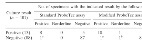
The standard ProbeTec assay was positive forM. tuberculosis
complex DNA for 8 of the 13 culture-positive specimens and negative for the remaining 5 (Table 1). Furthermore, the assay was positive for one culture-negative specimen, which was ob-tained from a patient in whom TBM was verified by culture with another specimen. The sensitivity and specificity were 61.5 and 98.8%, respectively, compared to the results of culture. After the discrepant results were examined by reviewing pa-tients’ previous laboratory data, the specificity increased to 100%.
The modified ProbeTec assay was positive for 10 of the 13
M. tuberculosiscomplex culture-positive specimens; 1 of the 3 culture-negative specimens had a high MOTA value of 1,989. The ProbeTec assay was negative for the remaining two spec-imens. Furthermore, the modified assay was positive for one culture-negative specimen and yielded a nearly positive MOTA value of 3,017 for one specimen. These specimens were from two different patients who had culture-verified TBM, which was determined with specimens collected previously. One of the specimens was taken during the third month of antituberculous treatment (Table 1). The overall sensitivity and specificity of the modified assay were 76.9 and 98.8%, respectively, compared to the results of culture. When the specificity was calculated on the basis of prior culture results as well, it improved to 100%. There was a clear difference in the magnitudes of the MOTA values for positive and true-negative specimens. The MOTA values for true-positive spec-imens obtained by the standard and the modified ProbeTec assays ranged from 3,894 to 80,611 (median, 10,983) and 5,256 to 73,413 (median, 30,470), respectively. The MOTA values for true-negative specimens obtained by the standard and the modified ProbeTec assays ranged from 0 to 400 (median, 6) and 0 to 307 (median, 15), respectively. If the cutoff value for a positive result for CSF was decreased to 1,000, the sensitivity of the modified assay increased to 84.6%, whereas the speci-ficity remained 100%. No differences in the sensitivities and the specificities of the standard procedure would be obtained by a decrease in the cutoff value for a positive result.
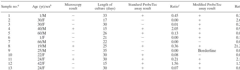
[image:2.603.301.544.89.158.2]The 13 culture-positive CSF specimens were from 12 differ-ent patidiffer-ents (Table 2). Two of the patidiffer-ents died (16.6%). One Danish patient died before the onset of treatment, as the results of both microscopy and the ProbeTec assay by both methods were negative. The culture result became positive 22 days after receipt of the specimen. The other patient who died had been admitted to a Turkish hospital and had received the correct treatment shortly after admission to the hospital. For this patient the results of both microscopy and the ProbeTec
TABLE 1. Results of culture and ProbeTec assays with different lysis procedures for CSF
Culture result (n⫽101)
No. of specimens with the indicated result by the following: Standard ProbeTec assay Modified ProbeTec assay
Positive Borderline Negative Positive Borderline Negative
Positive (13) 8 0 5 10 1 2
Negative (88) 1a 0 87 1a 1b 86 aThe specimen was from a patient from whom another specimen revealed
culture-verified TBM.
bThe specimen was from a patient with culture-verified TBM, as determined
with a previously collected specimen. This specimen was taken during the third month of antituberculous treatment.
on May 15, 2020 by guest
http://jcm.asm.org/
assays by both methods were positive. The ratio of the MOTA values for the specimens to the MOTA values for the IAC for the culture-positive specimens by the conventional ProbeTec assay ranged from 0.00 to 2.05 (median, 0.08), whereas the ratio by the modified procedure ranged from 0.00 to 21.28 (median, 0.37).
For the standard as well as the modified procedures, all negative results were valid, as no inhibition was detected. Re-sults were obtained in 3 to 4 h.
DISCUSSION
Delay or interruption of treatment for TBM were important predictors of death in a study with 434 patients with TBM in a country where the incidence of TBM is high (10). Another study has stressed the importance of rapid initiation of treat-ment, as a 3-day delay in the onset of treatment was a signif-icant indication for mortality (30). The application of rapid and
sensitive laboratory assays for the detection ofM. tuberculosis
complex organisms in CSF is necessary.
We aimed to assess the feasibility and reliability of the stan-dardized strand displacement amplification method with CSF specimens prospectively collected from both countries where the incidence of TBM is low and countries where the incidence of TBM is high. When the standard method was applied di-rectly to CSF, we obtained a 61.5% sensitivity compared to the results of culture. In order to obtain better performance and to diminish the loss of DNA during sample processing, we omit-ted the washing buffer step. This step is intended to remove inhibitors from the specimens. Even though the modified test was generally carried out with a smaller volume of CSF from each sample, we demonstrated that the modified test has a higher sensitivity (76.9%). As the ProbeTec assay is a semi-quantitative assay, the magnitudes of the MOTA scores are not directly indicative of the levels of organisms in the specimens. IAC is designed to validate negative amplification results. Thomsen et al. (27) demonstrated that the ratios of the MOTA
value forM. tuberculosiscomplex/MOTA value for IAC
cor-related better with both the smear and the culture results than
the absolute value for theM. tuberculosiscomplex by a
semi-quantitative PCR method. In the present study the ratios were considerably higher by the modified procedure than by the standard one. Both ProbeTec assay procedures yielded one false-positive result with the same specimen. The specimen was from a patient who was treated for culture-verified TBM, as confirmed with another specimen. These specimens were taken at the same time, which demonstrates the importance of ob-taining multiple samples. Furthermore, the modified assay pro-cedure yielded a high MOTA value for one specimen that was
culture negative for M. tuberculosiscomplex organisms. This
specimen was taken from a patient who was on his third month of antituberculous treatment and who was thus expected to be culture negative. This phenomenon was reported in earlier studies in which amplification methods were used (9, 12, 15, 27).
Since the system is approved for use with respiratory spec-imens, the cutoff value for a positive test result may be too high when the system is applied to certain nonrespiratory speci-mens, such as CSF, which contains unevenly distributed bacilli in much smaller amounts. In an earlier evaluation of the assay with both respiratory and nonrespiratory specimens (12), we found clear differences in the magnitudes of the MOTA values for true-positive and true-negative specimens, in line with the findings of the present study. If the cutoff MOTA value ob-tained by the modified method was decreased from 3,400 to 1,000, the sensitivity of this study would increase to 84.6% and the specificity would remain 100%. The method thus offers advantages for the fast confirmation of TBM, although a neg-ative result does not fully rule out a diagnosis of TBM. In a meta-analysis, Pai and coworkers (21) recently reviewed 49 studies evaluating both commercial and in-house nucleic acid amplification tests for the diagnosis of TBM, and the estimated values for sensitivity and specificity were 56 and 98%, respec-tively, for the 14 studies that used commercial tests.
[image:3.603.42.543.89.236.2]Omission of the washing step did not influence the rate of inhibition. A previous evaluation of the assay (12) found a level of inhibition of merely 0.3%. Certain specimens (i.e., blood, stool, and body fluids) contain inhibitors and thus require an intensive sample-washing step (7, 13, 14, 23, 26). During the
TABLE 2. Results of microscopy, times needed for culture, and results of ProbeTec assay with two different sample preparation procedures for culture-positive CSF
Sample no.a Age (yr)/sexb Microscopy
result culture (days)Length of Standard ProbeTecassay result Ratioc Modified ProbeTecassay result Ratio
1 1/M ⫺ 33 ⫹ 0.45 ⫹ 0.37
2 30/F ⫺ 17 ⫺ 0.00 ⫹ 2.64
3 30/F ⫺ 30 ⫺ 0.01 ⫹ 0.27
4 40/M ⫹ 15 ⫹ 2.05 ⫹ 0.93
5 60/M ⫺ 26 ⫹ 0.13 ⫹ 0.09
6 1/F ⫺ 21 ⫺ 0.00 ⫹ 0.16
7 66/M ⫺ 22 ⫺ 0.00 ⫺ 0.00
8 19/M ⫹ 25 ⫹ 0.36 ⫹ 21.28
9 25/M ⫺ 35 ⫺ 0.00 Borderline 0.02
10 22/F ⫺ 30 ⫹ 0.08 ⫺ 0.00
11 24/F ⫹ 30 ⫹ 0.21 ⫹ 2.16
12 42/F ⫺ 15 ⫹ 1.56 ⫹ 1.59
13 24/F ⫺ 30 ⫹ 0.07 ⫹ 0.80
aOnly two of the patients were born in Denmark; the remaining patients originated from areas where the incidence of TBM is high. Samples 1 to 7 were collected
in Denmark. Samples 2 and 3 were from the same patient.
bM, male; F, female.
cRatio, MOTA value for the specimen/MOTA value for the IAC.
on May 15, 2020 by guest
http://jcm.asm.org/
washing step, some of the bacterial DNA may be washed away. When amplification methods are applied to sources of speci-mens other than the respiratory tract, validation is crucial. We demonstrated that the washing buffer could be omitted when the ProbeTec assay is applied to CSF, and this may also be the case for other body fluids.
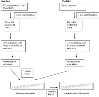
When amplification methods are applied to nonrespiratory specimens, optimal use of the available specimen is necessary. Such specimens are usually paucibacillary, and the numbers of samples that can be collected as well as sample amounts are limited. Thus, in order to improve the performance of the ProbeTec assay, modification of the sample preparation pro-cedure should be considered. Large volumes of the lysis and neutralization buffers are used, but the manufacturer
recom-mends that only 150l of the final 700l of DNA extract be
used for priming and amplification. The remaining 550l of
DNA lysate is not used, and this is difficult to justify when it comes to CSF. By the suggested modified method, the levels of the lysis and neutralization buffers were reduced 50% (Fig. 1). We applied this method in parallel with a standard procedure
to five microscopy-negative, M. tuberculosis complex
culture-positive, nonrespiratory clinical specimens from various sources. The standard ProbeTec assay was negative, but the modified method was positive in all five instances (data not shown). This procedure could not be applied to the CSF spec-imens due to a lack of material.
In conclusion, strand displacement amplification provides fast results in cases in which conventional methods may be insufficient for the diagnosis of TBM. The modified sample preparation step yields promising features and should be ap-plied to CSF samples. Lowering of the cutoff value could be considered. In the present study, lowering of the cutoff value improved the sensitivity without lowering the specificity. Fur-ther modification of the sample preparation procedure may increase the sensitivity even more.
ACKNOWLEDGMENTS
We thank Karin Øhrberg Lund and Birgit Svanholmer for skillful work in the laboratory.
REFERENCES
1. Ahuja, G. K., K. K. Mohan, K. Prasad, and M. Behari.1994. Diagnostic criteria for tuberculous meningitis and their validation. Tuber. Lung Dis. 75:149–152.
2. Bergmann, J. S., W. E. Keating, and G. L. Woods.2000. Clinical evaluation of the BDProbeTec ET system for rapid detection ofMycobacterium tuber-culosis. J. Clin. Microbiol.38:863–865.
3. Bidstrup, C., P. H. Andersen, P. Skinhoj, and A. B. Andersen.2002. Tuber-culous meningitis in a country with a low incidence of tuberculosis: still a serious disease and a diagnostic challenge. Scand. J. Infect. Dis.34:811–814. 4. Bonington, A., J. I. Strang, P. E. Klapper, S. V. Hood, W. Rubombora, M. Penny, R. Willers, and E. G. Wilkins.1998. Use of Roche AMPLICOR Mycobacterium tuberculosisPCR in early diagnosis of tuberculous meningitis. J. Clin. Microbiol.36:1251–1254.
5. Caws, M., S. M. Wilson, C. Clough, and F. Drobniewski.2000. Role of IS6110-targeted PCR, culture, biochemical, clinical, and immunological cri-FIG. 1. Work flows for standard and modified ProbeTec assays.
on May 15, 2020 by guest
http://jcm.asm.org/
[image:4.603.127.463.72.403.2]teria for diagnosis of tuberculous meningitis. J. Clin. Microbiol.38:3150– 3155.
6. Chedore, P., and F. B. Jamieson.2002. Rapid molecular diagnosis of tuber-culous meningitis using the Gen-probe Amplified Mycobacterium Tubercu-losis direct test in a large Canadian public health laboratory. Int. J. Tuberc. Lung Dis.6:913–919.
7. Ehlers, S., R. Ignatius, T. Regnath, and H. Hahn.1996. Diagnosis of ex-trapulmonary tuberculosis by Gen-Probe amplifiedMycobacterium tubercu-losisdirect test. J. Clin. Microbiol.34:2275–2279.
8. Ewer, K., J. Deeks, L. Alvarez, G. Bryant, S. Waller, P. Andersen, P. Monk, and A. Lalvani.2003. Comparison of T-cell-based assay with tuberculin skin test for diagnosis of Mycobacterium tuberculosis infection in a school tuber-culosis outbreak. Lancet361:1168–1173.
9. Hellyer, T. J., T. W. Fletcher, J. H. Bates, W. W. Stead, G. L. Templeton, M. D. Cave, and K. D. Eisenach.1996. Strand displacement amplification and the polymerase chain reaction for monitoring response to treatment in patients with pulmonary tuberculosis. J. Infect. Dis.173:934–941. 10. Hosoglu, S., M. F. Geyik, I. Balik, B. Aygen, S. Erol, T. G. Aygencel, A. Mert,
N. Saltoglu, I. Dokmetas, S. Felek, M. Sunbul, H. Irmak, K. Aydin, O. F. Kokoglu, H. Ucmak, M. Altindis, and M. Loeb.2002. Predictors of outcome in patients with tuberculous meningitis. Int. J. Tuberc. Lung Dis.6:64–70. 11. Johansen, I. S., B. H. Lundgren, J. P. Thyssen, and V. O. Thomsen.2002.
Rapid differentiation between clinically relevant mycobacteria in microscopy positive clinical specimens and mycobacterial isolates by line probe assay. Diagn. Microbiol. Infect. Dis.43:297–302.
12. Johansen, I. S., V. O. Thomsen, A. Johansen, P. Andersen, and B. Lundgren. 2002. Evaluation of a new commercial assay for diagnosis of pulmonary and nonpulmonary tuberculosis. Eur. J. Clin. Microbiol. Infect. Dis.21:455–460. 13. Kirschner, P., J. Rosenau, B. Springer, K. Teschner, K. Feldmann, and E. C. Bottger.1996. Diagnosis of mycobacterial infections by nucleic acid ampli-fication: 18-month prospective study. J. Clin. Microbiol.34:304–312. 14. Kolk, A. H., L. F. Kox, J. van Leeuwen, S. Kuijper, and H. M. Jansen.1998.
Clinical utility of the polymerase chain reaction in the diagnosis of extrapul-monary tuberculosis. Eur. Respir. J.11:1222–1226.
15. Kox, L. F., S. Kuijper, and A. H. Kolk.1995. Early diagnosis of tuberculous meningitis by polymerase chain reaction. Neurology45:2228–2232. 16. Lang, A. M., J. Feris-Iglesias, C. Pena, J. F. Sanchez, L. Stockman, P. Rys,
G. D. Roberts, N. K. Henry, D. H. Persing, and F. R. Cockerill III.1998. Clinical evaluation of the Gen-Probe amplified direct test for detection of Mycobacterium tuberculosiscomplex organisms in cerebrospinal fluid. J. Clin. Microbiol.36:2191–2194.
17. Lu, C. Z., J. Qiao, T. Shen, and H. Link.1990. Early diagnosis of tuberculous meningitis by detection of anti-BCG secreting cells in cerebrospinal fluid. Lancet336:10–13.
18. Mazurek, G. H., and M. E. Villarino.2003. Guidelines for using the Quan-tiFERON-TB test for diagnosing latentMycobacterium tuberculosis infec-tion. Morb. Mortal. Wkly. Rep. Recommun. Rep.52:15–18.
19. Mazzarelli, G., L. Rindi, P. Piccoli, C. Scarparo, C. Garzelli, and E. Tortoli.
2003. Evaluation of the BDProbeTec ET system for direct detection of Mycobacterium tuberculosisin pulmonary and extrapulmonary samples: a multicenter study. J. Clin. Microbiol.41:1779–1782.
20. Miorner, H., U. Sjobring, P. Nayak, and A. Chandramuki.1995. Diagnosis of tuberculous meningitis: a comparative analysis of 3 immunoassays, an im-mune complex assay and the polymerase chain reaction. Tuber. Lung Dis. 76:381–386.
21. Pai, M., L. L. Flores, N. Pai, A. Hubbard, L. W. Riley, and J. M. Colford, Jr. 2003. Diagnostic accuracy of nucleic acid amplification tests for tuberculous meningitis: a systematic review and meta-analysis. Lancet Infect. Dis.3:633– 643.
22. Pfyffer, G. E., P. Kissling, E. M. Jahn, H. M. Welscher, M. Salfinger, and R. Weber.1996. Diagnostic performance of amplifiedMycobacterium tubercu-losisdirect test with cerebrospinal fluid, other nonrespiratory, and respira-tory specimens. J. Clin. Microbiol.34:834–841.
23. Piersimoni, C., A. Callegaro, C. Scarparo, V. Penati, D. Nista, S. Bornigia, C. Lacchini, M. Scagnelli, G. Santini, and G. De Sio.1998. Comparative evaluation of the new Gen-ProbeMycobacterium tuberculosisamplified di-rect test and the semiautomated abbott LCxMycobacterium tuberculosis assay for direct detection ofMycobacterium tuberculosiscomplex in respira-tory and extrapulmonary specimens. J. Clin. Microbiol.36:3601–3604. 24. Piersimoni, C., C. Scarparo, P. Piccoli, A. Rigon, G. Ruggiero, D. Nista, and
S. Bornigia.2002. Performance assessment of two commercial amplification assays for direct detection ofMycobacterium tuberculosiscomplex from res-piratory and extrapulmonary specimens. J. Clin. Microbiol.40:4138–4142. 25. Porkert, M. T., M. Sotir, P. Parrott-Moore, and H. M. Blumberg.1997.
Tuberculous meningitis at a large inner-city medical center. Am. J. Med. Sci. 313:325–331.
26. Reischl, U., N. Lehn, H. Wolf, and L. Naumann.1998. Clinical evaluation of the automated COBAS AMPLICOR MTB assay for testing respiratory and nonrespiratory specimens. J. Clin. Microbiol.36:2853–2860.
27. Thomsen, V. O., A. Kok-Jensen, M. Buser, S. Philippi-Schulz, and H. J. Burkardt.1999. Monitoring treatment of patients with pulmonary tubercu-losis: can PCR be applied? J. Clin. Microbiol.37:3601–3607.
28. Thomson, R. B., Jr., and H. Bertram.2001. Laboratory diagnosis of central nervous system infections. Infect. Dis. Clin. N. Am.15:1047–1071. 29. Thwaites, G. E., T. T. Chau, K. Stepniewska, N. H. Phu, L. V. Chuong, D. X.
Sinh, N. J. White, C. M. Parry, and J. J. Farrar.2002. Diagnosis of adult tuberculous meningitis by use of clinical and laboratory features. Lancet 360:1287–1292.
30. Verdon, R., S. Chevret, J. P. Laissy, and M. Wolff.1996. Tuberculous men-ingitis in adults: review of 48 cases. Clin. Infect. Dis.22:982–988. 31. Watt, G., G. Zaraspe, S. Bautista, and L. W. Laughlin.1988. Rapid diagnosis
of tuberculous meningitis by using an enzyme-linked immunosorbent assay to detect mycobacterial antigen and antibody in cerebrospinal fluid. J. Infect. Dis.158:681–686.
32. Woods, G. L.2001. Molecular techniques in mycobacterial detection. Arch. Pathol. Lab. Med.125:122–126.