Copyright © 2001, American Society for Microbiology. All Rights Reserved.
Identification of
Ehrlichia
spp. and
Borrelia burgdorferi
in
Ixodes
Ticks in the Baltic Regions of Russia
ANDREY N. ALEKSEEV,1HELEN V. DUBININA,1INGRID VAN DE POL,2ANDLEO M. SCHOULS2* Zoological Institute Russian Academy of Sciences, St. Petersburg, Russia,1and Research Laboratory for Infectious
Diseases, National Institute of Public Health and the Environment, Bilthoven, The Netherlands2 Received 13 November 2000/Returned for modification 4 March 2001/Accepted 8 April 2001
The presence and distribution ofEhrlichiaspp. andBorrelia burgdorferisensu lato was demonstrated among
ixodid ticks collected in the Baltic regions of Russia, where Lyme borreliosis is endemic. A total of 3,426Ixodes
ricinus and 1,267 Ixodes persulcatus specimens were collected, and dark-field microscopy showed that 265
(11.5%)I. ricinusand 333 (26.3%)I. persulcatusticks were positive. From these samples, 472 dark-field-positive
and 159 dark-field-negative ticks were subjected to PCR and subsequent reverse line blot hybridization.
Fifty-four ticks (8.6%) carriedEhrlichiaspecies, and 4 (0.6%) carried ehrlichiae belonging to the Ehrlichia
phagocytophila complex, which includes the human granulocytic ehrlichiosis agent. The E. phagocytophila
complex and anEhrlichia-like species were detected only in I. ricinus whereas Ehrlichia muris was found
exclusively inI. persulcatus, indicating a possible vector-specific infection.Borrelia gariniiwas found
predom-inantly inI. persulcatus, butBorrelia afzeliiwas evenly distributed among the two tick species. Only twoI. ricinus
ticks carriedB. burgdorferisensu stricto, whileBorrelia valaisianaand a newly identifiedB. afzelii-like species
were found in 1.7 and 2.5% of all ticks, respectively. Of the dark-field-positive ticks, only 64.8% yielded a BorreliaPCR product, indicating that dark-field microscopy may detect organisms other thanB. burgdorferi sensu lato. These observations show that the agent of human granulocytic ehrlichiosis may be present in ticks in the Baltic regions of Russia and that clinicians should be aware of this agent as a cause of febrile disease.
Borrelia burgdorferisensu lato, the causative agent of Lyme disease, is frequently found in a variety of tick species through-out the world. The widespread distribution has made Lyme borreliosis the most prevalent tick-transmitted zoonotic dis-ease in humans (29). In Europe, five differentB. burgdorferi
sensu lato species are found, of whichBorrelia afzeliiand Bor-relia gariniiare present throughout the continent. In countries of central Europe, such as The Netherlands, Germany, Italy, and France,B. burgdorferisensu stricto andBorrelia valaisiana
are found (28).Borrelia lusitaniae is mainly found in Ixodes ricinus ticks in Portugal but has also been detected in ticks from the Czech Republic, Moldavia, Ukraine, and Belorussia (10). Previous surveys have shown 10 to 30% of theIxodesticks from the St. Petersburg and Kaliningrad regions of Russia carriedB. afzelii and B. garinii, but not B. burgdorferi sensu stricto (1). In a similar study it was shown that the prevalence ofB. burgdorferi sensu lato infection of I. ricinus ticks from nearby Helsinki, Finland, varied from 19 to 55% (7). In addi-tion, various other studies have shown that ticks in different regions of Russia are infected withB. burgdorferisensu lato (8, 11, 26).
Ehrlichioses are known as important tick-borne diseases in animals, particularly in wildlife and domestic ruminants (25). During the last decade, two previously unknownEhrlichia spe-cies have emerged as agents causing considerable public health problems in the United States (2, 5, 32). TheEhrlichiaspecies that was identified in 1986 as causing human monocytic ehr-lichiosis is designatedEhrlichia chaffeensis. Initially it was
as-sumed that this species was transmitted byAmblyomma ameri-canum only, but in a recent study researchers have also identified thisEhrlichiaspecies inIxodes ticks (9). Until now there have been no reports of confirmed cases of human mono-cytic ehrlichiosis outside the United States. More recently, the agent causing human granulocytic ehrlichiosis (HGE) was identified. This species belongs to theEhrlichiagenogroup 2, which includesEhrlichia phagocytophila, an organism isolated from ruminants like sheep, cattle, and deer, andEhrlichia equi, which is found in horses. These organisms, designated theE. phagocytophilacomplex, are closely related and may even rep-resent the same species. There have been several reports that described the presence of the HGE agent in ticks in Europe (3, 12, 20, 27). However, there seems to be a paucity of reports of confirmed cases of HGE in Europe, and most of these cases are found in Slovenia where Lyme borreliosis is highly preva-lent (14, 17, 19, 20, 30).
The aim of this study was to determine the prevalence of
Ehrlichia infection in ticks in a region where diseases like tick-borne encephalitis and Lyme borreliosis are highly preva-lent. For this reason,I. ricinusandIxodes persulcatusticks were collected from the Baltic regions of Russia and screened for the presence ofB. burgdorferisensu lato by dark-field micros-copy, followed by PCR and subsequent reverse line blot hy-bridization to identify the Borrelia and Ehrlichia species present in these ticks.
MATERIALS AND METHODS
Ticks.From 1997 to 1998, 4,693 ticks were collected by flagging in forested areas near Morskaja and Lisy Nos in the vicinity of St. Petersburg and along the Curonian Spit in the Kaliningrad enclave of Russia. Ticks were stored alive and analyzed by dark-field microscopy within hours after collection.
Dark-field examination of ticks.For dark-field inspection the contents of the gut from a dissected tick were ejected into a drop of saline. After being imme-diately covered with a thin cover glass, the slide was inspected under a micro-* Corresponding author. Mailing address: Research Laboratory for
Infectious Diseases, National Institute of Public Health and the Envi-ronment, P.O. Box 1, 3720 BA Bilthoven, The Netherlands. Phone: 31302742121. Fax: 31302744449. E-mail: LM.Schouls@rivm.nl.
2237
on May 15, 2020 by guest
http://jcm.asm.org/
scope for the presence of live spirochetes, with 250 fields viewed. The remainder of the tick was stored in 70% ethanol at 4°C for PCR analysis.
Preparation of DNA extracts from ticks.Ticks were processed as described before (23). Briefly, ticks were taken from the 70% ethanol solution, briefly
dried, and boiled for 20 min in 100l of 0.7 M ammonium hydroxide to free the
DNA. After being cooled, the vial with the lysate was left open and incubated for 20 min at 90°C to evaporate the ammonia. The tick lysate was either used directly
for PCR or stored at⫺20°C until use.
Construction of spike DNA.To assess the efficiency of the PCRs we con-structed plasmids that carried the sequences complementary to the primer
se-quences and used these as internal spikes in theEhrlichiaandBorreliaPCRs. For
construction of theEhrlichiaspike, primers were designed that carried a hybrid
of the sequences of a restriction site, theEhrlichia16S rRNA priming sites, and
primer sequences encompassing a 508-bp region of thetmpBgene ofTreponema
pallidum. The restriction sites were incorporated into the primers to facilitate cloning and subcloning of the PCR products but were not used in this study. For
the construction of a spike control for theBorreliaPCR, a similar approach was
used. In the latter case, hybrids between the sequences of a restriction site, the
Borreliaprimer sequences, and primer sequences encompassing a 589-bp region
of thetmpAgene ofT. pallidumwere used. PCRs were run withT. pallidum
DNA, and the PCR products were cloned directly into a TA-TOPO vector (Invitrogen, Groningen, The Netherlands). The plasmids were isolated and pu-rified with the Qiagen Plasmid Minikit (Hilden, Germany) and used as spike
controls with theEhrlichia(tmpBspike) andBorrelia(tmpAspike) PCR. The
appropriate spike concentration was determined by titrating the amount of spike
in a PCR with serial dilutions ofEhrlichiaandBorreliaDNA. The amount of
spike DNA used allowed the detection of a single copy of the specific target. The composition of the spike construction primers and the spike probes is presented in Table 1.
PCR amplifications.PCR amplifications were performed with an Omnigene thermal cycler (Hybaid, Ltd., Teddington, United Kingdom). DNA amplification
was done with 50-l reaction volumes. For the amplification ofEhrlichiaDNA,
each reaction mixture contained 10 fg oftmpBspike DNA, 80 pmol of primers
16S8FE and B-GA1B, 1.2 U of SuperTaq DNA polymerase (HT Biotechnology,
Ltd., Cambridge, United Kingdom), 0.26g of the TaqStart antibody (Clontech
Laboratories, Palo Alto, Calif.), 0.1 U of uracil-DNA glycosylase (UDG) (GibcoBRL, Life Technologies B.V., Breda, The Netherlands), deoxynucleoside
triphosphates (100M dUTP, 100M dTTP, 200M dATP, 200M dGTP,
and 200M dCTP), and SuperTaq buffer (10 mM Tris-HCl [pH 9.0], 50 mM
KCl, 1.5 mM MgCl2, 0.01% stabilizer, 0.1% Triton X-100). A 25-l overlay of
paraffin oil was added to the tubes, followed by 5l of the tick DNA extract. The
mixture was incubated for 3 min at 37°C to promote UDG activity, followed by a 10-min incubation at 94°C to inactivate the UDG. To minimize nonspecific amplification a touchdown-up PCR program was used: two cycles of 20 s at 94°C, 30 s at 65°C, and 30 s at 72°C; and then two cycles with conditions identical to the previous cycles, but with an annealing temperature of 63°C. During the subse-quent two cycle sets, the annealing temperature was lowered by 2°C until it reached 55°C. Then, an additional 20 cycles, each 20 s at 94°C, 30 s at 55°C, and 30 s at 72°C, and 20 cycles, each 20 s at 94°C, 30 s at 63°C, and 30 s at 72°C, followed the touchdown program. The PCR was ended by an extra incubation for 7 min at 72°C and held at 65°C to keep the UDG inactive. For the amplification ofB. burgdorferisensu lato DNA, similar conditions were used. However, in this
PCR, 1 fg oftmpAspike DNA, 20 pmol of the primers 23SBor and B-5SBor, 2.5
U of SuperTaq, and 0.55g of TaqStart were used. In addition, the DNA was
amplified in a regular touchdown PCR ranging from 60 to 50°C, with 40 cycles at the touchdown temperature.
To monitor for the occurrence of false-positive PCR results, negative controls were included during extraction of the tick samples: one control sample for each five tick samples with a minimum of two controls. In addition, each time that PCR was performed, negative- and positive-control samples were included. To minimize contamination, the reagent setup, the extraction and sample additions, and the PCR and sample analyses were performed in three separate rooms, the first two rooms of which were kept at positive pressure and had airlocks.
Reverse line blot hybridization.The reverse line blotting technique was per-formed as described before (23, 24) with some modifications. Briefly, solutions
with 5⬘-amino-linked oligonucleotide probes ranging from 6 to 800 pmol were
coupled covalently to an activated Biodyne C membrane in a line pattern with a miniblotter (Immunetics, Cambridge, Mass.). After the oligonucleotide probes
were bound, the membrane was taken from the miniblotter, washed in 2⫻SSPE
(360 mM NaCl, 20 mM Na2HPO4䡠H2O, 2 mM EDTA) with 0.1% sodium
do-decyl sulfate (SDS) at 60°C and placed in the miniblotter again with the oligo-nucleotide lines perpendicular to the slots. Ten microliters of the biotin-labeled
PCR product was diluted in 150l of 2⫻SSPE–0.1% SDS, denatured for 10 min
at 99°C, and cooled rapidly on ice. The slots of the miniblotter were filled with the denatured PCR product, and hybridization was performed for 1 h at 42°C. The membrane was removed from the miniblotter and washed twice for 10 min
in 2⫻SSPE–0.1% SDS at 51°C. Subsequently, the membrane was incubated for
30 min at 42°C with streptavidin-peroxidase (Boehringer GmbH, Mannheim,
Germany), diluted 1:4,000 in 2⫻SSPE–0.5% SDS, and washed twice for 10 min
in 2⫻SSPE–0.5% SDS. Hybridization was visualized by incubating the
mem-brane with ECL detection liquid (Amersham International plc, Den Bosch, The Netherlands) and exposing an X-ray film (Hyperfilm; Amersham) to the
mem-brane. For species identification the biotinylated EhrlichiaPCR product was
hybridized with seven different oligonucleotide probes in the reverse line blot
assay. Similarly, the biotinylated spacer fragment of theBorreliaPCR was
hy-bridized with fiveB. burgdorferigenospecies-specific oligonucleotide probes. All
primers and probes are displayed in Table 1.
DNA sequencing and data analysis.PCR products, used for DNA sequencing, were purified with Qiaquick PCR purification kits (Qiagen). For DNA sequenc-ing reactions, the fluorescence-labeled dideoxynucleotide technology was used. The sequenced fragments were separated, and the data was collected with ABI 377 and ABI 3700 automated DNA sequencers (PE Biosystems, Nieuwerkerk aan de Ijssel, The Netherlands). The collected sequences were assembled, edited, and analyzed with the DNAStar package (Madison, Wis.). The phylogenetic tree and multiple alignments were constructed with the alignment and clustering modules in the Bionumerics package (Applied Maths, Kortrijk, Belgium).
Nucleotide sequence accession numbers.The partial 16S rRNA gene sequence ofEhrlichia murisand the 5S-23S intergenic spacer region of theB. afzelii-like organism found in this study are available in the GenBank database under accession numbers AF312907 and AF312906, respectively.
RESULTS
PCR detection ofBorreliaandEhrlichia DNAs in ticks.In
this study 2,305 I. ricinus and 1,267I. persulcatus ticks were collected by flagging, and screening by dark-field microscopy for the presence of spirochetes showed that 265 (11.5%) of the
I. ricinusticks and 333 (26.3%) of theI. persulcatusticks were dark-field positive. By random choice, 215 dark-field-positive and 80 dark-field-negativeI. ricinusticks and 257 dark-field-positive and 79 dark-field-negative I. persulcatus ticks were selected and analyzed by PCR and subsequent reverse line blot hybridization to detect and identifyBorreliaandEhrlichia spe-cies. In 338 (53.6%) of 631 ticks analyzed,BorreliaDNA was detected whereasEhrlichiaDNA was found in 54 (8.6%) ticks (Table 2). In 296 samples (46.9%), only Borrelia DNA was found, and in 12 samples (1.9%) only Ehrlichia DNA was detected. Forty-two (6.7%) extracts of all 631 ticks carried both
BorreliaandEhrlichiaDNA.
Comparison of dark-field results and detection of Borrelia
and Ehrlichiaby PCR.In 209 of the 257 (81.3%) dark-field-positiveI. persulcatusticks,B. burgdorferisensu lato DNA was detected (Table 2). In contrast, only 97 of the 215 (45.1%) dark-field-positive I. ricinusticks carried B. burgdorferisensu lato DNA. Fifteen of the 80 (18.8%) dark-field-negative I. ricinus and 17 of the 79 (21.5%) dark-field-negativeI. persul-catus ticks carried B. burgdorferi sensu lato DNA. Ehrlichia
DNA was detected in 29 (11.2%) dark-field-positiveI. persul-catusticks and in 24 (11.1%) dark-field-positiveI. ricinusticks. Only one (1.3%) dark-field-negativeI. ricinustick carried Ehr-lichia, and none of the dark-field-negativeI. persulcatusticks carried detectableEhrlichiaDNA.
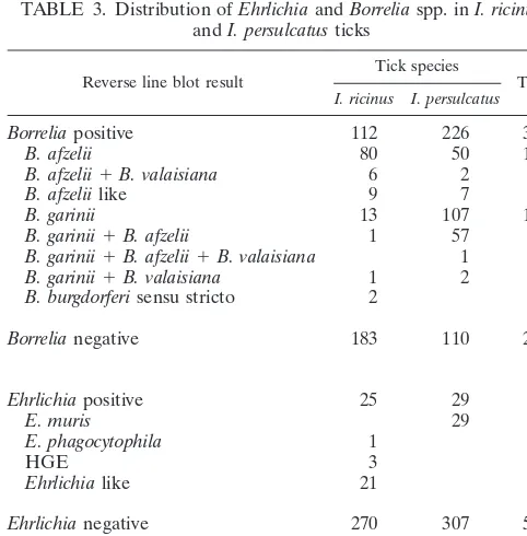
Distribution ofBorreliaandEhrlichiaspecies in ticks.The
majority of theBorrelia-positive ticks carriedB. afzeliiand/orB. gariniiDNA. There was a remarkable difference in distribution of the various Borreliaspecies between I. persulcatus and I. ricinus (Table 3). Of the 226 Borrelia-positive I. persulcatus
ticks 107 (47.3%) contained B. garinii, 50 (22.1%) carriedB.
on May 15, 2020 by guest
http://jcm.asm.org/
TABLE 1. Oligonucleotide primers and probes used in PCR and hybridization assays Oligonucleotide primer or probe Sequence Target organism Target gene Nucleotide position Reference or source Borrelia -specific primers B-5SBor 5 ⬘ biotin-GAGTTCGCGGGAGAGTAGGTTATT B. burgdorferi sensu lato 5S rRNA ( rrfA ) 82–105 This study 23SBor TCAGGGTACTTAGATGGTTCACTT B. burgdorferi sensu lato 23S rRNA ( rrlB ) 208–185 This study Probes SL 5 ⬘ amino-CTTTGACCATATTTTTATCTTCCA B. burgdorferi sensu lato 23S-5S spacer a 182–167 24 SS 5 ⬘ amino-AACACCAATATTTAAAAAACATAA B. burgdorferi sensu stricto 23S-5S spacer 59–36 24 GA 5 ⬘ amino-AACATGAACATCTAAAAACATAAA B. garinii 23S-5S spacer 58–35 24 AF 5 ⬘ amino-AACATTTAAAAAATAAATTCAAGG B. afzelii 23S-5S spacer 44–21 24 VS 5 ⬘ amino-CATTAAAAAAATATAAAAAATAAATTTAAGG B. valaisiana 23S-5S spacer 51–21 24 Ruski 5 ⬘ amino-GAATAAAACATTCAAATAATATAAAC B. afzelii like 23S-5S spacer 67–42 This study Ehrlichia -specific primers 16S8FE GGAATTCAGAGTTGGATCMTGGYTCAG Eubacteria 16S rRNA gene 8–27 27 B-GA1B 5 ⬘ biotin-CGGGATCCCGAGTTTGCCGGGACTTCTTCT Ehrlichia genus 16S rRNA gene 476–456 27 Probes A-EhrAll 5 ⬘ amino-TTATCGCTATTAGATGAGCC Ehrlichia genus 16S rRNA gene 203–222 27 A-HGE 5 ⬘ amino-GCTATAAAGAATAGTTAGTGG HGE agent 16S rRNA gene 87–107 27 A-Phago 5 ⬘ amino-TTGCTATAAAGAATAATTAGTGG E. phagocytophila 16S rRNA gene 85–107 27 A-D-HGE 5 ⬘ amino-GCTATGAAGAATAGTTAGTG HGE agent variant 16S rRNA gene 87–106 27 A-D-Phago 5 ⬘ amino-TTGCTATGAAGAATAATTAGTG E. phagocytophila variant 16S rRNA gene 87–106 27 A-ESchot 5 ⬘ amino-GCTGTAGTTTACTATGGGTA Ehrlichia like 16S rRNA gene 76–95 27 A-ECan 5 ⬘ amino-TCTGGCTATAGGAAATTGTTA Ehrlichia canis 16S rRNA gene 85–105 27 A-EChaf 5 ⬘ amino-ACCTTTTGGTTATAAATAATTGTTA E. chaf feensis 16S rRNA gene 83–107 27 A-EmurisC 5 ⬘ amino-GCTATAGGTTCGCTATTAG E. muris C variant 16S rRNA gene 85–103 This study A-EmurisT 5 ⬘ amino-AGCTATAGGTTTGCTATTAGT E. muris T variant 16S rRNA gene 86–104 This study Spike primers 23SBortmpA CGGGATCCC-TCAGGGTACTTAGATGGTTCACTT-ACCCCGAACTTTTCTCC b T. pallidum tmpA 199–215 This study 5SBortmpA GGAATT-CGAGTTCGCGGGAGAGTAGGTTATT-CTTCCCCAGTTCTGTGTAGA T. pallidum tmpA 725–706 This study EhrFtmpB GCTCTAGAG-AGAGTTTGATCCTGGCTCAG-CGCCTATCCGACCGAGTAT T. pallidum tmpB 308–326 This study EhrRtmpB CGGGATCCC-GAGTTTGCCGGGACTTCTTCTA-CCACGTAGTCACCTTGTATT T. pallidum tmpB 756–736 This study Probes A-TmpABor 5 ⬘ amino-TACAATCTAAGATCCAGACT T. pallidum tmpA 346–365 This study A-TmpBEhr 5 ⬘ amino-GAAAACTCGAAGAACAAAGAA T. pallidum tmpB 502–522 This study a The SL probe targets the spacer and the first eight bases of the 23S gene sequence ( rrlB ). b The dashes in the spike primers indicate the separation between the restriction site sequences, the Borrelia or Ehrlichia sequence, and the tmpA -o r tmpB -specific sequence.
on May 15, 2020 by guest
http://jcm.asm.org/
[image:3.612.108.501.73.720.2]afzelii, and 57 (25.2%) carried bothB. afzeliiandB. garinii. In contrast, 80 (71.4%) out of 112Borrelia-positiveI. ricinusticks carriedB. afzelii, 13 (11.6%) carriedB. gariniiDNA, and only 1 tick (0.9%) was infected with bothB. afzeliiand B. garinii. Only oneI. persulcatustick was triple infected byB. afzelii,B. garinii, and B. valaisiana, and two I. ricinus ticks carried B. burgdorferisensu stricto DNA.
Sixteen of allBorrelia-positive ticks reacted with theB. burg-dorferi sensu lato probe only. Therefore, we sequenced the 5S-23S intergenic spacer of six of these ticks and found that they carried Borreliaspecies with identical spacer sequences which were similar to but distinct from the spacer ofB. afzelii
(Fig. 1). We therefore designated this species asB. afzeliilike, designed a new probe (Ruski) for use with the reverse line blot, and hybridized the PCR products of all 16 samples with the new probe. As expected, all 16 samples reacted with this probe,
and no cross-hybridization with other species-specific probes was seen.
Of the 25Ehrlichia-positiveI. ricinusticks, 3 carried HGE DNA, 1 contained E. phagocytophila DNA, and 21 carried DNA from an organism that was identified before in Dutch ticks and designated as anEhrlichia-like organism (27). In 29I. persulcatusticks, DNA fromEhrlichiaspecies was found that initially reacted with the Ehrlichiagenus-specific probe only. From these samples the PCR products were sequenced, and comparison showed that their sequences were identical. Align-ment with DNA sequences in the GenBank data bank revealed a close similarity (⬎99%) with E. muris16S rRNA gene se-quences. We thereafter designed twoE. muris-specific probes that differed only in one residue targeting position 95 of the 16S rRNA gene and hybridized all 29 above-mentioned PCR products with these probes. All samples reacted with the E. murisC probe but not with theE. murisT probe.
There was no significant difference betweenBorreliaor Ehr-lichiainfection rates in the different sexes or stages of theI. ricinusticks investigated (Table 4). However, inI. persulcatus
ticks approximately 75% of the adults and only 35.5% of the nymphs were infected with Borrelia. Similarly, about 10% of adultI. persulcatusticks carriedEhrlichiaDNA, whereas only 1.6% of nymphs did. None of the 24 larvae carried any detect-ableBorreliaorEhrlichiaDNA.
DISCUSSION
In the study presented here we have shown thatEhrlichia
species were found in 8.6% of ixodid ticks collected from vegetation in the Baltic region of Russia. Members of the granulocyticE. phagocytophilagroup, including some reacting with our HGE agent probe, were found inI. ricinusbut not in
[image:4.612.53.553.84.243.2]I. persulcatus. However, the rate of HGE infection in these ticks was low (1%). This prevalence of infection is comparable to that found by several other research groups in Europe (13, 16, 18, 21). However, other European studies have reported infection rates ranging from 3.2% in Slovenia to 28.9% in The Netherlands (6, 20, 27, 31), and some studies from the United States even reported a prevalence of infection inIxodes scapu-larisup to 50% (4, 15). The explanation for these large differ-ences in prevalence of infection needs to be established with comparative studies. About 7.1% of the I. ricinusticks were TABLE 2. Comparison of dark-field microscopy and reverse line blot detection ofBorreliaandEhrlichiaDNA in ticks
Tick species and result by dark-field
microscopy No. of ticks
Reverse line blota
Borrelia Ehrlichia
Positive Negative Positive Negative
I. ricinus
Positive 215 97 118 24 191
Negative 80 15 65 1 79
Total 295 112 183 25 270
I. persulcatus
Positive 257 209 48 29 228
Negative 79 17 62 0 79
Total 336 226 110 29 307
Total 631 338 293 54 577
aValues are numbers of ticks.
TABLE 3. Distribution ofEhrlichiaandBorreliaspp. inI. ricinus
andI. persulcatusticks
Reverse line blot result Tick species Total
I. ricinus I. persulcatus
Borreliapositive 112 226 338
B. afzelii 80 50 130
B. afzelii⫹B. valaisiana 6 2 8
B. afzeliilike 9 7 16
B. garinii 13 107 120
B. garinii⫹B. afzelii 1 57 58
B. garinii⫹B. afzelii⫹B. valaisiana 1 1
B. garinii⫹B. valaisiana 1 2 3
B. burgdorferisensu stricto 2 2
Borrelianegative 183 110 293
Ehrlichiapositive 25 29 54
E. muris 29 29
E. phagocytophila 1 1
HGE 3 3
Ehrlichialike 21 21
Ehrlichianegative 270 307 577
Total no. of ticks 295 336 631
on May 15, 2020 by guest
http://jcm.asm.org/
[image:4.612.52.293.483.728.2]infected with anEhrlichia-like organism previously identified in I. ricinus ticks found in The Netherlands and Italy (27). Remarkably, this species was not found inI. persulcatusticks from the same regions, suggesting a vector specificity of this
Ehrlichia species. Although the true nature of this species remains unknown, phylogenetic analysis based on the 16S rRNA gene sequence indicates a close relationship with the monocytic group of ehrlichiae. Nearly 9% of theI. persulcatus
ticks were infected with anEhrlichiaspecies with a 16S rRNA gene sequence nearly identical to one of the publishedE. muris
sequences. This monocyticEhrlichiaspecies was identified only inI. persulcatusticks, again suggesting a vector-specific Ehr-lichia infection. In a recent study, I. persulcatus ticks from Perm, Russia (region of Ural Mountains), were tested for the presence ofEhrlichiaby PCR usingEhrlichia-specific primers (22). None of the ticks carried detectable HGE DNA. How-ever, 5 out of 35 ticks tested yielded a 16S rRNA PCR product of which the DNA sequence was shown to be identical to that ofE. muris. This result confirms our finding thatE. muriswas the onlyEhrlichiaspecies found inI. persulcatusticks.
Analysis of the ticks for the presence of Borrelia species corroborated earlier findings on infection rates of ticks in the Baltic region. Speciation by reverse line blot hybridization showed that many ticks (10.6%) carried more than oneBorrelia
species. The majority of the ticks were infected withB. afzelii
and/orB. garinii;B. burgdorferisensu stricto was found in only two tick extracts. In 2.5% of the ticks aB. burgdorferispecies was found which carried a 5S-23S rRNA spacer sequence sim-ilar to, but distinct from, that of B. afzelii. ThisB. afzelii-like species was found both inI. persulcatusandI. ricinus. We have not investigated whether coinfection of this species with other
B. burgdorferisensu lato species occurs. Furthermore, we have no data that indicates that this species is pathogenic for hu-mans. There was no significant difference between infection rates ofI. persulcatusandI. ricinuswithB. afzelii. In contrast, of the 182 ticks infected withB. gariniionly 15 (8.2%) wereI. ricinusticks. As with the distribution of theEhrlichiaspecies this suggests a vector-specific infection. It is uncertain whether this represents true vector specificity or a relationship between the vector and its host range. To determine whether an asso-ciation between pathogen and tick species exists, additional studies, including studies of the vector hosts such as large and small mammals and birds, are required.
In our study, none of the larvae contained any detectable
BorreliaorEhrlichiaDNA, but the number of larvae included in this study is too low to draw any conclusions from this result. However, a more significant observation was thatI. persulcatus
adults are infected with either Borrelia or Ehrlichia species more than twice as often as nymphs. This was true if either the dark-field examination or the PCR result was used as an indi-cator ofBorreliainfection. Such a stage-dependent prevalence of infection was not seen in theI. ricinus ticks. In fact, 60 to 80% of theI. ricinusadults and nymphs included in the study were dark-field positive, and 38 to 43% of the same ticks were positive with theBorreliaPCR.
[image:5.612.61.547.71.147.2]The latter finding showed that in a large proportion of the dark-field-positive ticks noBorreliaDNA was detected. A pos-sible explanation for this observation is that dark-field micros-copy is more sensitive thanBorreliaPCR in detectingBorrelia. However, the detection ofBorreliaDNA in 18% of the dark-field-negative samples argues against that. Furthermore, we have shown in previous studies that PCR, followed by the FIG. 1. Multiple aligment and clustering of the 23S-5S intergenic spacer region ofB. burgdorferisensu lato and reverse line blot result using the amplified spacer sequences. Multiple alignment and clustering were performed with the complete 174- to 182-bp spacer region and are only partially displayed. The coordinates of theB. burgdorferisensu stricto spacer sequence are indicated above the sequences, and the probe regions are denoted by grey boxes. Reverse line blot probes are indicated as follows: VS,B. valaisiana; RS,B. afzeliilike; AF,B. afzelii; GA,B. garinii; SS,
B. burgdorferisensu stricto; SL,B. burgdorferisensu lato.
TABLE 4. Distribution of infection type, stratified by tick species, sex, and stage
Infection status Tick species Sex Stage Total
Female Male Nymph Larva
Infected⫹noninfected I. ricinus 107 89 77 22 295
I. persulcatus 154 118 62 2 336
Borreliainfected I. ricinus 47 35 30 0 112
I. persulcatus 112 92 22 0 226
Ehrlichiainfected I. ricinus 13 4 8 0 25
I. persulcatus 15 13 1 0 29
Double infected I. ricinus 8 4 6 0 18
I. persulcatus 13 11 0 0 24
on May 15, 2020 by guest
http://jcm.asm.org/
[image:5.612.54.551.591.727.2]reverse line blot method, is both sensitive and specific. False-negative results due to inhibition of the PCR were excluded by the use of internal spike controls. There is a possibility that the DNA of theBorreliaspecies was degraded during storage, but earlier experiments in our lab have shown thatBorreliaDNA in ticks stored in ethanol is extremely stable. In addition, the fact that there is a significant difference inBorreliaDNA detection between I. persulcatus and I. ricinus dark-field-positive ticks also suggests that the microorganisms seen in the dark-field analysis may represent species other thanB. burgdorferisensu lato and that this species is found more frequently inI. ricinus
than inI. persulcatus. If this hypothesis is correct, studies that use dark-field microscopy as the sole method of determining the rate of Borrelia infection in ticks may yield an overesti-mated prevalence of infection. Further PCR studies using primers that will amplify the 16S rRNA gene of a broad range of bacteria and subsequent sequence analyses will have to corroborate this theory and identify the species.
To our knowledge, there have been no reports until now in the scientific literature in English of cases of ehrlichiosis in any part of Russia. However, in the city of St. Petersburg, many cases of Lyme disease are reported (250 cases per year; 5 cases per 100,000 inhabitants), and the majority of these cases are patients who sustained tick bites in the suburban region of the city. It is this region where we collected the Borrelia- and
Ehrlichia-infected ticks used in this study. It is therefore con-ceivable that some people in the St. Petersburg region may have been infected with the HGE agent. Due to the lack of sufficient diagnostic tools and the lack of awareness, these cases of human ehrlichiosis may have gone unnoted. However, the rate ofEhrlichiainfection in the ticks is about 10- to 20-fold lower than the rate of Borreliainfection. This would suggest that the chance of infection with Ehrlichiaafter a tick bite is significantly lower than the chance of being infected with Bor-reliabut that such exposure may still account for 10 to 25 cases of ehrlichiosis in the St. Petersburg area. Therefore, awareness of possible cases of ehrlichiosis remains important in tick-infested areas like the suburban region of St. Petersburg.
ACKNOWLEDGMENTS
The research described in this paper was made possible in part by grant N98-04-49899 from the Russian Basic Research Foundation and in part by grant 9600864 from the Danish Research Councils.
REFERENCES
1.Alekseev, A. N., H. V. Dubinina, L. P. Antykova, T. I. Dzhivanyan, S. G. Rijpkema, N. V. Kruif, and M. Cinco.1998. Tick-borne borrelioses pathogen
identification inIxodesticks (Acarina, Ixodidae) collected in St. Petersburg
and Kaliningrad Baltic regions of Russia. J. Med. Entomol.35:136–142.
2.Anderson, B. E., J. E. Dawson, D. C. Jones, and K. H. Wilson.1991.Ehrlichia chaffeensis, a new species associated with human ehrlichiosis. J. Clin.
Micro-biol.29:2838–2842.
3.Baumgarten, B. U., M. Rollinghoff, and C. Bogdan.1999. Prevalence of
Borrelia burgdorferiand granulocytic and monocytic ehrlichiae inIxodes
ric-inusticks from southern Germany. J. Clin. Microbiol.37:3448–3451.
4.Chang, Y. F., V. Novosel, C. F. Chang, J. B. Kim, S. J. Shin, and D. H. Lein.
1998. Detection of human granulocytic ehrlichiosis agent andBorrelia
burg-dorferiin ticks by polymerase chain reaction. J. Vet. Diagn. Investig.10:56–59. 5.Chen, S. M., J. S. Dumler, J. S. Bakken, and D. H. Walker.1994.
Identifi-cation of a granulocytotropicEhrlichia species as the etiologic agent of
human disease. J. Clin. Microbiol.32:589–595.
6.Cinco, M., D. Padovan, R. Murgia, M. Maroli, L. Frusteri, M. Heldtander, K. E. Johansson, and E. O. Engvall.1997. Coexistence ofEhrlichia phago-cytophilaandBorrelia burgdorferisensu lato inIxodes ricinusticks from Italy
as determined by 16S rRNA gene sequencing. J. Clin. Microbiol.35:3365–3366.
7.Junttila, T., M. Peltomaa, H. Soini, M. Marjamaki, and M. K. Viljanen.
1999. Prevalence ofBorrelia burgdorferiinIxodes ricinusticks in urban
rec-reational areas of Helsinki. J. Clin. Microbiol.37:1361–1365.
8.Kovalevskii, Y. V., and E. I. Korenberg.1995. Differences inBorrelia
infec-tions in adultIxodes persulcatusandIxodes ricinusticks (Acari: Ixodidae) in
populations of north-western Russia. Exp. Appl. Acarol.19:19–29.
9.Kramer, V. L., M. P. Randolph, L. T. Hui, W. E. Irwin, A. G. Gutierrez, and D. J. Vugia.1999. Detection of the agents of human ehrlichioses in ixodid
ticks from California. Am. J. Trop. Med. Hyg.60:62–65.
10. Lefleche, A., D. Postic, K. Girardet, O. Peter, and G. Baranton.1997.
Char-acterization ofBorrelia lusitaniaesp. nov. by 16S ribosomal DNA sequence
analysis. Int. J. Syst. Bacteriol.47:921–925.
11. Lesnyak, O., E. Laikovskaya, I. Kufko, H. Bruinink, N. Baranova, and S. Rijpkema.1998. Clinical features of Lyme borreliosis in the middle Urals
and distribution ofBorrelia burgdorferisensu lato species in localIxodes
persulcatusticks. Zentralbl. Bakteriol.288:111–119.
12. Leutenegger, C. M., N. Pusterla, C. N. Mislin, R. Weber, and H. Lutz.1999.
Molecular evidence of coinfection of ticks withBorrelia burgdorferisensu lato
and the human granulocytic ehrlichiosis agent in Switzerland. J. Clin.
Mi-crobiol.37:3390–3391.
13. Liz, J. S., L. Anderes, J. W. Sumner, R. F. Massung, L. Gern, B. Rutti, and M. Brossard.2000. PCR detection of granulocytic ehrlichiae inIxodes ricinus
ticks and wild small mammals in western Switzerland. J. Clin. Microbiol.
38:1002–1007.
14. Lotric-Furlan, S., M. Petrovec, T. Avsic-Zupanc, W. L. Nicholson, J. W. Sumner, J. E. Childs, and F. Strle.1998. Human ehrlichiosis in central
Europe. Wien. Klin. Wochenschr.110:894–897.
15. Magnarelli, L. A., K. C. Stafford III, T. N. Mather, M.-T. Yeh, K. D. Horn, and J. S. Dumler.1995. Hemocytic rickettsia-like organisms in ticks: sero-logic reactivity with antisera to ehrlichiae and detection of DNA of agent of
human granulocytic ehrlichiosis by PCR. J. Clin. Microbiol.33:2710–2714.
16. Ogden, N. H., K. Bown, B. K. Horrocks, Z. Woldehiwet, and M. Bennett.
1998. GranulocyticEhrlichiainfection in ixodid ticks and mammals in
wood-lands and upwood-lands of the U. K. Med. Vet. Entomol.12:423–429.
17. Oteo, J. A., J. R. Blanco, V. Martinez de Artola, and V. Ibarra.2000. First report of human granulocytic ehrlichiosis from southern Europe (Spain).
Emerg. Infect. Dis.6:430–432.
18. Parola, P., L. Beati, M. Cambon, P. Brouqui, and D. Raoult.1998. Ehrlichial
DNA amplified fromIxodes ricinus(Acari: Ixodidae) in France. J. Med.
Entomol.35:180–183.
19. Petrovec, M., S. Lotric Furlan, T. A. Zupanc, F. Strle, P. Brouqui, V. Roux, and J. S. Dumler.1997. Human disease in Europe caused by a granulocytic
Ehrlichiaspecies. J. Clin. Microbiol.35:1556–1559.
20. Petrovec, M., J. W. Sumner, W. L. Nicholson, J. E. Childs, F. Strle, J. Barlic, S. Lotric-Furlan, and T. Avsic Zupanc.1999. Identity of ehrlichial DNA
sequences derived fromIxodes ricinusticks with those obtained from patients
with human granulocytic ehrlichiosis in Slovenia. J. Clin. Microbiol.37:209–210.
21. Pusterla, N., J. B. Huder, H. Lutz, and U. Braun.1998. Detection of Ehr-lichia phagocytophilaDNA inIxodes ricinusticks from areas in Switzerland
where tick-borne fever is endemic. J. Clin. Microbiol.36:2735–2736.
22. Ravyn, M. D., E. I. Korenberg, J. A. Oeding, Y. V. Kovalevskii, and R. C. Johnson.1999. Monocytic Ehrlichia inIxodes persulcatusticks from Perm,
Russia. Lancet353:722–723.
23. Rijpkema, S., D. Golubic, M. Molkenboer, N. Verbeek De Kruif, and J. Schellekens.1996. Identification of four genomic groups ofBorrelia
burgdor-ferisensu lato inIxodes ricinusticks collected in a Lyme borreliosis endemic
region of northern Croatia. Exp. Appl. Acarol.20:23–30.
24. Rijpkema, S. G., M. J. Molkenboer, L. M. Schouls, F. Jongejan, and J. F. Schellekens.1995. Simultaneous detection and genotyping of three genomic
groups ofBorrelia burgdorferisensu lato in Dutch Ixodes ricinusticks by
characterization of the amplified intergenic spacer region between 5S and
23S rRNA genes. J. Clin. Microbiol.33:3091–3095.
25. Rikihisa, Y.1991. The tribeEhrlichieaeand ehrlichial diseases. Clin.
Micro-biol. Rev.4:286–308.
26. Sato, Y., K. Miyamoto, A. Iwaki, T. Masuzawa, Y. Yanagihara, E. I. Koren-berg, N. B. Gorelova, V. I. Volkov, L. I. Ivanov, and R. N. Liberova.1996.
Prevalence of Lyme disease spirochetes inIxodes persulcatusand wild
ro-dents in far eastern Russia. Appl. Environ. Microbiol.62:3887–3889.
27. Schouls, L. M., I. Van De Pol, S. G. T. Rijpkema, and C. S. Schot.1999.
Detection and identification ofEhrlichia,Borrelia burgdorferisensu lato, and
Bartonellaspecies in DutchIxodes ricinusticks. J. Clin. Microbiol.37:2215– 2222.
28. Smith, M., G. Gettinby, M. Granstrom, J. S. Gray, E. C. Guy, C. Revie, J. N. Robertson, and G. Stanek.1998. The European union concerted action
world wide web site for Lyme borreliosis. Zentralbl. Bakteriol.287:266–269.
29. Steere, A. C.1989. Lyme disease. N. Engl. J. Med.321:586–596. 30. van Dobbenburgh, A., A. P. van Dam, and E. Fikrig.1999. Human
granu-locytic ehrlichiosis in western Europe. N. Engl. J. Med.340:1214–1216.
31. von Stedingk, L. V., M. Gurtelschmid, H. S. Hanson, R. Gustafson, L. Dotevall, E. O. Engval, and M. Granstrom.1997. The human granulocytic
ehrlichiosis (HGE) agent in Swedish ticks. Clin. Microbiol. Infect.3:573–574.
32. Walker, D. H., and J. S. Dumler.1996. Emergence of the ehrlichioses as
human health problems. Emerg. Infect. Dis.2:18–29.