International Journal of Emerging Technology and Advanced Engineering
Website: www.ijetae.com (ISSN 2250-2459, ISO 9001:2008 Certified Journal, Volume 8, Issue 1, January 2018)
142
An Integrated Framework for Identifying Multiple Cancer
Condition from MRI Images
Parvati Angadi
1, Dr. M Nagendra
21Research Scholar, Rayalaseema University, Kurnool, India
2Professor & HOD, Dept of Computer Science & Technology, S K University, Anantapuramu, India
Abstract— Magnetic Resonance Imaging (MRI) is one of the frequently adopted radiological procedures for diagnosis of critical disease conditions like cancer. However, MRI is not always perfect owing to its sensitivity issues giving rise to false positives and hence it calls for more number of expensive tests. We reviewed the existing literatures to find that they are highly symptomatic in nature where the applicability of presented schemes are never being claimed to work on different disease diagnosis. Therefore, we introduce a very novel and yet simple technique that bears the capability to perform accurate identification of regions infected with carcinoma. The novel contribution of this paper is that it offers a single framework with capability to offer accurate diagnosis of three different cancers e.g. brain cancer, lung nodules, and breast cancer. The study outcome also shows a good correlation of machine learning and their potential impact on diagnosis process of MRI images.
Keywords—Cancer Detection, Medical Imaging, Magnetic Resonance Imaging, Accuracy, Machine Learning.
I. INTRODUCTION
With the significant improvement in medical image processing, the diagnosis of the critical disease condition like cancer is becoming easy. At present, various forms of sophisticated devices are utilized in order to acquire radiological images e.g. ultrasound image, x-rays, magnetic resonance imaging (MRI), positron emission tomography (PET) scan etc [1]. Out of all the forms of radiological imaging, MRI is considered to be highly accurate as well as expensive too when it comes to higher degree of complexity in diagnosis [2]. According to clinical society, MRI has the potential to offer early detection of various disease condition especially breast cancer [3] and its inferences are also found to be quite reliable. However, usage of MRI also causes identification of lesions of secondary type to be called as benign state of cancer. These forms of outcomes are quite misleading and at present it is avoided by considering multiple slices of MRI images in order to observe the infected regions more closely. Such process is not only expensive but also consumes maximum time in order to come to a conclusion about the diagnosis.
Hence the pitfalls of MRI are i) it is highly sensitive in nature that definitely gives rise to outcomes of false positive nature, ii) there is specificity factor in MRI and hence it is not capable of detecting the critical lesions with sufficient degree of specificity without any forms of dependencies and inclusion of more number of expensive imaging procedure or biopsies. At present, there have been various research-based techniques in order to address this problem of diagnosis especially related to cancer [4]-[8]; however, no such technique has been ever found to be in practice in ground reality. This evidently proves that detection of cancer from radiological images includes various research challenges that are yet to be solved. Apart from this, existing mechanism to solve such cancer diagnosis problems are highly specific in nature, which means that solution to one problem is actually not meant for solving other problem even to a minor extent. This gives rise to constraint optimization problems in medical diagnosis [9]. It has also been explored that machine learning and classification techniques has greater deal of contribution in diagnosing disease condition like cancer; however, none of the existing techniques has been ever claimed to be computationally efficient. With the rise of different forms of disease analytical tools running over low-end device, computational efficiency should be given a secondary priority whereas accuracy is always the primary priority. Irrespective of using different image capturing tools on different forms of cancer, it is only the images which have to be subjected to certain framework for effective disease diagnosis. At present, there are no such tools or no such investigation carried out to find if different forms of radiological images are actually going to be treated equally by a computational model that bears the capability of identifying the exact location of the cancer.
International Journal of Emerging Technology and Advanced Engineering
Website: www.ijetae.com (ISSN 2250-2459, ISO 9001:2008 Certified Journal, Volume 8, Issue 1, January 2018)
143 Section I.A discusses about the existing literature where different techniques are discussed for detection of brain cancer, lung cancer, and breast cancer followed by discussion of research problems in brief in Section I.B. Proposed solution towards addressing the identified problems is briefed in I.C. Section II discusses about research methodology being implemented with flow of the different processes for assisting in disease detection followed by discussion of result analysis for all the three scenarios of cancer images in the form of MRI in Section
III. Finally, the conclusive remarks are provided in Section IV.
A. Background
This section presents a brief contribution of various techniques being evolved in recent times towards identification of cancer using medical imaging. Our prior work has presented certain techniques to address such problems too [10]-[13]. We identify that cancer related to brain, lungs and breast are associated with complication and hence we explore more about existing solution. The work carried out by Ambily et al. [14] have used neural network in order to localize the brain tumor. Adoption of segmentation techniques using AdaBoost classifier and wavelets has been seen in the work of Islam et al. [15] and Demirhan et al. [16]. The segmentation process is also enhanced along with seed selection process and graph theory for analyzing brain tumor during radio surgical procedure as seen in the work of Hamamci et al. [17]. Usage of k-means clustering for identification of brain tumor has been noticed in the work of Malathi and Kamal [18]. Sheejakumari and Gomathi [19] have integrated neural network with swarm intelligence for enhancing the detection performance. Convolution approach can significantly enrich the deep learning process that offers better segmentation process in brain tumor identification as reported in the work of Pereira et al.[20]. Similar adoption of convolution neural network is seen in the work of Yu et al. [21] emphasizing on analyzing lung cancer. Discrete forms of predictive techniques are also reported to assist in identification of lung cancer as reported in the by Chauhan and Jaiswal [22]. The joint technique of fuzzy logic and neural network also offers a better classification process in analyzing lung cancer as reported by Deshmukh and Shinde [23]. Kureshi et al. [24] have introduced a predictive framework for investigating decision factor and its impact on personalized medicine for lung cancer. Santhanam et al. [25] have constructed a framework that offers three dimensional visualization of the lung tumor using augmental reality.
Literatures have also different signal-based investigations towards analyzing lung cancer e.g. Trans et al. [26]. It is also found that microwave imaging potentially assists in analyzing lung cancer considering its frequency domain as reported in the work of Zamani et al. [27]. We also find that there are some significant work carried out in breast cancer detection using neural network as discussed by Kanojia and Abraham [28]. Literatures have also witnessed adoption of Fuzzy C-means clustering that has significant contribution towards better accuracy of breast cancer detection as put forward by Al-Ayyoub et al. [29]. Vidya and Mathew [30] have also used fuzzy logic as well as homomorphic filtering for identification of breast cancer. Popular search optimization technique e.g. genetic algorithm is also found to be used along side with neural network for solving identification problem of breast cancer as seen in the work of Singh et al. [31]. K-nearest neighbor technique is also reported to have better detection performance in breast cancer as discussed by Al-hadidi et al. [32]. Reis et al. [33] have presented an involuntary classification process using local binary pattern while genomic-based research approach is reported by Stfakianakis et al. [34] to facilitate solving identification of breast cancer problems. There are good numbers of existing techniques towards identification of specific cancer condition. The next section outlines the problems associated with existing system that has not been addressed in the existing techniques.
B. Research Problem
Some of the potential problems associated with the existing research techniques are:
Absence of a common framework or scheme that could address identification of region inflicted with different forms of cancer.
Existing approaches doesn’t offer any evidence of benchmarking when it comes to adoption of machine learning approach and thereby fails to state any significant effective factors in it.
Adoption of existing classifiers as well as other associated learning-based techniques is potentially associated with computational burden, which is not addressed in existing system.
International Journal of Emerging Technology and Advanced Engineering
Website: www.ijetae.com (ISSN 2250-2459, ISO 9001:2008 Certified Journal, Volume 8, Issue 1, January 2018)
144 Therefore, the problem statement of the proposed study can be stated as “Constructing a unique identification-based framework that can perform identification of multiple cases of potential cancer regions jointly with better computational efficiency.”
C. Proposed Solution
The previous section outlines the research problems associated with existing techniques for identification of major cancers. This section further provides supporting information about the mechanism adopted with justification behind the planning of implementation. The proposed technique adopts an analytical research methodology where implication of various machine learning process and classifiers were investigated towards identification process of three different case studies based on cancer medical images. The adoption of the case studies is essential as different form of radiological images may possess different set of challenges that may hinder the progress of detection as well as diagnosis process of the cancer. This section briefs about the original work being carried out towards achieving the cumulative research goal in terms of research methodology being adopted in each case studies. Fig.1 highlights the overall scheme of methodology, where it is seen that ANN is considered as frequently used in the form of classifier as compared to others e.g. SVM, KNN, etc. Hence, the proposed study formulates three different case studies of MRI image of brain, lung, and breast cancer. This part of the case study formulation uses multiple approaches e.g. segmentation, histogram, classifier, blocking, etc in order to perform an analysis for impact of different classifier on detection accuracy.
Explore Different Classifiers
Frequently Used Classifier Less Frequently Used Classifier
ANN SVM, KNN, others
Formulation of case Studies
Brain MRI Image Lung MRI Image Breast MRI Image
Draw Inference
[image:3.612.50.289.507.639.2]Case Specific methodology (Apply ANN)
Figure 1 Overall Scheme of Adopted Research Methodology
II. RESEARCH METHODOLOGY
This section discusses about an integrated technique to perform identification of carcinomic region associated with brain, breast, and lung.
The analysis of different case study is basically meant for assessing impact of ANN on accuracy values on different case studies. Irrespective of different set of mechanism applied for feature extraction, all the case studies uses ANN to find that it doesn’t offer higher accuracy every time for different case studies. This conclusion of case study investigation is also in agreement with the different nature of complications in different case studies. It is also explored that ANN generates higher accuracy in majority of cases with faster convergence rate. Therefore, this analysis considers ANN to be a still better form of classifier with some degree of pitfalls, which is required to be modified.
A. Methodology for Identification of Brain Cancer The methodology involved in designing the first case study is as briefly discussed below:
Rationale behind Selection of Brain MRI Image: The first part of our study chooses to work on brain MRI images owing to the complication of the type of oncological cancer. Brain cancer is still the most challenging cancerous condition with increased mortality rate and hence accuracy in diagnosis is highly required. We believed that owing to complicated form of the brain MRI images, there are possibilities to perform inaccurate feature extraction that may call for false alarm. Therefore, owing to the criticality of the disease condition, we choose to consider brain MRI image as our first case study. Dataset used: MR-TIP database [35].
Process Involved in Design Methodology: The
International Journal of Emerging Technology and Advanced Engineering
Website: www.ijetae.com (ISSN 2250-2459, ISO 9001:2008 Certified Journal, Volume 8, Issue 1, January 2018)
145
Input Brain MRI Image
Gradient magnitude
Watershed Transform
Mark Foreground
Remove dark Spots Regional
Maxima Superimpose Mark
Background Watershed(Seg
mentation)
Construct DAG Construct BN Histogram
Calculation Region Merging
Extract Features ANN Training
[image:4.612.319.565.129.241.2]ANN Detection
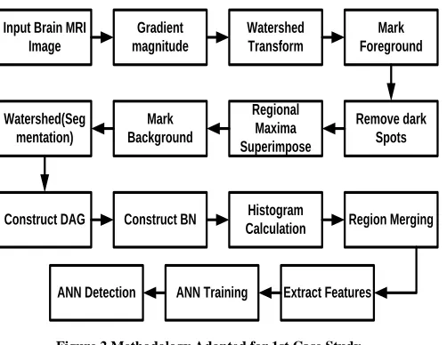
Figure 2 Methodology Adopted for 1st Case Study
Finally, the detection process is carried out to detect the location of cancer and the study outcome is assessed using conventional accuracy parameters e.g. True/false- positive/negative, Sensitivity, Recall rate, Specificity, Precision, F1-Score.
B. Methodology for Identification of Lung Cancer
The methodology involved in designing the second case study is as briefly discussed below:
Rationale behind Selection of Brain MRI Image: According to medical science, the rate of metastasis in Lung cancer is quite higher than any other form of cancer. Hence, this is also one of the disease conditions that demands early stage of detection. Dataset used: Lung Image Database Consortium image
collection (LIDC-IDRI) [36]
Process Involved in Design Methodology: In this case, the proposed study performs multiple blocking on the lung MRI images followed by entropy calculation that further assists in filtering nodules in order to perform feature extraction. Neural network is used for performing training that finally assists in performing identification of the region of the lung infected by cancer. The prime motive of this case study was to perform precise identification using less number of processes in order to retain maximum computational efficiency.
Input Lung MRI Image (Dicom)
Specify Block Size
Calculate Entropy
Filter Nodule Candidate
Feature Extraction Classify Nodes
Train Classifier Perform
Detection
Figure 3 Methodology Adopted for 2nd Case Study
C. Methodology for Identification of Breast Cancer
The methodology involved in designing the third case study is as briefly discussed below:
Rationale behind Selection of Brain MRI Image: The third case study of the proposed system is considered to be breast cancer imaging. The reason behind its selection process is due to the urgency of the cancer to be detected in the early stage. An efficient diagnosis of the breast cancer always calls for early detection and hence a better mechanism is required to perform efficient detection. Dataset used: Breast-MRI-NACT-Pilot [37]
Process Involved in Design Methodology: The input to the proposed system is basically an MRI image of breast cancer in DICOM format that is subjected to the homomorphic filter that has the capability to minimize the extent of multiplicative noise from the MRI image. The next step is to apply discrete wavelet transform for the purpose of extracting the two dimensional coefficient out of which the coefficient with lower value (LL) is extracted and gradient magnitude value is obtained from it. By doing this, it offers better mechanism for performing segregation of foreground to background object resulting in precise detection accuracy. The processed image from gradient magnitude is further subjected to watershed algorithm in order to achieve the perfect object. Neural network is applied as a part of training that is found to significantly assist in detection of the region infected by cancer.
Input Breast MRI Image
(Dicom)
Apply Homomorphic
Filter
Apply DWT Extract low
Coefficient
Obtain Gradient Magnitude Watershed
Algorithm Train Classifier
[image:4.612.51.302.131.325.2]Perform Detection
[image:4.612.323.580.586.693.2]International Journal of Emerging Technology and Advanced Engineering
Website: www.ijetae.com (ISSN 2250-2459, ISO 9001:2008 Certified Journal, Volume 8, Issue 1, January 2018)
146 While performing analysis of the above three case studies, the focus was mainly laid towards improving the applicability of the neural network towards accuracy. However, our investigation shows that different problems of cancer detection and diagnosis cannot be solved uniformly by the neural network. We have also tested with other forms of classifiers e.g. KNN, SVM, etc, where we find that accuracy is more for them as compared to neural network. However, applying neural network maintains better balance between computational demands as well as accuracy. This evaluation assists in forming conclusion that ANN is always the cost effective algorithm for assisting in precise detection with lower involvement of computational burden unlike other classifiers who just offers slightly increased accuracy but at the cost of computational burden. This outcome significantly assists in evolving up in an optimized framework where the detection performance is enhanced by the ANN with faster convergence rate.
III. RESULT ANALYSIS
[image:5.612.332.556.126.505.2]This section discusses about the outcomes obtained from the implementation of the proposed system that initiates with i) identification of brain cancer, ii) identification of lung cancer, and iii) identification of breast cancer. All the proposed techniques make use of Artificial Neural Network as a classified in order to perform identification of presence of tumour initiating with brain MRI image. The proposed system consists of series of techniques and methods formulated analytically in order to perform precise extraction of features. Fig.5 highlights the sample visual outcomes of this framework using ANN. The visual outcome shows how the input grayscale image (Fig.5(a)) is processed to obtain the gradient magnitude (Fig.5(b)) followed by watershed transform technique (Fig.5(c)). This process further assists in marking the foreground (Fig.5(d)) of given image which is further subjected to map with Bayesian network (Fig.5(e)) followed by histogram analysis (Fig.5(f)). Finally, region merging operation (Fig.5(g)) assists to perform final detection of tumor (Fig.5(h)).
Figure 5 Visual Outcomes of Identification of Brain Tumor
In the next process, we consider to change the input of lung MRI image where the image format is basically DICOM (Fig.6). The system allows multiple blocking operations i.e. 8x8, 16x16, till 32x32 (Fig.6(b)) followed by block splitting operation and computation of entropy (Fig.6(c)). The next significant step is to perform the filtering process of the lung nodule (Fig.6(d)) followed by feature extraction. Depending upon the selection of experimental technique based on machine learning, the proposed system finally performs classification of nodules (Fig.6(e)) and thereby computes the outcome by displaying the identified tumor (Fig.6(f)).
e) Bayesian N/W
f) Histogram
International Journal of Emerging Technology and Advanced Engineering
Website: www.ijetae.com (ISSN 2250-2459, ISO 9001:2008 Certified Journal, Volume 8, Issue 1, January 2018)
[image:6.612.109.224.131.328.2]147
Figure 6 Visual Outcomes of Identification of Lung Cancer
Figure 7 Visual Outcomes of Identification of Breast Cancer
The last phase of the outcome analysis considers breast cancer MRI image where the grayscale image is taken as an input. The transformation operation is applied over the filtered image in order to obtain the lower coefficient and gradient magnitude. Finally, watershed technique is applied in order to obtain the outcome of either benign or malignant case of breast cancer.
[image:6.612.365.526.199.312.2]For better analysis, we carry out a comparative analysis of the proposed technique with the most frequently adopted scheme of other similar approaches i.e. K-Nearest neighborhood (KNN) algorithm and Support Vector Machine (SVM).
Figure 8 Comparative Analysis
Fig.8 highlights the comparative analysis of proposed system with existing system with respect to various performance parameters e.g. true positive (TP), true negative (TN), sensitivity, specificity, precision, and F1 Score. This analysis is carried out on lung MRI images, which shows that nearly all the techniques offer similar performance of accuracy. Hence, there cannot be significant conclusion drawn with respect to the quality of an image in this regards. Interestingly, we find that usage of ANN offers much smaller computational time of 0.3776 seconds whereas KNN and SVM offers the computational time of 0.7241 seconds and 0.7881 seconds respectively. Hence, ANN proves to be the computationally efficient algorithm when it comes to solve the identification problem in lung cancer. Shorter response time is also in compliance with the supportability of the cost effective computational approach for analyzing various lung MRI images that usually comes in higher numbers and hence it also offers good scalability by exhibiting uniform performance over multiple images.
[image:6.612.96.237.354.583.2] [image:6.612.360.532.558.680.2]International Journal of Emerging Technology and Advanced Engineering
Website: www.ijetae.com (ISSN 2250-2459, ISO 9001:2008 Certified Journal, Volume 8, Issue 1, January 2018)
148
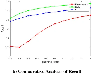
[image:7.612.88.247.142.268.2]b) Comparative Analysis of Recall Figure 9 Comparative Analyses of Precision & Recall
The study finding shows that SVM offers better outcome with respect to precision and recall rate, which is a clear indication to prove that applicability of ANN is not same for different medical condition. It is also explored that SVM and KNN has nearly similar performance of accuracy. A closer look into the graphical trends exhibited in Fig.9 shows that SVM offer significant advantage of superior precision and recall rate for increasing number of training ratio expressed in terms of probability. Owing to inclusion of regularization attributes in SVM, it doesn’t exhibit lower value of objective function of precision and recall and hence show steady growth in the graphical trend. On the other hand, irrespective of better classification behavior of malicious and benign case of tumor for breast images, KNN technique is very much dependent on value of K that cannot take autonomous decision and is highly dependent on user input. Another significant pitfall of KNN is significant computational complexity associated with the identification process. Although, the cumulative accuracy performance of ANN is found lower than SVM and KNN, but the graphical trend shows extremely good increment of accuracy that cannot be seen in SVM or KNN. Hence, ANN could be used for better accuracy in identification process whereas SVM could be used for cost-effective computational process inclusion.
IV. CONCLUSION
The commercial usage of MRI has never called up any issue as after the MRI is performed then the final clinical inference is carried out by attending physician. Hence, there are no notable problems since then and it depends completely on the expertise of medical professional. This also lowers the rate of faster diagnosis process as the process of inferring the actual disease condition is still manual where MRI just offers a magnified view of the disease section.
However, it is serious problem when the future calls for designing a computational model with a capability to diagnose the disease condition. In such case, we can’t afford to make any errors in the diagnosis problems. The common MRI itself suffers from false positive and hence there is no reliability of its outcome if the system of disease diagnosis is automated. Moreover, we find that existing techniques are too much focused on treating the MRI images depending on the case studies. Hence, we remove such dependencies by introducing a universal framework where different forms of radiological inputs can be given and yet it can show specific outcomes pertaining to disease. We also solve the constraint optimization problems using three different classifiers to find that each classifier plays a unique role in different cases.
REFERENCES
[1] Sabine Weckbach, Incidental Radiological Findings, Springer, 2017 [2] Andrew W. Wood, Non-ionizing Radiation Protection: Summary of
Research and Policy Options
[3] Alan B. Hollingsworth, M.D., Mammography and Early Breast Cancer Detection: How Screening Saves Lives, McFarland, 2016 [4] H. Qi and N. A. Diakides, "Thermal infrared imaging in early breast
cancer detection-a survey of recent research," Proceedings of the 25th Annual International Conference of the IEEE Engineering in Medicine and Biology Society (IEEE Cat. No.03CH37439), 2003, pp. 1109-1112 Vol.2.
[5] M. S. Islam, N. Kaabouch and W. C. Hu, "A survey of medical imaging techniques used for breast cancer detection," IEEE International Conference on Electro-Information Technology , EIT 2013, Rapid City, SD, 2013, pp. 1-5.
[6] L. Kapoor and S. Thakur, "A survey on brain tumor detection using image processing techniques," 2017 7th International Conference on Cloud Computing, Data Science & Engineering - Confluence, Noida, 2017, pp. 582-585
[7] G. Niranjana and M. Ponnavaikko, "A Review on Image Processing Methods in Detecting Lung Cancer Using CT Images," 2017 International Conference on Technical Advancements in Computers and Communications (ICTACC), Melmaurvathur, 2017, pp. 18-25. [8] R. Trigui, J. Miteran, L. Sellami, P. Walker and A. Ben Hamida, "A
classification approach to prostate cancer localization in 3T multi-parametric MRI," 2016 2nd International Conference on Advanced Technologies for Signal and Image Processing (ATSIP), Monastir, 2016, pp. 113-118.
[9] Anke Meyer-Baese, Volker J. Schmid, Pattern Recognition and Signal Analysis in Medical Imaging, Elsevier, 2014
[10] P. N. Angadi and P. K. Srimani, “Cumulative Techniques for Early Detection of Breast Cancer: A Review”, International Association of Scientific Innovation and Research, vol.8, No.6, 2014
[11] P. N. Angadi and P. K. Srimani, “Automatic MASS Classification OF Breast Cancer using ANN”, International Journal of Current Research, 2015
International Journal of Emerging Technology and Advanced Engineering
Website: www.ijetae.com (ISSN 2250-2459, ISO 9001:2008 Certified Journal, Volume 8, Issue 1, January 2018)
149 [13] P. N. Angadi and P. K. Srimani, "DODOR: Dual Optimization for
Detection of Oncological Region in radiological image," 2015 International Conference on Emerging Research in Electronics, Computer Science and Technology (ICERECT), Mandya, 2015, pp. 198-203.
[14] P.K. Ambily, S.P. James, R.R.Mohan, “Brain Tumor Detection using Image Fusion and Neural Network”, International Journal of Engineering Research and General Science, Vol. 3, Issue. 2, 2015 [15] A. Islam, S. M. S. Reza and K. M. Iftekharuddin, "Multifractal
Texture Estimation for Detection and Segmentation of Brain Tumors," in IEEE Transactions on Biomedical Engineering, vol. 60, no. 11, pp. 3204-3215, Nov. 2013.
[16] A. Demirhan, M. Törü and İ. Güler, "Segmentation of Tumor and Edema Along With Healthy Tissues of Brain Using Wavelets and Neural Networks," in IEEE Journal of Biomedical and Health Informatics, vol. 19, no. 4, pp. 1451-1458, July 2015.
[17] A. Hamamci, N. Kucuk, K. Karaman, K. Engin and G. Unal, "Tumor-Cut: Segmentation of Brain Tumors on Contrast Enhanced MR Images for Radiosurgery Applications," in IEEE Transactions on Medical Imaging, vol. 31, no. 3, pp. 790-804, March 2012.
[18] R. Malathi, A.R.N. Kamal, “Brain Tumor Detection and Identification Using K-Means Clustering Technique”, Special Issue Published in Int. Jnl. Of Advanced Networking and Applications, pp. 14-18, 2015
[19] V. Sheejakumari, S. Gomathi, “Brain tumor detection from MRI images using histon based segmentation and modified neural network”, Biomedical Research, 2016
[20] S. Pereira, A. Pinto, V. Alves and C. A. Silva, “Brain Tumor Segmentation Using Convolutional Neural Networks in MRI Images”, IEEE Transactions on Medical Imaging, Vol. 35, No. 5, 2016
[21] D. Yu et al., "Convolutional neural networks for predicting molecular profiles of non-small cell lung cancer," 2017 IEEE 14th International Symposium on Biomedical Imaging (ISBI 2017), Melbourne, VIC, 2017, pp. 569-572.
[22] D. Chauhan and V. Jaiswal, "An efficient data mining classification approach for detecting lung cancer disease," 2016 International Conference on Communication and Electronics Systems (ICCES), Coimbatore, 2016, pp. 1-8.
[23] S. Deshmukh and S. Shinde, "Diagnosis of Lung Cancer using Pruned Fuzzy Min-Max Neural Network," 2016 International Conference on Automatic Control and Dynamic Optimization Techniques (ICACDOT), Pune, 2016, pp. 398-402.
[24] N. Kureshi, S. S. R. Abidi and C. Blouin, "A Predictive Model for Personalized Therapeutic Interventions in Non-small Cell Lung Cancer," in IEEE Journal of Biomedical and Health Informatics, vol. 20, no. 1, pp. 424-431, Jan. 2016.
[25] A. P. Santhanam et al., "A Display Framework for Visualizing Real-Time 3D Lung Tumor Radiotherapy," in Journal of Display Technology, vol. 4, no. 4, pp. 473-482, Dec. 2008.
[26] V. H. Tran et al., "Breath Analysis of Lung Cancer Patients Using an Electronic Nose Detection System," in IEEE Sensors Journal, vol. 10, no. 9, pp. 1514-1518, Sept. 2010.
[27] A. Zamani, S. A. Rezaeieh and A. M. Abbosh, "Lung cancer detection using frequency-domain microwave imaging," in Electronics Letters, vol. 51, no. 10, pp. 740-
[28] M. G. Kanojia and S. Abraham, "Breast cancer detection using RBF neural network," 2016 2nd International Conference on Contemporary Computing and Informatics (IC3I), Noida, 2016, pp. 363-368.
[29] M. Al-Ayyoub, S. M. AlZu'bi, Y. Jararweh and M. A. Alsmirat, "A GPU-based breast cancer detection system using Single Pass Fuzzy C-Means clustering algorithm," 2016 5th International Conference on Multimedia Computing and Systems (ICMCS), Marrakech, 2016, pp. 650-654.
[30] V. K. Vidya and S. Mathew, "An accurate method of breast cancer detection from ultra sound images using probabilistic fuzzy clustering algorithm," 2016 International Conference on Communication Systems and Networks (ComNet), Thiruvananthapuram, 2016, pp. 231-235
[31] I. Singh, K. Sanwal and S. Praveen, "Breast cancer detection using two-fold genetic evolution of neural network ensembles," 2016 International Conference on Data Science and Engineering (ICDSE), Cochin, 2016, pp. 1-6.
[32] M. R. Al-Hadidi, A. Alarabeyyat and M. Alhanahnah, "Breast Cancer Detection Using K-Nearest Neighbor Machine Learning Algorithm," 2016 9th International Conference on Developments in eSystems Engineering (DeSE), Liverpool, 2016, pp. 35-39.
[33] S. Reis et al., "Automated Classification of Breast Cancer Stroma Maturity From Histological Images," in IEEE Transactions on Biomedical Engineering, vol. 64, no. 10, pp. 2344-2352, Oct. 2017. [34] S. Sfakianakis, E. S. Bei, M. Zervakis, D. Vassou and D.
Kafetzopoulos, "On the Identification of Circulating Tumor Cells in Breast Cancer," in IEEE Journal of Biomedical and Health Informatics, vol. 18, no. 3, pp. 773-782, May 2014.
[35] “'Magnetic Resonance Imaging MRI'”, http://www.mr-tip.com/serv1.php?type=db1&dbs=Magnetic%20Resonance%20Imag ing%20MRI, retrieved on 09-Jan-2017
[36] “LIDC-IDRI”,
https://wiki.cancerimagingarchive.net/display/Public/LIDC-IDRI, retrieved on 09-Jan-2017
[37] “Breast-MRI-NACT-Pilot”,