International Journal of Emerging Technology and Advanced Engineering
Website: www.ijetae.com (ISSN 2250-2459,ISO 9001:2008 Certified Journal, Volume 3, Issue 8, August 2013)
398
Detection of Melamine Using IB Group Metallic Nanoparticles
Amy Magdalene Paul
1, Drithin Chakravarthy
2, Thanuja V.
31Assistant Professor, Department of Nanotechnology, SRM University, Kattankulathur, Chennai. 2, 3 Department of Nanotechnology SRM University
Abstract— Melamine is an organic base and a trimer of
cyanamide, with a 1,3,5-triazine skeleton. Excess quantity of melamine when consumed leads to kidney and renal failure. It eventually damages the organs of the body and leads to death. So it is necessary to detect melamine in early stage. Conventional techniques are expensive, time-consuming and cumbersome. Nanotechnology comes to the rescue in this regard. Metallic nanoparticles of IB group elements detect melamine at exponentially lower cost with fast detection. Gold nanoparticles are highly instrumental in detection of melamine. The expenses can be reduced even further by using silver and copper nanoparticles. Colorimetric properties indicate the presence of melamine. Change in the color of the metallic nanoparticle solution indicates the presence of melamine in the respective solvent. This process of detection of low concentration of melamine using IB group metallic nanoparticles of Gold, Silver and Copper can be immobilised on Lab-on-a-chip device.
Keywords—Melamine , AuNP , AgNP , CuNP , Lab on chip
I. INTRODUCTION
Melamine is an organic base and a trimer of cyanamide, with a 1,3,5-triazine skeleton. Like cyanamide, it contains 67% nitrogen by mass and, if mixed with resins, has fire retardant properties due to its release of nitrogen gas when burned or charred, and has several other industrial uses. Melamine is also a metabolite of cyromazine, a pesticide. It is formed in the body of mammals that have ingested cyromazine. It has been reported that cyromazine can also be converted to melamine in plants. Melamine combines with cyanuric acid and related compounds to form melamine cyanurate and related crystal structures, which have been implicated as contaminants or biomarkers in chinese protein adulterations.
Figure 1.1
Melamine is combined with formaldehyde to produce melamine resin, a very durable thermosetting plastic used in Formica, melamine dinnerware, laminate flooring and dry erase boards. Melamine foam is used as insulation, soundproofing material and in polymeric cleaning products such as Magic Eraser.
Melamine is one of the major components in Pigment Yellow 150, a colorant in inks and plastics. Melamine is involved in the fabrication of melamine poly-sulfonate used as superplasticizer for making high-resistance concrete. Sulfonated melamine formaldehyde (SMF) is a polymer used as cement admixture to reduce the water content in concrete while increasing the fluidity and the workability of the mix during its handling and pouring. It results in concrete with a lower porosity and a higher mechanical strength, exhibiting an improved resistance to aggressive environments and a longer life-time.
The use of melamine as fertilizer for crops had been envisaged during the 1950s and 1960s because of its high nitrogen content (2/3). However, melamine is much more expensive to produce than are other common nitrogen fertilizers, such as urea. To be effective as a fertilizer, it is essential that the plant nutrients are released or made available in a manner that matches the needs of the growing crop. The nitrogen mineralization process for melamine is extremely slow, making this product both economically and scientifically impractical for use as a fertilizer. Melamine dinnerware, Melamine and its salts are used as fire-retardant additives in paints, plastics, and paper. Melamine derivatives of arsenical drugs are potentially important in the treatment of African trypanosomiasis. Melamine use as non-protein nitrogen (NPN) for cattle was described in a 1958 patent. In 1978, however, a study concluded that melamine ―may not be an acceptable non-protein N source for ruminants‖ because its hydrolysis in cattle is slower and less complete than other nitrogen sources such as cottonseed meal and urea.
International Journal of Emerging Technology and Advanced Engineering
Website: www.ijetae.com (ISSN 2250-2459,ISO 9001:2008 Certified Journal, Volume 3, Issue 8, August 2013)
399 There is an instrument (SPRINT) developed by the company CEM Corp that allows the determination of protein content directly in some applications; this cannot be fooled by adding melamine in the sample. Melamine is also used as a nitrogen and carbon source for N-doped carbon nanotube. N-CNT’s can be prepared via Chemical Vapor Deposition (CVD) method by pyrolysizing melamine under an Ar atmosphere in a horizontal glass tube. A thin film of iron (5 nm) is first deposited on a Si/SiO2 wafer. N-CNT synthesis occurs at furnace temperatures between 800 and 980 °C
II. EXPERIMENTAL
MATERIAL USED : Hydro Chloro Auric Acid
(HAuCl4) , Silver Nitrate, Copper Sulphate Penta Hydrate,
Tri Sodium Citerate , Sodium Boro Hydrate , Melamine, Acetic Acid , Distilled Water , Iso Propylene Alchohal, Toulene .
METHODS:
A] Synthesis of Gold Nanoparticles
Au NPs were prepared by the reduction of HAuCl4 with
trisodium citrate. Typically, 25 ml of trisodium citrate (38.8
mM) was rapidly injected into a boiling solution of HAuCl4
[image:2.612.105.233.493.579.2](250mL, 1mM) and the mixed solution was further refluxed for another 15 min into a wine-red suspension. The suspension was gradually cooled to room temperature under stirring, and then filtered through a 0.2 mm Millipore membrane. The filtrate was stored in refrigerator at 4°C for further use.
Figure 2.1 B] Synthesis of Silver Nanoparticles
Ag NPs was prepared by the borohydride reduction method according to the reference with some modifications (Zhao et al., 2009). All glassware used in the following procedure was soaked in a freshly prepared aqua regia for 24 hours and rinsed thoroughly in water and oven-dried prior to use. Ag NPs were synthesized according to the following procedures.
First, 20 ml mixed solution of silver nitrate (0.25 M) and sodium citrate (0.25 M) was placed into a 250 ml round-bottom flask with three necks, then 10 ml of sodium borohydride(10 mM) was added dropwise to the 250 ml round bottom flask with three necks with high speed stirring for 10 min, then the yellow solution was obtained, the mixture was then cooled to room temperature and stewed for 8 hours, finally the yellow solution of Ag NPs
was stored at 4o C for furthur use.
Figure 2.2 C] Synthesis of Copper nanoparticles
Cu NPs was prepared by the borohydride reduction method according to the reference with some modifications (Zhao et al., 2009). All glassware used in the following procedure was soaked in a freshly prepared aqua regia for 24 hours and rinsed thoroughly in water and oven-dried prior to use. Cu NPs were synthesized according to the following procedure. First, 20 ml mixed solution of copper sulphate penta hydrate (0.25 M) and sodium citrate (0.25 M) was placed into a 250 ml round-bottom flask with three necks, then 10 ml of sodium borohydride (10 mM) was added drop wise to the 250 ml round bottom flask with three necks with high speed stirring for 10 min, then the brown solution was obtained, the mixture was then cooled to room temperature and stewed for 8 hours, finally the green color solution of Cu NPs was stored at 4°C for furthur use.
International Journal of Emerging Technology and Advanced Engineering
Website: www.ijetae.com (ISSN 2250-2459,ISO 9001:2008 Certified Journal, Volume 3, Issue 8, August 2013)
400 D] Preparation standard samples of melamine
A 100ml of distilled water was taken in a beaker. 1 gram of melamine was added to the beaker, thus making the concentration 1gram/1ml. Four 1ml containers were taken. Each filled with distilled water. Using a microliter pipette 10 microliters, 20 microliters, 30 microliters and 1 microliter of water solution containing melamine is taken from the beaker. Each 1ml container is added with different concentrations of melamine. Now the concentration of Melamine in each 1ml container is 10 microliter per ml, 20 microliter per ml, 30 microliter per ml and 1 microliter per ml.
E] Detection of Melamine
16 one milliliter plastic containers were taken .They are filled with IB group metallic nanoparticles to accommodate four concentrations of melamine in four containers of each of elemental metallic nanoparticle solution. Different concentrations of melamine were added into each of the plastic containers till the color change was observed.
III. RESULTS AND DISSCUSION
SEM images:
Gold Nanoparticle
Figure 3.1
[image:3.612.59.277.424.589.2]Figure 3.2
Figure 3.3 Silver Nanoparticles :
International Journal of Emerging Technology and Advanced Engineering
Website: www.ijetae.com (ISSN 2250-2459,ISO 9001:2008 Certified Journal, Volume 3, Issue 8, August 2013)
[image:4.612.54.284.125.601.2]401 Figure 3.5
Figure 3.6 Copper Nanoparticle :
Figure 3.7
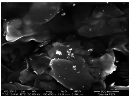
SEM analysis gives the distribution of particles and size of the particle. Excess amount of reducing agent caused dispersion of particles. Average size of the particle was found to be 20 nm. SEM images of gold, silver and copper nanoparticles were given in the figures 4.1 to 4.7.
UV Spectrophotometer Analysis:
Figure 3.8
Figure 3.8
[image:4.612.324.565.137.716.2]International Journal of Emerging Technology and Advanced Engineering
Website: www.ijetae.com (ISSN 2250-2459,ISO 9001:2008 Certified Journal, Volume 3, Issue 8, August 2013)
402 Figure 3.11
Figure 3.12
[image:5.612.50.571.98.695.2]Figure 3.13
Figure 3.14
Figure 3.15
International Journal of Emerging Technology and Advanced Engineering
Website: www.ijetae.com (ISSN 2250-2459,ISO 9001:2008 Certified Journal, Volume 3, Issue 8, August 2013)
[image:6.612.47.289.119.708.2]403 Figure 3.17
Figure 3.18
Figure 3.19
UV Spectrophotometer analysis plays a vital role in
detection of melamine using IB group metallic
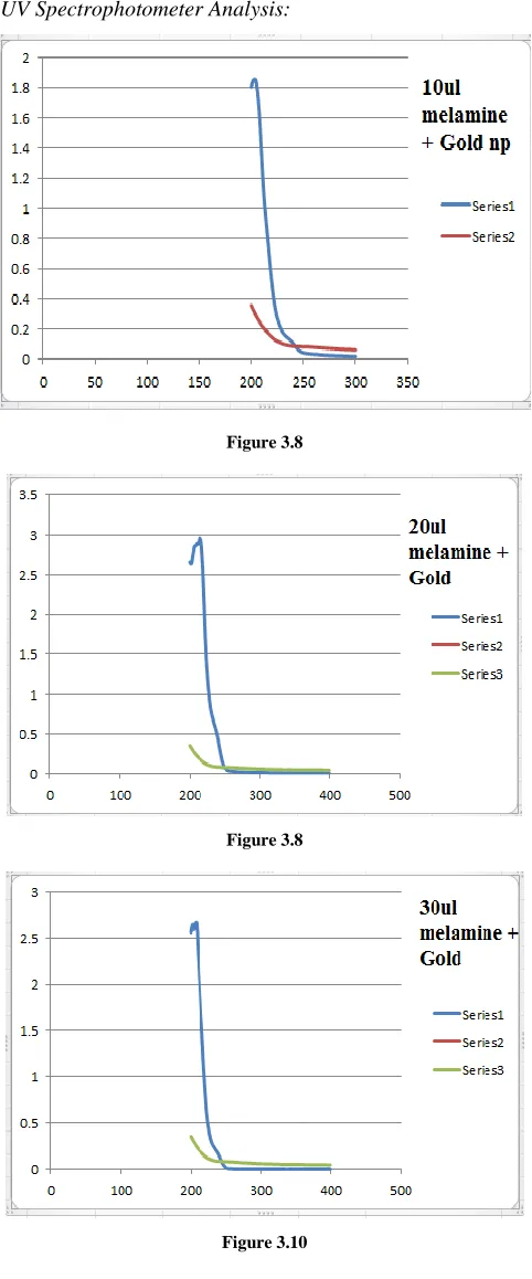
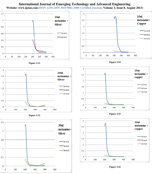
nanoparticles. Figures 4.8-4.16 shows the dampening peak of the solution containing various concentrations of melamine i.e. 10 microliter/ml, 20 microliter/ml and 30 microliter/ml. This is due to the process called Photo bleaching.
Photo bleaching is the photochemical destruction of a dye or a fluorophore. In microscopy, photo bleaching may complicate the observation of fluorescent molecules, since they will eventually be destroyed by the light exposure necessary to stimulate them to produce fluorescence. This is difficult to obtain in time-lapse microscopy.
However, photo bleaching may also be used prior to
adding the (primarily antibody-linked) fluorescent
molecules, in an attempt to quench self fluorescence. This can help to improve signal-to-noise ratio.
Photobleaching may also be exploited to study the motion and/or diffusion of molecules, for example via the FRAP or FLIP techniques.
Loss of activity caused by photo bleaching can be controlled by reducing the intensity or time-span of light exposure, by increasing the concentration of fluorophores, by reducing the frequency and thus the photon energy of the input light, or by employing more robust fluorophores that are less prone to bleaching (e.g. Alexa Fluors or DyLight Fluors). To a reasonable approximation, a given molecule will be destroyed after a constant exposure (intensity of emission X emission time X number of cycles) because, in a constant environment, each absorption-emission cycle has an equal probability of causing photobleaching.
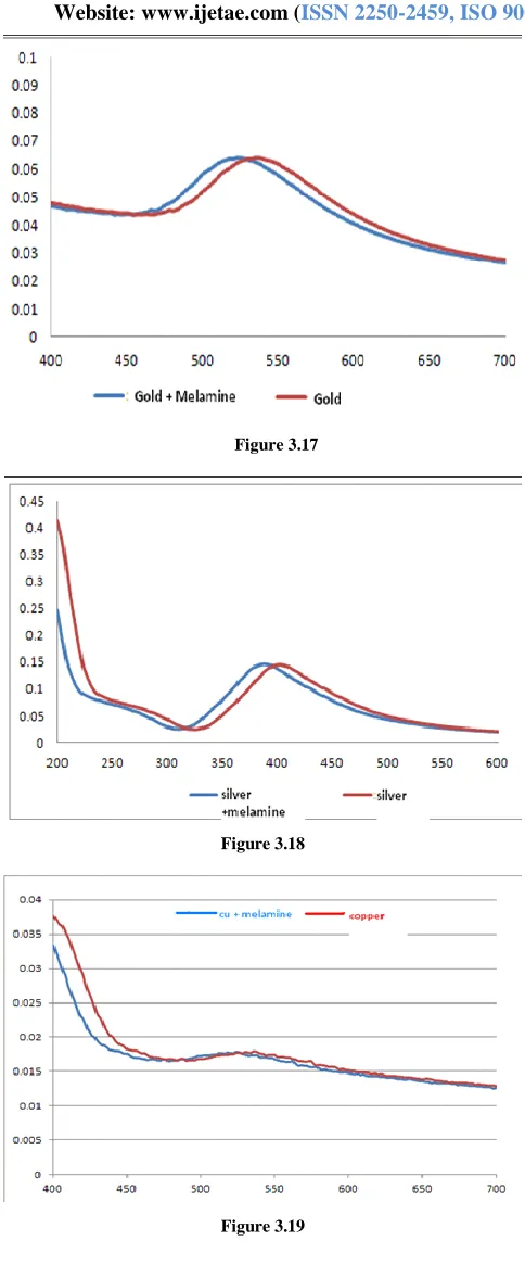
So the concentration has been reduced by ten times. 1 microliter/ml concentration of melamine was made to interact with nanoparticles of IB group elements. UV Spectroscopy of the corresponding solutions was taken. Figures 4.17-4.19 clearly indicate the peak shift.
Figures 4.17-4.19 were depicted in such a way that they were compared with the actual peaks of gold, silver and copper nanoparticle. In case of GoldNP there is blue-shift which indicates blue colour, in case of silver there is peak shift in UV region, indicating detection internally, in case of copper there is a red-shift making solution colourless.
Acknowledgement
International Journal of Emerging Technology and Advanced Engineering
Website: www.ijetae.com (ISSN 2250-2459,ISO 9001:2008 Certified Journal, Volume 3, Issue 8, August 2013)
404 IV. CONCLUSION
Melamine is an organic base and a trimer of cyanamide, with a 1,3,5-triazine skeleton. Excess quantity of melamine when consumed leads to kidney and renal failure. It eventually damages the organs of the body and leads to death. It is necessary to detect melamine in early stage. Nanotechnology is one of the emerging technology which helps in detecting the melamine in early stage . Metallic nanoparticles of IB group elements detect melamine at exponentially lower cost with fast detection technique. Gold nanoparticles are highly instrumental in detection of melamine. Silver and Copper nanoparticle are also helps in the detection of melamine. The expenses can be reduced even further, by using silver and copper nanoparticle . Colorimetric properties indicate the presence of melamine. Change in the color of the metallic nanoparticle solution indicates the presence of melamine in the respective solvent. Thus making the detection of Melamine cheap and quick. This process of detection of various concentrations of melamine using IB group metallic nanoparticles of Gold, Silver and Copper can be installed on Lab-on-a-chip device.
REFERENCES
[1] "Pet food recall". AVMA. Retrieved 2012-06-20.
[2] Scott McDonald, "Nearly 53,000 Chinese children sick from milk", Associated Press (22 September 2008)
[3] Gang et al. (1 September 2009). "The risk of melamine-induced nephrolithiasis in young children starts at a lower intake level than recommended by the WHO". Pediatric Nephrology. Retrieved 2009-10-08.
[4] .E. A. E. Garber, J. Food Prot., 2008, 71, 590–594.
[5] J. P. Toth and P. C. Bardalaye, J. Chromatogr., A, 1987, 408, 335– 340.
[6] T. M. Vail, P. R. Jones and O. D. Sparkman, J. Anal. Toxicol., 2007,31, 304–312.
[7] (a) J.M. Nam, S. J. Park and C. A. Mirkin, J. Am. Chem. Soc., 2002, 124, 3820–3821; (b) P. M. Tessier, J. Jinkoji, Y.-C. Cheng,J. L. Prentice and A. M. Lenhoff, J. Am. Chem. Soc., 2008, 130,3106– 3112.
[8] R. Elghanian, J. J. Storhoff, R. C. Mucic, R. L. Letsinger and C. A. Mirkin, Science, 1997, 277, 1078–1081; (b) J. S. Lee, A. K. R. Lytton-Jean, S. J. Hurst and C. A. Mirkin, Nano Lett., 2007,7,2112– 2115.
[9] J. S. Lee, M. S. Han and C. A. Mirkin, Angew. Chem., Int. Ed., 2007, 46, 4093–4096; (b) D. Li, A. Wieckowska and I. Willner,Angew. Chem., Int. Ed., 2008, 47, 3927–3931.
[10] Y. Jiang, H. Zhao, N. Zhu, Y. Lin, P. Yu and L. Mao, Angew.Chem., Int. Ed., 2008, 47, 8601–8604; (b) M. S. Han, A. K. R. Lytton-Jean, B. Oh, J. Heo and C. A. Mirkin, Angew.Chem., Int. Ed., 2006, 45, 1807–1810.
[11] Chi H, Liu B H, Guan G J, et al. A simple, reliable and sensitive colorimetric visualization of melamine in milk by unmodified Gold Nanoparticles. Analyst,2010,135:1070-1075.
[12] K. C. Grabar, R. G. Freeman, M. B. Hommer and M. J. Natan, Anal. Chem., 1995, 67, 735–743.
![Figure 2.1 B] Synthesis of Silver Nanoparticles](https://thumb-us.123doks.com/thumbv2/123dok_us/8726702.885652/2.612.105.233.493.579/figure-b-synthesis-of-silver-nanoparticles.webp)