5079
Introduction
Stem cells are characterized by their ability to self-renew and to produce numerous differentiated daughter cells. These two special properties enable stem cells to play a central role in generating and maintaining most adult tissues in higher organisms. Embryonic stem cells give rise to all types of tissue stem cells, whereas tissue stem cells are directly responsible for generating and maintaining specific tissues in an organ. Malignant proliferation of stem cells is the leading cause of cancer, whereas under-proliferation of stem cells leads to abnormalities such as tissue dystrophy, anemia, immunodeficiency or infertility. Current research on stem cells has focused on how they self-renew and produce differentiated daughter cells. However, an equally important question is how the fate of stem cells in a tissue is initially established during development. At present, this question remains largely unexplored.
Germline development in Drosophila provides an excellent opportunity to study the establishment of the stem cell fate in individual tissues. This is because the germline in Drosophila is well characterized as a tissue lineage. Moreover, germline development typifies the development of a stem cell-derived tissue. The Drosophila germline originates from pole cells that form at the posterior pole of the syncytial blastoderm. Pole cells then migrate into the abdominal region of the embryo,
where some of them coalesce with somatically derived gonadal mesodermal cells to form two embryonic gonads (Williamson and Lehmann, 1996). The gonadal pole cells are now generally called primordial germ cells (PGCs). During larval development, both PGCs and somatic gonadal cells proliferate but remain relatively undifferentiated. At the larval-pupal transition, ovarian morphogenesis takes place, transforming the larval gonad into the functional adult ovary (Godt and Laski, 1995; King, 1970). During this transition, a subset of PGCs acquire the stem cell fate; they initiate asymmetric divisions to produce differentiated daughter cells called cystoblasts, which eventually develop into an egg chamber (King, 1970; Zhu and Xie, 2003). The continued self-renewing division of germline stem cells (GSCs) in pupal and adult stages leads to the growth and maintenance of the adult ovarian germline as a large and dynamic tissue. Thus, the ovarian germline is a typical stem cell-derived tissue, with stem cells established from a subset of their embryonic precursors at the larval-pupal transition.
Like stem cells in mammalian tissues, GSCs in Drosophila reside in a defined locale, called the stem cell niche, in the tissue (Lin, 2002). In differentiating early pupal ovaries and differentiated adult ovaries, GSCs are located in the most anterior tip of the ovariole, the functional unit of the ovary, in a specialized structure called the germarium. Here, GSCs are in direct contact with somatic cap cells (Deng and Lin, 1997;
A fundamental yet unexplored question in stem cell biology is how the fate of tissue stem cells is initially determined during development. In Drosophila, germline stem cells (GSCs) descend from a subset of primordial germ cells (PGCs) at the onset of oogenesis. GSC determination may occur at the onset of oogenesis when a subset of PGCs is induced to become GSCs by contacting niche cells. Alternatively, the GSC fate could be predetermined for a subset of PGCs before oogenesis, due to either their interaction with specific somatic cells in the embryonic/ larval gonads, or their inherently heterogeneous potential in becoming GSCs, or both. Here, we show that anterior somatic cells in the embryonic gonad already differ from posterior somatic cells and are likely to be the precursors of niche cells in the adult ovary. Furthermore, only pole cells in the anterior half of the embryonic gonad give rise to the PGCs that frequently acquire contact with nascent
niche cells in the late larval ovary. Eventually, only these contacting PGCs become GSCs, whereas non-contacting PGCs directly differentiate into cystoblasts. The strong preference of these ‘anterior PGCs’ towards contacting niche cells does not require DE-cadherin-mediated adhesion and is not correlated with either orientation or rate of their divisions. These data suggest that the GSC fate is predetermined before oogenesis. The predetermination probably involves soma/pole-cell interaction in the anterior half of the embryonic gonad, followed by an active homing mechanism during PGC proliferation to maintain the contact between the ‘anterior PGCs’ and anterior somatic cells.
Key words: Stem cell, Germline, Primordial germ cell, Somatic signalling, Drosophila
Summary
Germline stem cells in the Drosophila ovary descend from pole
cells in the anterior region of the embryonic gonad
Miho Asaoka* and Haifan Lin†
Department of Cell Biology, BOX 3709, Duke University Medical Center, Durham, NC 27710, USA *Present address: The National Institute of Genetics, Mishima, Shizuoka 411-8540, Japan
†Author for correspondence (e-mail: h.lin@cellbio.duke.edu)
Accepted 14 July 2004
Development 131, 5079-5089
Published by The Company of Biologists 2004 doi:10.1242/dev.01391
King, 1970; Lin and Spradling, 1993; Lin and Spradling, 1997; Zhu and Xie, 2003). GSCs divide asymmetrically with regard to cap cells, so that the daughter GSC remains in contact with cap cells while the cystoblast becomes displaced one cell away (Deng and Lin, 1997). Recent studies have shown that signals from cap cells and their neighboring terminal filament cells are essential for GSC maintenance (Chen and McKearin, 2003; Cox et al., 1998; Cox et al., 2000; King and Lin, 1999; King et al., 2001; Song et al., 2004; Song et al., 2002; Xie and Spradling, 1998; Xie and Spradling, 2000). These somatic cells thus form the stem cell niche that ensures the self-renewing ability of GSCs. The self-renewing ability of GSCs also requires interplay between cell-autonomous genes that promote stem cell division and those that drive differentiation (Cox et al., 2000; Forbes and Lehmann, 1998; Lin and Spradling, 1997; McKearin and Spradling, 1990; Parisi and Lin, 1999; Wang and Lin, 2004).
Although much is known about mechanisms that govern GSC maintenance, how the stem cell fate is initially determined during germline development remains unexplored. It is known that four to seven PGCs are partitioned into each germarium during larval-pupal transition (King, 1970; Parisi and Lin, 1999). However, only two to three PGCs in direct contact with cap cells become GSCs in pupal and adult ovaries (Deng and Lin, 1997; King, 1970; Zhu and Xie, 2003), and it remains elusive how these two to three PGCs are selected to contact cap cells and become stem cells. It is possible that all PGCs have an identical potential to become stem cells. In this case, the stem cell fate would not be determined until the onset of oogenesis during the larval-pupal transition, when niche cells start to induce their adjacent PGCs to become GSCs (herein called the late induction hypothesis). Alternatively, stem cell fate determination could occur before the onset of oogenesis (herein called the predetermination hypothesis). The predetermination could be caused by interaction between a subset of PGCs with specific somatic cells in the embryonic/ larval gonads (herein called predetermination by induction), or by the inherent heterogeneity of pole cells in developmental potential, so that some of them are already destined to stem cell fate when they are formed (herein called predetermination by lineage). Finally, both mechanisms may participate in the predetermination process. Among these possibilities, both the late induction and predetermination-by-induction hypotheses are supported by the role of the stem cell niche in GSC maintenance (Lin, 2002), whereas the predetermination-by-lineage hypothesis is consistent with the heterogeneity of pole cells in polar granule content, mitotic rate, and splicing activity towards P-transposase pre-mRNA (Kobayashi et al., 1993; Sonnenblick, 1950; Technau and Campos-Ortega, 1986). Here, we report experiments indicating that the GSC fate is specified by predetermination and not the late induction mechanism. Furthermore, our data indicate the potential involvement of somatic induction in the predetermination process.
Materials and methods
Drosophila culture and stocksThe lacZ enhancer-trap line 3914 was as described previously (Asaoka et al., 1998). EGFP-vas (Sano et al., 2002) and y w flies were used as sources of donor embryos and hosts for pole-cell transplantation, respectively. FRT42DshgR69/CyO (Godt and Tepass,
1998) and w hs-FLP; FRT42Darm-lacZ flies were used for germline clonal analyses. All stocks were maintained at 25°C on standard Drosophila medium.
‘Single’ pole cell transplantation
Pole-cell transplantation was conducted at 22°C as described (Kobayashi et al., 1996). Donor eggs and host eggs were collected at 50-minute intervals, and then allowed to develop to 150-200 minutes after egg laying. Pole cells were isolated from donor embryos at the cellular blastoderm stage and injected into the posterior pole of same stage host embryos. Only one to three donor pole cells (average 2.2 pole cells) from a single embryo were injected to a host embryo. This number of transplanted pole cells ensures that each host receives a single donor pole cell in its gonads at a high frequency. The injected embryos were kept at 18°C until embryonic gonads were formed (stage 14-15). Each of these stage 14-15 embryos were transferred into a small drop of silicone oil on the microscope slide, covered with a coverslip, and then examined under the fluorescent microscope to locate the donor pole cell in the host embryonic gonad. After examination, the host embryos were kept at 18°C in a moist chamber until they hatched into larvae. The hatched larvae were collected and divided into different groups according to the number and location of labeled pole cells in the gonads at stage 14-15. Each group of larvae was transferred to standard Drosophila medium in individual 35-mm culture dishes (Becton Dickinson Labware, Franklin Lakes, NJ) and incubated at 25°C until pupation. The culture dishes were supplemented with TM6-bearing larvae so that total number of the larvae was 15-17 per dish, as too few larvae in a dish causes low viability. The pupae were transferred into vials with fresh Drosphila medium and raised at 25°C. The TM6-bearing larvae and the adults were recognized by their Tubby phenotype and the wild-type eye color, respectively, and were eliminated. Ovaries and testes were dissected from each host animal at the late third instar larval/prepupal stage, or at the adult stage (within 24 hours after eclosion), and then fixed and stained separately, as described below.
Dissection of larval and adult ovaries and testes
The dissection and fixation of larval and adult ovaries and testes was performed as previously described for the adult ovary (Lin et al., 1994). After the 3-minute fixation and several washes, the adult samples were stained with 1 µg/ml 4′,6-diamidino-2-phenyindole (DAPI) in 13PBS for 10 minutes, then mounted in 50% glycerol in 13PBS and 2% anti-quenching agent DABCO, and examined by fluorescent microscopy to identify GFP-labeled cells. For larval gonads, the GFP signal was weak, so the GFP expression was detected by indirect immunofluorescence microscopy (see below).
Histochemical and immunological staining
Confocal images were collected using a Zeiss LSM510-META confocal microscope.
For ovary staining with anti-DE-cadherin antibody, ovaries were dissected in 13EBR (Lin et al., 1994) and fixed in 3.7% formaldehyde in PCM (100 mM PIPES pH 6.9, 1 mM CaCl2, 2 mM MgSO4) for 15 minutes. The staining procedures and image collection were the same as described above, except that a PBT solution containing 0.4% of Triton X-100 was used instead of the standard PBT solution.
Germline clone analysis
The shotgun (shg) germline clone was generated using the FLP-DFS technique, as described previously (Chou and Perrimon, 1996). w hs-FLP /Y; FRT42Darm-lacZ males were mated with FRT42DshgR69/CyO virgin females. Egg collection was performed in the culture tubes for 12 hours. The culture tubes containing the first instar larvae (34-46 hours after egg laying) were heat shocked for 1 hour in a 37°C water bath to induce mitotic recombination. The ovaries of the heat-shocked larvae were dissected at late third instar larval stage, and processed for immunostaining and confocal microscope analysis. shg mutant clones and siblings were identified as negative and lacZ-overexpressed PGCs, respectively.
Results
3914 is a marker for anterior somatic cells in both female and male embryonic gonads
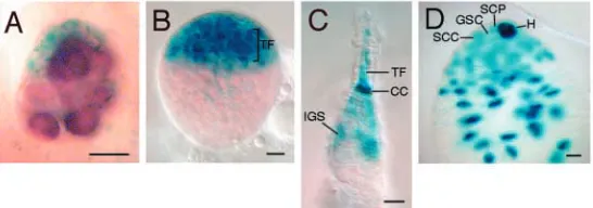
[image:3.612.294.567.514.610.2]To distinguish between the late induction and predetermination hypotheses, we reasoned that the latter would predict certain differences among PGCs that would correlate with their fate as GSCs in the adult ovary. Such differences could occur as early as in the embryonic stage. In this case, if predetermination involves somatic induction, one would expect that the embryonic gonad would be composed of different subset of somatic cells, so that only those pole cells adjacent to a particular subset of somatic cells were induced to become GSCs. By contrast, if predetermination is solely due to a heterogeneous lineage mechanism, one would expect to see at least two types of pole cells in the embryonic gonad, with their positions in the gonad bearing no correlation with a particular subset of somatic gonadal cells. Previous studies have shown that somatic cells in the anterior region of the gonad are different from posterior somatic cells, and that, in the testis, the anterior tip of somatic gonads appears to become hub cells that are equivalent to terminal filament and cap cells in the ovary (Boyle and DiNardo, 1995; DeFalco et al., 2003; Gönczy et al., 1992; Kiger and Fuller, 2001; Russell et al., 1992). Here, we describe an enhancer-trap marker, 3914, that is expressed specifically in somatic cells in the anterior half of both female and male embryonic gonads. As shown in Fig. 1, the enhancer-trap marker 3914 starts to be expressed in somatic cells of the embryonic gonads immediately following gonad formation (stage 14) (Asaoka et al., 1998). In 95% of stage 14-15 embryos, 3914 is expressed in the anterior half of the somatic gonad (Fig. 1A). Expression in the posterior half of the somatic gonad is detectable only in 11% of embryos (n=38). Even in these embryos, the anterior expression is always much stronger than the posterior expression. The anterior-specific expression of 3914 is observed in both sexes, as confirmed by staining for expression of the Sxl protein, which reveals the sex identity of individual embryos (data not shown). These results confirm that,
even in the newly formed embryonic gonad, anterior somatic cells are already different from posterior somatic cells.
The strong anterior expression of 3914 persists in both male and female gonads through to the end of embryogenesis (stage 17; 100% of embryos, n=27). At this stage, although 80% of female embryos expand 3914 expression posteriorly to cover two-thirds of the gonad, the posterior somatic cells only display weak expression. In all male embryos, 3914 expression is also expanded posteriorly to cover two-thirds of the gonad. However, unlike in female gonads, all of these cells maintain a uniformly strong expression, with the strongest expression confined to the anterior tip area (data not shown). This pattern may reflect the fact that male gonads initiate spermatogenesis by the end of embryogenesis (Cooper, 1950), and thus contain 3914-positive somatic cyst progenitor cells in the posterior region (cf. Fig. 1D, and see below).
Anterior somatic cells in the embryonic gonad are likely to be the precursors of GSC niche cells in the ovaries and testes
In adult ovaries and testes, anterior somatic cells constitute a stem cell niche that plays a crucial role in maintaining GSCs (Lin, 2002). In ovaries, niche cells include terminal filament and cap cells, and possibly inner germarial sheath cells (Fig. 1C). These cell types form in the anterior region of ovaries at the larval-pupal transition stage (Godt and Laski, 1995; King, 1970). In testes, the niche is composed of hub cells, somatic cyst progenitor cells, and, possibly, early somatic cyst cells (Fig. 1D). Testicular niche morphogenesis begins near the end of embryogenesis (Cooper, 1950). To determine whether the 3914 enhancer activity correlates with the signaling ability of somatic cells, we examined 3914 expression in larval and adult ovaries, as well as in the adult testis.
In late third instar larval ovaries, the 3914 enhancer trap is expressed in the anterior somatic region, where forming terminal filament and cap cells reside (Fig. 1B). Particularly, strong expression was detected in developing terminal filament and cap cells. However, expression was never detected in the medial region that contains PGCs and somatic interstitial cells,
or in the posterior region that includes the precursors of basal stalk cells.
In the adult ovary, 3914 is expressed strongly in cap cells, the central component of the niche, with weaker expression in terminal filament and inner germarial sheath cells (Fig. 1C). The expression pattern of 3914 in embryonic gonads, larval and adult ovaries suggests that the anterior somatic cells in the embryonic gonad are likely to be the precursors of stem cell niche cells in the larval and adult ovaries, and that they may also have a signaling function.
In the adult testis, 3914 was also expressed in the niche cells, with strong expression in hub cells, the central component of the niche, and weaker expression in somatic cyst progenitor cells and their daughter cells, the somatic cyst cells (Fig. 1D). This corroborates a previous enhancer-trap study that suggests that the hub precursors are a subset of cells located at the anterior tip of the embryonic gonad (Gönczy et al., 1992). Moreover, the broader expression pattern of 3914, covering the entire anterior half of the embryonic gonad, suggests that these anterior somatic cells not only give rise to hub
cells, but also to somatic cyst progenitor cells and their daughter cyst cells that form the niche in the apical region of the testis.
Anterior and posterior pole cells in the embryonic gonad give rise to GSCs and cystoblasts, respectively
Our analysis of 3914 expression raises the possibility that anterior somatic cells of the embryonic gonad are involved in determining the GSC fate, and that this fate could be determined as early as in the early embryonic gonad. To test this possibility, we investigated whether only those pole cells in contact with the anterior somatic cells in the embryonic gonad give rise to GSCs in the adult ovary. To this end, we traced the position and fate of single pole cells from early embryonic gonadal stage to the adult stage using a vasa-EGFP gene that is specifically and continuously expressed in the germline (Sano et al., 2002). The following strategy was used for the lineage tracing (see Materials and methods). A single EGFP-marked wild-type pole cell was transplanted into the pole cell region of a wild-type recipient embryo at the cellular blastoderm stage. The recipient embryos then continued to develop to stage 14-15, at which time the position of the GFP-marked pole cells in the newly formed embryonic gonad was examined and recorded. The embryo was then allowed to develop to adulthood. Its ovaries or testes were then dissected and analyzed to determine the fate of the EGFP-marked pole cell.
We transplanted EGFP-marked pole cells into 1425 cellular blastoderm embryos, 694 of which incorporated a marked pole cell in their gonads following gonadal formation (stages 14-15; Fig. 2A-D). Eventually, 45 of such marked embryos developed into pupal-adult females. Of these, 19 females were developed from embryos with the marked pole cell located in the anterior half of the
embryonic gonad (anterior pole cells; Fig. 2A,B). Seven such females still retained EGFP-marked germ cells in the ovary. Twenty-four females developed from embryos with a marked pole cell in the posterior half region of the embryonic gonad (posterior pole cell; Fig. 2C,D). Ten such females still retained EGFP-marked germ cells in the ovary. Thus, the survival rates for the transplanted anterior and posterior pole cells are very similar (37% versus 42%, P>0.1). Finally, two females developed from embryos with a marked pole cell on the exact midline of the embryonic gonad were excluded from this analysis.
Despite their similar survival rates, the fates of anterior and posterior pole cells are very different. Out of seven labeled anterior pole cells, six gave rise to EGFP-labeled GSCs (86%, Fig. 2E-H); only one directly differentiated into a cystoblast (Fig. 3). The presence of an EGFP-marked GSC in contact with cap cells is obvious (Fig. 2E-H). As expected, it had produced multiple EGFP-labeled germline cysts and developing egg chambers by the adult stage. These labeled germline cysts
Fig. 2. Pole cells located in the anterior half of the embryonic gonad develop into GSCs in the adult ovary. (A-D) Embryonic gonads viewed as superimposed DIC and EGFP images, showing representative locations of an EGFP-marked pole cell in the embryonic gonad. All gonads are oriented with anterior to the left and posterior to the right. The outline of the gonad is evident in DIC, with arrowheads pointing to the gonadal border to assist viewing. A marked pole cell (white spheres) is located at the anterior tip (A), anterior (B), posterior (C), or posterior tip (D). Dotted lines show the midline of the gonads. (E,G) An ovariole with one EGFP-labeled GSC (white arrow in G), as shown in DIC (E) and corresponding EGFP (G) images, from a female developed from an embryo with a marked pole cell located in the anterior half of the gonad. The EGFP-labeled GSC has produced multiple labeled germline cysts and egg chambers, which form an alternating pattern of labeled germline cysts and egg chambers in the ovariole. (F,H) An ovariole with both GSCs labeled by EGFP (white arrows in H), as shown in DIC (F) and corresponding EGFP (H) images, from a female developed from an embryo with a marked pole cell located in the anterior half of the gonad. The EGFP-marked GSCs have produced a
[image:4.612.254.562.299.552.2]and egg chambers were interspersed with their unlabeled counterparts derived from the unlabeled GSC(s) in the same germarium, forming an ovariole consisting of a string of alternating ‘bright’ and ‘dark’ egg chambers (Fig. 2E,G). When a single labeled pole cell gives rise to both (or all three) GSCs in a germarium, then the entire ovariole is composed of uniformly ‘bright’ egg chambers (Fig. 2F,H).
By contrast, among 10 labeled posterior pole cells, nine directly differentiated into cystoblasts (90%, Fig. 3). Only one generated a single EGFP-labeled GSC (P<0.005, versus anterior pole cells). The cystoblast fate can also be easily discerned, because the labeled cystoblasts, by the adult stage, have further developed into germline cysts (Fig. 3A,C), or even egg chambers (Fig. 3B,D). In these ovarioles, only a small fraction of cysts and/or chambers were labeled (1-7, average number/ovariole= 2.22, number of ovarioles examined=74). Taken together, the above observations indicate a strong tendency for anterior pole cells in the embryonic gonad to give rise to GSCs, whereas the posterior pole cells tend to give rise to PGCs in the larval ovary that directly differentiate into cystoblasts.
PGCs derived from a single pole cell are not anchored to each other or to specific somatic cells during proliferation
How can an anterior pole cell in the embryonic gonad give rise to GSCs? Three recent findings suggest that anterior pole cells are anchored to anterior somatic cells in the embryonic gonad and that this anchorage is maintained during larval development so that a pole cell gives rise to a clone of PGCs that are partitioned only into a few adjacent germaria during the larval-pupal transition. First, E-cadherin is required in both pole cells and somatic gonadal precursor cells for the formation
of embryonic gonad (Jenkins et al., 2003). Second, in the early pupal ovary, a PGC in contact with newly formed cap cells tends to divide along the plane of cap cells, generating two to three GSCs that all contact cap cells within a germarium [‘clonal expansion’ theory (Zhu and Xie, 2003)]. Third, in the adult ovary, E-cadherin is essential for anchoring GSCs to cap cells to maintain their stem cell fate (Song et al., 2002).
To examine whether PGCs derived from an anterior pole cell adhere to each other and to anterior somatic cells during their proliferation, we first looked at the distribution of daughter cells of a single, labeled anterior pole cell in larval/prepupal ovaries. We transplanted EGFP-labeled pole cells into 520 blastoderm embryos, recorded the position of the single, labeled pole cell in the newly formed gonad of the recipient embryos (stage 14-15), allowed the embryo to develop to the late third instar larval or prepupal stage, and then isolated ovaries for immunofluorescence analysis (see Materials and methods). Such ovaries were stained with anti-GFP antibody to identify PGCs derived from the marked pole cell, and with anti-1B1 antibody, both to outline the developing terminal filament and cap cells and to label spectrosomes (Deng and Lin, 1997).
To our surprise, only 14% of ovaries contained a clone of labeled germ cells in contact with one another (n=14). By contrast, in 72% of ovaries, labeled cells were dispersed widely along both the anteroposterior and mediolateral axes, with some PGCs even passing across the midline into the posterior region (17% of ovaries, Fig. 4C, Fig. 5A). In particular, in 43% of ovaries, all labeled daughter cells were highly dispersed, whereas a small number of labeled cells were associated in another 29% of ovaries. Even in these 29% of ovaries, most PGCs were dispersed several cells away from each other. The remaining 14% of ovaries had only one labeled daughter cell. These results indicate that PGCs derived from an anterior pole cell do not adhere to each other or to anterior somatic cells during their proliferation. Instead, they become dispersed and intermixed with other pole cell progeny during larval development. Furthermore, as there was no difference in the extent of dispersion between daughter cells derived from anterior versus posterior pole cells, it can be surmised that PGCs derived from a single pole cell are dispersed during proliferation irrespective of the original position of the pole cell in the embryonic gonad.
Similarly, in adult ovaries, GSCs derived from one pole cell were widely distributed to multiple niches. Single-labeled anterior pole cells each produced 3-12 labeled GSCs (average 7.3) that were distributed into 1-10 ovarioles (average 4.8) within an adult ovary [n(ovariole)=122; n(ovary)=6]. Among these ovarioles, 83% of them contained only one labelled GSC, as evident by the alternating pattern of the labeled egg chambers (Fig. 2E,G). In only 17% of ovarioles were the two to three GSCs all labeled (Fig. 2F,H).
[image:5.612.49.298.74.220.2]A similar frequency of dispersion was observed for PGCs derived from posterior pole cells, as inferred from the labeling pattern in the adult ovary. A single, labeled posterior pole cell produced 2-35 differentiated daughter cells (average 16.4) that were distributed into 1-13 ovarioles (average 7.4), which represents, on average, 31% of ovarioles in an ovary [n(ovariole)=236; n(ovary)=10]. In 45% of these ovarioles, only one germline cyst or egg chamber was labeled (n=74). These results indicate that PGCs derived from a single posterior pole cell are also dispersed during ovarian morphogenesis.
PGCs derived from anterior pole cells preferentially enter nascent stem cell niches
As only anterior pole cells give rise to GSCs, we would expect their daughter PGCs to be much more accessible to nascent stem cell niches in larval/prepupal ovaries, even
[image:6.612.48.293.143.521.2]though they are not anchored to anterior somatic cells in the gonad. To test this hypothesis, we determined whether only PGCs derived from anterior pole cells would contact developing cap cells in late third instar larval and prepupal ovaries. As expected, 90% of PGCs derived from anterior pole cells were in contact with developing cap cells [Fig. 4A,A′; n(labelled PGC)=19; n(ovary)=6]. Only 10% of the PGCs were not in contact with developing cap cells. This
Fig. 4. PGCs derived from anterior pole cells preferentially associate with the nascent cap cells. (A,A′) A late third instar larval ovary derived from an embryonic gonad containing a labeled anterior pole cell viewed under low and high magnification, respectively. The ovary, with the anterior pole up, is stained with anti-1B1 antibody (red) to outline somatic cells and spectrosomes in germ cells, and with anti-GFP antibody (green) to identify PGCs derived from the marked pole cell. Note that the PGC marked by an arrow is in direct contact with the base of two forming terminal filaments (TF). (B,B′) A late third instar larval ovary derived from an embryonic gonad containing a labeled posterior pole cell viewed under low and high magnification, respectively. The ovary, with the anterior pole up, is stained with anti-1B1 (red) and anti-GFP (green) antibodies. Note that the PGC marked by an arrow is approximately six cells away from the base of the forming terminal filaments (TF). (C) Summary of the location of PGCs (green) derived from marked anterior or posterior pole cells (green). The fractional numbers at the lower right corner of each third instar larval or prepupal (3°L/PP) ovary indicates the number of ovaries with shown location of PGCs versus the total number of this class of 3°L/PP ovaries examined. Yellow cells are nascent TFs. Scale bars: in A, 20 µm for A,B; in A′, 10 µm for A′,B′.
[image:6.612.333.537.177.454.2]frequency corresponds well with the observation that 14% of anterior pole cells produce only differentiating germline cyst or egg chambers in the adult ovaries (see above). By contrast, 92% of PGCs derived from posterior pole cells were in not contact with developing cap cells in the larval/prepupal ovaries [Fig. 4B,B′; n(labelled PGC)=38; n(ovary)=8]. The difference between the frequencies of anterior and posterior pole cells in giving rise to cap cell-contacting PGCs (90% versus 8%, respectively) is highly significant (P<0.0001). These results show that PGCs derived from anterior pole cells have a strong tendency to become stem cells because they preferentially enter the nascent stem cell niche in the larval/ pupal transitional ovaries.
DE-cadherin is not required for PGCs to enter the nascent stem cell niche
Then, how do PGCs derived from anterior pole cells preferentially enter the nascent stem cell niche? It may be due to one of the following four mechanisms, or a particular combination of them. (1) Anterior pole cells and their progeny PGCs are anchored to the anterior somatic gonadal cells and their progeny via static cell-cell adhesion, such as adherens or desmosomes junctions (static anchorage mechanism). (2) PGC divisions occur more frequently along the niche cell/PGC interface, owing to somatic induction or mechanical constraints of the ovary (differential mitotic orientation mechanism). (3) PGCs derived from an anterior pole cell divide at a slower rate so that their progeny is more confined to the anterior region (differential mitotic rate mechanism). (4) The dispersed PGCs derived from the anterior pole cells actively home back to the niche cell/PGC interface in response to somatic induction (active homing mechanism).
The static anchorage mechanism is unlikely to play a significant role, because, as described above, the progeny of a single pole cell are widely dispersed in the larval gonad both laterally and anteroposteriorly, irrespective of the position of the original pole cell (see above; Fig. 4C, Fig. 5A). Thus, even though cell adhesion molecules such as E-cadherin are required for the association of pole cells and somatic cells in the embryonic gonad, and for the later anchoring of GSCs to cap cells in adult ovaries (Jenkins et al., 2003; Van Doren et al., 2003; Song et al., 2002), such adhesion may not function during PGC proliferation in the larval gonad. In support of this reasoning, DE-cadherin and β-catenin only start to accumulate at the interface between nascent cap cells and future GSCs at the late third instar larval stage (Song et al., 2002).
To determine whether DE-cadherin is required for PGCs even to enter the nascent stem cell niches, we used a FLP-mediated mitotic recombination technique to induce the production of PGCs that are null for the DE-cadherin gene shotgun (shg) in the shg–/+ first instar larvae (see Materials and methods). The shg–/shg–PGCs and their +/+ sibling PGCs are marked by zero and two copies of the lacZ gene, respectively (Fig. 5). We then compared the locations of the marked DE-cadherin-deficient PGCs and their wild-type sibling PGCs in late third instar larval ovaries. The marked DE-caderin-deficient PGCs and their wild-type sibling PGCs show similar distributions in the anterior and posterior regions of the ovary, with the wild-type PGCs actually distributed somewhat more in the posterior region (Fig. 5E). In particular, 16.8±4.6% of DE-cadherin-deficient PGCs were in contact with cap cells, as
compared with 6.1±3.2% of their siblings (P=0.066, t test, Fig. 5E). These data indicate that DE-cadherin is not required for PGCs to enter the nascent niches, and that it might constrain PGCs from actively reaching cap cells.
The preferential niche occupancy of PGCs derived from anterior pole cells is not due to differential orientation or rate of divisions
We next examined whether PGCs derived from anterior pole cells preferentially enter the nascent stem cell niche as a result of differential mitotic orientation or rate. In third instar larval ovaries, the divisional orientation of PGCs can be easily observed at the prolonged telophase/G1 phase, when the dividing GSC pair displays dumbbell morphology with an elongated spectrosome located in the neck region (Fig. 6A-D). Moreover, the frequency of the prolonged telophase/G1 phase GSC pairs reflects the frequency of PGC divisions. The mitotic orientation of cap cell-contacting PGCs displays a random distribution that is very similar to anterior PGCs that are not in contact with cap cells and posterior PGCs (Fig. 6). These results show that the strongly preferential entry of PGCs derived from anterior pole cells into the nascent stem cell niche is not due to the differential orientation mechanism.
It is also unlikely that a slower mitotic rate of PGCs derived from anterior pole cells confine them to the niche cell/PGC interface. First, these PGCs are widely dispersed throughout the larval ovary irrespective of their mitotic rate (see above; Fig. 4C, Fig. 5A). Second, the mitotic rate of these PGCs is very similar to that of other PGCs in the same larval ovary. In the third instar ovary, the frequency of telophase/G1 PGCs in contact with cap cells is 8.9±7.9% [n(PGC)=181; n(ovary)=13], very similar to that of the remaining PGCs in the ovary [5.7±2.3%; n(PGC)=787; n(ovary)=13; P=0.173]. These results rule out the existence of the differential mitotic mechanism in GSC determination.
Both anterior and posterior pole cells can give rise to GSCs in the testis
Fig. 6. PGCs proliferate with random divisional orientation. (A-D) Late third instar larval ovaries containing dividing PGCs (outlined by thick dotted lines). The ovaries were double stained with anti-Vasa antibody (green) to label all PGCs, and anti-1B1 antibody (red) to visualize spectrosome orientation in PGCs and to outline somatic cells. Dividing PGCs in contact with TF/CpCs (outlined by thin dotted lines) are oriented perpendicularly (A,C) or in parallel (B), or approximately 45° (D) to the cap cell-PGC interface. PGCs in contact with posterior somatic cells also divide perpendicularly (C), or in parallel (A,B), to the soma-germline interface. (E) A quantitative comparison of the divisional orientation of PGCs contacting CpCs, of those in the anterior half but not contacting cap cells (anterior PGCs), and of those in the posterior half (posterior PGCs). The CpC-PGC interface is assigned a value of 90°. Each red dot in the diagram represents an individual sample (n=63).
[image:8.612.120.484.420.631.2]Discussion
A fundamental question in stem cell biology concerns how the fate of tissue stem cells is determined during development. GSCs in the Drosophila ovary represent an excellent model to explore this essentially uncharted territory. It has been documented that functional GSCs are established at the onset of oogenesis during larval-pupal transition (King, 1970; Zhu and Xie, 2003). However, when and how the GSC fate is initially determined during pre-oogenic development remains elusive. The study reported here represents the first effort to investigate the process of GSC fate determination.
The GSC fate is predetermined prior to GSC establishment
The process of cell fate determination is known to precede the establishment of the determined cell type in a few studied systems. For example, the initial cue for the determination of the anteroposterior axis of the Drosophila embryo lies in its remote ancestor, the 16-cell germline cyst that resides in the germarium of the ovary of the mother (Manseau and Schüpbach, 1989). The posterior location of the oocyte in the cyst determines the anteroposterior axis of the future embryo. As another example, the initial determining cue for the oocyte fate within the 16-cell germline cyst appears to be already present in its four-generation-removed precursor cell, the cystoblast (Lin and Spradling, 1995). The asymmetric retention of the spectrosomal material in the daughter ‘cystoblast’ during the subsequent four rounds of division determines its fate as an oocyte. Here, we have shown that the GSC fate is predetermined prior to oogenesis, with the predetermination apparently occurring as a continuous process (see below), starting at least as early as in the newly formed embryonic gonad; the location of a subset of pole cells in the anterior half of the gonad provides the first positional cue that predetermines the strong preference of their progeny for entering nascent stem cell niches in the late third instar larval ovaries, and eventually their fate as GSCs.
Embryonic gonad exerts somatic induction in GSC fate predetermination
Then, how does this positional cue determine the fate of future GSCs? Our study suggests that, at least, a somatic induction mechanism is involved in the process. Even if a subset of pole cells is destined to become GSCs upon their formation as a result of the heterogeneous lineage mechanism, specific interactions must occur between these ‘privileged’ pole cells and anterior somatic cells. Otherwise, such pole cells could locate to any region of the gonad. This signaling function of the anterior somatic cells in the embryonic gonad is consistent with their being the likely precursor cells of the future niche cells.
The signaling from the anterior somatic cells to anterior pole cells must be via direct cell-cell contact, because every somatic cell in the embryonic gonads extends its cellular processes to envelop individual pole cells (Jenkins et al., 2003; Van Doren et al., 2003). Therefore somatic signalling via direct cell-cell contact would ensure that anterior pole cells receive the signal, while preventing posterior pole cells from receiving the signal.
Somatic induction may exist in the larval gonad for PGCs to enter the nascent niche by an active homing mechanism
[image:9.612.47.560.487.722.2]Even though somatic induction in the embryonic gonad, either alone or together with a heterogeneous lineage mechanism, is likely to be responsible for initiating the GSC predetermination process, this embryonic mechanism alone is not sufficient for GSC fate determination. This is because different PGCs derived from the same pole cells can adapt different fates. We found that approximately 17% of single anterior pole cells gave rise to both PGCs that are in contact with cap cells in the late third instar larval ovary and PGCs that are distant to cap cells (Fig. 4C). Correspondingly, approximately 14% of anterior pole cells gave rise to both GSCs and cystoblasts in the adult
Fig. 8. The somatic induction model for GSC fate determination. For details, see text.
s C S G f o t n e m h s i l b a t s E . 3 p e t S s C G P f o t n e m t r o s s A e v i t c A . 2 p e t S s C G P o t e u C l a n o i t i s o P 1 p e t S
(embryonicgonad) (larvalgonad) (prepupalgonad)
ovary (data not shown). These observations suggest that predetermination in the embryonic gonads is somewhat flexible. Only those PGCs in contact with nascent niche cells in the late third instar larval ovary appear to become GSCs.
How, then, do PGCs derived from anterior pole cells maintain their contact with anterior somatic cells during larval development? Our study rules out any significant role of static anchorage, differential mitotic orientation or differential mitotic rate in the process (see Results). One possibility is that, even though PGCs derived from an anterior pole cell become passively dispersed during proliferation, they tend to populate the anterior region. Our data do not rule out this mechanism. However, these data favor the active homing mechanism. Passively dispersed PGCs derived from an anterior pole cell are selectively sorted out into the niche cell/PGC interface in the larval gonad as a result of niche signaling, where they become anchored to newly formed cap cells during the larval-pupal transition. Such somatic signaling could start in embryonic gonads. Consistent with this, a gap junction protein, Drosophila Innexin 4, is present in pole cells in embryonic gonads, even though its function at this stage is not clear (Tazuke et al., 2002). Regardless of when the somatic induction is initiated, it probably plays an important role in ensuring that the progeny of anterior pole cells in the embryonic gonad will sustain their proximity to the future stem cell niche.
Potential role of the heterogeneous lineage mechanism in GSC fate determination
This study does not directly address the role of the heterogeneous lineage mechanism in initial GSC fate determination during embryogenesis because we did not distinguish individual donor pole cells for their intrinsic properties, such as polar granule content or their initial position in the embryo. However, our study suggests that the lineage mechanism is unlikely to play any significant role in the maintenance phase of the predetermination process during larval development, because a labeled pole cell, either anterior or posterior, can produce both cap cell-contacting and non-contacting PGCs in the larval ovary, as well as both GSC and cystoblasts in the adult ovary (see Results). These results suggest that the lineage mechanism does not govern the fate of pole cell progeny in the larval ovary.
A model for the determination of stem cell fate in the female germline
The discussion above allows us to propose a somatic induction hypothesis for GSC fate determination during female germline development (Fig. 8). In this model, the anterior positioning of pole cells in the embryonic gonad provides the initial positional cue that predetermines the fate of future GSCs. Such predetermination is initiated by cell-cell interactions between somatic cells and pole cells in the anterior half of the embryonic gonad, and is maintained by signalling from anterior somatic cells in the larval gonad. Such somatic signaling would lead to the active homing of PGCs derived from the anterior pole cells to maintain their contact with the signaling cells in the larval gonad. At the late third instar larval stage, the developing terminal filament and cap cells express DE-cadherins and other molecules to anchor the adjacent PGCs, transforming them into GSCs. Finally, it is possible that pole cells are heterogeneous in their potential upon their
formation, which might affect their initial choice of position in the embryonic gonad. If so, the lineage and somatic induction mechanisms would act in sequence to determine the GSC fate.
Difference between female and male GSC fate establishment
In male gonads, both anterior and posterior pole cells can give rise to GSCs, which suggests that the male embryonic gonad has a larger niche for GSC fate establishment than the female gonad. This is consistent with the finding that the 3914 enhancer trap becomes quite uniformly expressed in the anterior two-thirds of the male gonad towards the end of embryogenesis. As male GSCs are established by the end of embryogenesis, the GSC niche must have been established by this point. Specifically, hub cells, somatic cyst progenitor cells, and possibly their daughter cyst cells, have formed (Cooper, 1950). As somatic cyst progenitor cells and their daughter cyst cells express 3914 (Fig. 1D), and occupy the posterior half of the embryonic gonad, male gonads show posterior 3914 staining. The fact that some posterior pole cells are also destined to become GSCs suggests that the hub cells extend to the posterior half to contact some of the posterior pole cells during embryogenesis.
Sex incompatibility of transplantation
As we did not select donor and host embryos according to their sex for transplantation, 50% of transplanted embryos should have received pole cells of the opposite sex. Previous studies have shown that XY pole cells in female gonads never enter oogenesis. They can sometimes survive to enter spermatogenesis, producing germline cysts containing 30-1000 cells. These cells include undifferentiated germ cells, spermatocytes and many degenerating cells (Steinmann-Zwicky et al., 1989). In our experiments, however, no such cyst was detected, suggesting that the transplanted male pole cells have degenerated in female hosts before the adult stage. This degeneration could be due to the difference of hosts: Steinmann-Zwicky et al. (Steinmann-Zwicky et al., 1989) transplanted multiple pole cells into germlineless embryos, whereas we transplanted only a few pole cells into wild-type embryos containing a normal complement of endogenous pole cells. Thus, XY germ cells may be effectively out-competed by the XX poles cells in the female gonads.
XX pole cells in male gonads can also enter an abortive spermatogeneic pathway. However, most of them degenerate before adult stage, even when they were transplanted into an agametic gonad (Steinmann-Zwicky et al., 1989). As expected, we saw no oogenic cells in the testis.
References
Asaoka, M., Sano, H., Obara, Y. and Kobayashi, S. (1998). Maternal Nanos regulates zygotic gene expression in germline progenitors of Drosophila
melanogaster. Mech. Dev. 78, 153-158.
Asaoka-Taguchi, M., Yamada, M., Nakamura, A., Hanyu, K. and Kobayashi, S. (1999). Maternal Pumilio acts together with Nanos in germline development in Drosophila embryos. Nat. Cell Biol. 1, 431-437.
Boyle, M. and DiNardo, S. (1995). Specification, migration and assembly of the somatic cells of the Drosophila gonad. Development 121, 1815-1825. Chen, D. and McKearin, D. (2003). Dpp signaling silences bam transcription
directly to establish asymmetric divisions of germline stem cells. Curr. Biol. 13, 1786-1791.
Chou, T. B. and Perrimon, N. (1996). The autosomal FLP-DFS technique for generating germline mosaics in Drosophila melanogaster. Genetics 144, 1673-1679.
Cooper, K. W. (1950). Normal spermatogenesis in Drosophila. In Biology of
Drosophila, (ed. M. Demerec), pp. 1-61. New York, NY: John Wiley & Sons.
Cox, D. N., Chao, A., Baker, J., Chang, L., Qiao, D. and Lin, H. (1998). A novel class of evolutionarily conserved genes defined by piwi are essential for stem cell self-renewal. Genes Dev. 12, 3715-3727.
Cox, D. N., Chao, A. and Lin, H. (2000). piwi encodes a nucleoplasmic factor whose activity modulates the number and division rate of germline stem cells.
Development 127, 503-514.
DeFalco, T. J., Verney, G., Jenkins, A. B., McCaffery, J. M., Russell, S. and van Doren, M. (2003). Sex-specific apoptosis regulates sexual dimorphism in the Drosophila embryonic gonad. Dev. Cell 5, 205-216.
Deng, W. and Lin, H. (1997). Spectrosomes and fusomes anchor mitotic spindles during asymmetric germ cell divisions and facilitate the formation of a polarized microtubule array for oocyte specification in Drosophila. Dev. Biol. 189, 79-94.
Forbes, A. and Lehmann, R. (1998). Nanos and Pumilio have critical roles in the development and function of Drosophila germline stem cells. Development 125, 679-690.
Godt, D. and Laski, F. A. (1995). Mechanisms of cell rearrangement and cell recruitment in Drosophila ovary morphogenesis and the requirement of bric a
brac. Development 121, 173-187.
Godt, D. and Tepass, U. (1998). Drosophila oocyte localization is mediated by differential cadherin-based adhesion. Nature 395, 387-391.
Gönczy, P., Viswanathan, S. and DiNardo, S. (1992). Probing spermatogenesis in Drosophila with P-element enhancer detectors. Development 114, 89-98. Hardy, R. W., Tokuyasu, K. T., Lindsley, D. L. and Garavito, M. (1979). The
germinal proliferation center in the testis of Drosophila melenogaster. J.
Ultrastruct. Res. 69, 180-190.
Hay, B., Jan, L. Y. and Jan, Y. N. (1990). Localization of Vasa, a component of Drosophila polar granules, in maternal-effect mutants that alter embryonic anteroposterior polarity. Development 109, 425-433.
Jenkins, A. B., McCaffery, J. M. and van Doren, M. (2003). Drosophila E-cadherin is essential for proper germ cell-soma interaction during gonad morphogenesis. Development 130, 4417-4426.
Kiger, A. A. and Fuller, M. T. (2001). Male Germ-line Stem Cells. In Stem Cell
Biology (ed. D. Gottlieb), pp. 149-187. Cold Spring Harbor, NY: Cold Spring
Harbor Laboratory Press.
King, R. C. (1970). Ovarian development in Drosophila melanogaster. New York, NY: Academic Press.
King, F. J. and Lin, H. (1999). Somatic signaling mediated by fs(1)Yb is essential for germline stem cell maintenance during Drosophila oogenesis. Development 126, 1833-1844.
King, F. J., Szakmary, A., Cox, D. N. and Lin, H. (2001). Yb modulates the divisions of both germline and somatic stem cells through piwi- and hh-mediated mechanisms in the Drosophila ovary. Mol. Cell 7, 497-508. Kobayashi, S., Kitamura, T., Sasaki, H. and Okada, M. (1993). Two types of
pole cells are present in the Drosophila embryo, one with and one without splicing activity for the third P-element intron. Development 117, 885-893. Kobayashi, S., Yamada, M., Asaoka, M. and Kitamura, T. (1996). Essential
role of the posterior morphogen nanos for germline development in Drosophila.
Nature 380, 708-711.
Lin, H. (2002). The stem-cell niche theory: lessons from flies. Nat. Rev. Genet. 3, 931-940.
Lin, H. and Spradling, A. C. (1993). Germline stem cell division and egg chamber development in transplanted Drosophila germaria. Dev. Biol. 159, 140-152.
Lin, H. and Spradling, A. C. (1995). Fusome asymmetry and oocyte determination. Dev. Genetics 16, 6-12.
Lin, H. and Spradling, A. C. (1997). A novel group of pumilio mutations affects the asymmetric division of germline stem cells in the Drosophila ovary.
Development 124, 2463-2476.
Lin, H., Yue, L. and Spradling, A. C. (1994). The Drosophila fusome, a germline-specific organelle, contains membrane skeletal proteins and functions in cyst formation. Development 120, 947-956.
Manseau, L. and Schüpbach, T. (1989). The egg came first, of course! Trends
Genet. 5, 400-405.
McKearin, D. M. and Spradling, A. C. (1990). bag-of-marbles: a Drosophila gene required to initiate both male and female gametogenesis. Genes Dev. 4, 2242-2251.
Parisi, M. and Lin, H. (1999). The Drosophila pumilio gene encodes two functional protein isoforms that play multiple roles in germline development, gonadogenesis, oogenesis and embryogenesis. Genetics 153, 235-250. Russell, J., Gennissen, A. and Nusse, R. (1992). Isolation and expression of two
novel Wnt/wingless gene homologues in Drosophila. Development 115, 475-485.
Sano, H., Nakamura, A. and Kobayashi, S. (2002). Identification of a transcriptional regulatory region for germline-specific expression of vasa gene in Drosophila melanogaster. Mech. Dev. 112, 129-139.
Song, X., Zhu, C. H., Doan, C. and Xie, T. (2002). Germline stem cells anchored by adherens junctions in the Drosophila ovary niches. Science 296, 1855-1857.
Song, X., Wong, M. D., Kawase, E., Xi, R., Ding, B. C., McCarthy, J. J. and Xie, T. (2004). Bmp signals from niche cells directly repress transcription of a differentiation-promoting gene, bag of marbles, in germline stem cells in the
Drosophila ovary. Development 131, 1353-1364.
Sonnenblick, B. P. (1950). The early embryology of Drosophila melanogaster. In Biology of Drosophila (ed. M. Demerec), pp. 62-167. New York, NY: Wiley. Steinmann-Zwicky, M., Schmid, H. and Nothiger, R. (1989). Cell-autonomous and inductive signals can determine the sex of the germ line of drosophila by regulating the gene Sxl. Cell 57, 157-166.
Tazuke, S. I., Schulz, C., Gilboa, L., Fogarty, M., Mahowald, A. P., Guichet, A., Ephrussi, A., Wood, C. G., Lehmann, R. and Fuller, M. T. (2002). A germline-specific gap junction protein required for survival of differentiating early germ cells. Development 129, 2529-2539.
Technau, G. M. and Campos-Ortega, J. A. (1986). Lineage analysis of transplanted individual cells in embryos of Drosophila melanogaster. III. Commitment and proliferative capabilities of pole cells and midgut progenitors.
Rouxs Arch. Dev. Biol. 195, 489-498.
Van Doren, M., Mathews, W. R., Samuels, M., Moore, L. A., Broihier, H. T. and Lehmann, R. (2003). fear of intimacy encodes a novel transmembrane protein required for gonad morphogenesis in Drosophila. Development 130, 2355-2364.
Wang, Z. and Lin, H. (2004). Nanos maintains germline stem cell self-renewal by preventing differentiation. Science 303, 2016-2019.
Williamson, A. and Lehmann, R. (1996). Germ cell development in Drosophila.
Annu. Rev. Cell Dev. Biol. 12, 365-391.
Xie, T. and Spradling, A. C. (1998). decapentaplegic is essential for the maintenance and division of germline stem cells in the Drosophila ovary. Cell 94, 251-260.
Xie, T. and Spradling, A. C. (2000). A niche maintaining germ line stem cells in the Drosophila ovary. Science 290, 328-330.