Amendment history:
Corrigendum
(February 2009)
Endocrine functions of bone in mineral
metabolism regulation
L. Darryl Quarles
J Clin Invest.
2008;
118(12)
:3820-3828.
https://doi.org/10.1172/JCI36479
.
Given the dramatic increase in skeletal size during growth, the need to preserve skeletal
mass during adulthood, and the large capacity of bone to store calcium and phosphate,
juxtaposed with the essential role of phosphate in energy metabolism and the adverse
effects of hyperphosphatemia, it is not surprising that a complex systems biology has
evolved that permits cross-talk between bone and other organs to adjust phosphate balance
and bone mineralization in response to changing physiological requirements. This review
examines the newly discovered signaling pathways involved in the endocrine functions of
bone, such as those mediated by the phosphaturic and 1,25(OH)
2D-regulating hormone
FGF23, and the broader systemic effects associated with abnormalities of calcium and
phosphate homeostasis.
Science in Medicine
Find the latest version:
Biological importance of phosphate regulation
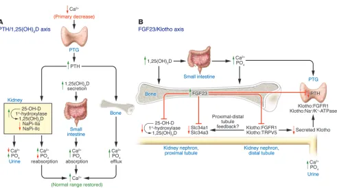
Intracellular phosphate is involved in intermediary metabolism and other essential cellular functions (1), whereas extracellular phosphate is necessary for matrix mineralization (2). Since both extremes of hypophosphatemia and hyperphosphatemia have negative effects (1, 3), adaptive mechanisms have evolved to pro-tect organisms from hypophosphatemia and hyperphosphatemia and to coordinate the changing phosphate needs for bone min-eralization and phosphate homeostasis. Historically, phosphate homeostasis has been viewed from the perspective of the parathy-roid hormone/1,25-dihydroxy vitamin D [PTH/1,25(OH)2D] axis, which regulates both systemic calcium and phosphate homeo-stasis (Figure 1A). In response to hypocalcemia, the parathyroid gland (PTG) increases the production and secretion of PTH, which targets the renal distal tubule to decrease renal calcium excretion and the proximal tubule to inhibit phosphate reabsorption and to stimulate 1,25(OH)2D production. Action of 1,25(OH)2D on the small intestines increases active calcium and phosphate trans-port (4). PTH also has direct effects on bone via PTH receptors in osteoblasts, resulting in increased calcium and phosphate efflux from the exchangeable bone fluid compartment (5) and through RANKL-dependent, osteoclast-mediated bone resorption of min-eralized bone (6). The direct kidney and bone effects of PTH, along with the concomitant actions of 1,25(OH)2D, restore serum cal-cium levels to normal. The phosphaturic actions of PTH offset vitamin D–mediated gastrointestinal phosphate absorption (4) and PTH-dependent phosphate efflux from bone (7), thereby pre-venting the development of hyperphosphatemia.
Recently, a novel hormonal cascade involving FGF23 and Klotho has been identified that principally regulates phosphate, vitamin D homeostasis, and mineralization of bone (Figure 1B) (8–13).
FGF23 and Klotho participation in a bone-kidney axis
FGF23 is a 32-kDa protein with an N-terminal region, contain-ing the FGF-homology domain and a novel 71–amino acid C terminus (8, 9). FGF23 is phylogenetically grouped with FGF19 (mouse FGF15) and FGF21 gene products (8, 10), members of a subfamily of FGFs that act as hormones/systemic factors due to their ability to interact with FGF receptor (FGFR) in the presence of members of the Klotho family of proteins. FGF23 binds to
Klotho (KL), which encodes a type I membrane, β-glycosidase–like protein (11, 12) that is an essential cofactor for FGF23 binding to FGFRs (12–14). In vitro studies indicate that the N-terminal region of FGF23 binds to and activates FGFR1, -3, and -4 at phys-iological concentrations only in the presence of Klotho, which binds to FGFR and the C terminus of FGF23 to convert the canonical FGFRs to a specific receptor for FGF23 (14–16). This is in contrast with the more typical paracrine/local functions of other FGFs that require extracellular acidic glycosaminoglycans (e.g., heparin) for receptor activation (17).
FGF23 principally functions as a phosphaturic factor (9, 18, 19) and counter-regulatory hormone for 1,25(OH)2D produc-tion (20) via binding to KL:FGFR complexes in the kidney (14, 15). Excess FGF23 causes hypophosphatemia via inhibition of solute carrier family 34, member 1–dependent (SLC34A1-depen-dent) and SLC34A2-dependent phosphate transport (also known as sodium phosphate cotransporter 2 [NPT2a] or NaPi-IIa and NPT2c or NaPi-IIc, respectively). Excess FGF23 also suppresses 1,25(OH)2D via inhibition of 25-hydroxyvitamin D-1α –hydrox- ylase (CYP27B1) that is converted to 1,25(OH)2D and stimula-tion of 24-hydroxylase (CYP24) that inactivates 1,25(OH)2D in the proximal tubule of the kidney (9, 18, 21, 22) (Figure 1B). In contrast, deficiency of FGF23 results in the opposite renal pheno-type, consisting of hyperphosphatemia and elevated production of 1,25(OH)2D (19, 23–27). In addition, Fgf23 -null mice have soft-tissue calcifications, severe growth retardation, abnormalities of bone mineralization, and a shortened lifespan (25–27). Inactivat-ing mutations or deletion of Kl results in end-organ insensitivity
Endocrine functions of bone in mineral
metabolism regulation
L. Darryl Quarles
The Kidney Institute and Division of Nephrology, University of Kansas Medical Center, Kansas City, Kansas, USA.
Given the dramatic increase in skeletal size during growth, the need to preserve skeletal mass
dur-ing adulthood, and the large capacity of bone to store calcium and phosphate, juxtaposed with the
essential role of phosphate in energy metabolism and the adverse effects of hyperphosphatemia,
it is not surprising that a complex systems biology has evolved that permits cross-talk between
bone and other organs to adjust phosphate balance and bone mineralization in response to
chang-ing physiological requirements. This review examines the newly discovered signalchang-ing pathways
involved in the endocrine functions of bone, such as those mediated by the phosphaturic and
1,25(OH)
2D-regulating hormone FGF23, and the broader systemic effects associated with abnormalities of calcium
and phosphate homeostasis.
Nonstandard abbreviations used: ADHR, autosomal dominant hypophosphatemic rickets; ARHR, autosomal recessive hypophosphatemic rickets; ASARM, acidic, serine- and aspartic acid–rich motif; BMP1, bone morphogenetic protein 1; DMP1, dentin matrix acidic phosphoprotein 1; FGFR, FGF receptor; HRH, hypophosphatemic rickets and hyperparathyroidism; KL, Klotho ; MEPE, matrix extracellular phospho-glycoprotein with ASARM motif; NPT2, sodium phosphate cotransporter 2; OGD, osteoglophonic dysplasia; 1,25(OH)2D, 1,25-dihydroxy vitamin D; PHEX,
phosphate-regulating gene with homologies to endopeptidases on the X chromo- some; PTG, parathyroid gland; PTH, parathyroid hormone; sFRP4, secreted frizzled- related protein 4; SLC34A1, solute carrier family 34, member 1; XLH, X-linked hypo-phosphatemic rickets.
science in medicine
to FGF23, which is characterized by hyperphosphatemia, elevated 1,25(OH)2D levels, early mortality, and soft-tissue calcifications (13, 28–30), a phenotype that resembles that of Fgf23-null mice (31). In contrast, FGF21 stimulates lipolysis and glucose uptake and FGF19 regulates bile acid secretion via binding to another member of the Klotho family, β-Klotho (32–35).
Klotho also has other functions that are distinct from its role in mediating FGF23 effects (36), including regulation of calcium absorption in the kidney distal convoluted tubule, through stabi-lizing the membrane expression of the transient receptor potential cation channel, subfamily V, member 5 (36), and regulation of PTH secretion (37), by facilitating the recruitment of an Na+/K+-ATPase to the parathyroid chief cell membrane (38). A circulating form of Klotho containing the N-terminal extracellular domain is derived either from a separate Kl gene transcript generated through alter-native transcriptional termination that encodes a secreted protein (39) or by membrane shedding, involving the proteases ADAM10 or ADAM17 (40). Cleavage and secretion of Klotho is regulated by both extracellular calcium fluctuation and insulin. Circulat-ing Klotho has a plethora of functions, including effects on the intracellular insulin/insulin-like growth factor 1–signaling cas-cade (12, 41) and adipocyte differentiation (42).
Organs that produce FGF23
FGF23 is mainly produced and secreted by osteocytes in bone (19, 27) (Figure 1B), but it is also expressed in pericyte-like cells sur- rounding the venous sinuses in the bone marrow, in the ventro-lateral thalamic nucleus in the brain and in thymus, and in lymph nodes (19). The relative contribution of these sites to circulating FGF23 levels is not known, and this is an important problem need- ing investigation. Nevertheless, the high levels of FGF23 expres-sion in osteocytes, the most abundant cell in bone, suggests that the source of circulating FGF23 is mainly bone (19).
FGF23 target organs
[image:3.585.48.539.78.353.2]The target organs for FGF23 are theoretically defined by the coex-pression of the membrane form of Klotho and FGFR1c, -3c, and -4 (14, 15). Klotho is expressed in high levels in PTGs, kidney, testis, ovary, brain, pituitary, and apical plasma membrane of ependy-mal cells in the choroid plexus but not in bone, lung, liver, skin, spleen, small intestines, or adrenal glands. The limited pattern of Klotho expression corresponds to the target tissues function-ally defined by upregulation of the gene early growth-responsive 1 (Egr-1) after administration of FGF23 to mice (14, 15). The kidney, however, is the principal physiologically defined target for FGF23,
Figure 1
Interrelationships among FGF23, PTH, 1,25(OH)2D, and Klotho. (A) The PTH/1,25(OH)2D axis. The principal function of the PTH/1,25(OH)2D
axis is to regulate calcium homeostasis. Decrements in serum calcium levels stimulate PTH secretion by the PTG, which targets the kidney to reduce urinary calcium excretion, stimulate 1α-hydroxylase activity, and enhance the fractional excretion of phosphate (PO4), and targets bone to
increase the efflux of calcium and phosphate. The resulting increase in 1,25(OH)2D targets the gastrointestinal tract to increase dietary
absorp-tion of calcium, which suppresses PTH. (B) The FGF23/Klotho axis. FGF23 produced by bone principally targets the kidney, leading to reductions
in serum phosphate and 1,25(OH)2D levels by stimulating the fractional excretion of phosphate and reducing 1α-hydroxylase activity. The
in which this circulating factor inhibits phosphate reabsorption and production of 1,25(OH)2D (9, 25) (Figure 1B). The precise segments and the physiologically relevant receptor(s) for FGF23 in the kidney are not entirely clear. Although FGF23 can bind to FGFR3, FGFR4, and FGFR1 in vitro, neither deletion of FGFR3 or FGFR4 impairs the phosphaturic actions of FGF23 in mice, suggesting that the remaining in vitro target, FGFR1, is the physi- ologically relevant receptor for FGF23 in the kidney (43). More-over, the highest levels of the FGFR1:Klotho complex are present in the distal tubules, whereas the biological actions of FGF23 are in the proximal tubules (44) (Figure 1B). Ex vivo studies of proxi-mal tubular segments or cell lines have demonstrated variable effects of exogenously added FGF23 to inhibit sodium-dependent phosphate transport (9, 45). Interpretation of these findings are confounded by the use of nonphysiological amounts of FGF23 and the authenticity of the proximal tubular phenotype in cell culture models, which may be contaminated with distal tubu-lar cells and/or may have undergone dedifferentiation (45–49). Alternatively, FGF23 actions on the proximal tubule may be indi-rect, possibly through FGF23 stimulation of the distal tubule and release of paracrine factors that regulate proximal tubule function (i.e., a distal-to-proximal tubular feedback mechanism). Identification of the tubular segments in the kidney that are the direct target for FGF23 is another important unresolved issue. In addition, FGF23 decreases the expression of Klotho by the kidney, thereby creating complex feedback pathways for regulating phos-phate and calcium metabolism (36, 50).
The PTG also expresses FGFR and Klotho and is a target for FGF23 as evidenced by the effects of recombinant FGF23 to stimu- late increments in Egr-1 expression in the PTG of mice (14) (Fig- ure 1B), but there are some discrepancies about whether FGF23-dependent pathways stimulate or inhibit PTH secretion (37, 51). Elevated FGF23 levels in human diseases and mouse models are associated with hyperparathyroidism (22, 52), but FGF23-medi- ated reductions in 1,25(OH)2D production and secondary incre-ments in PTH might explain this association. Conversely, FGF23 has recently been shown to inhibit expression of PTH mRNA and secretion of PTH from parathyroid cells (37, 53). Adding to the complexity of this issue is the observation that elevated Klotho levels are associated with hyperparathyroidism in humans, poten- tially via a direct effect to regulate PTH secretion through its main-tenance of cell surface Na+/K+-ATPase activity (36).
Additional abnormalities are observed in association with FGF23 excess and deficiency, such as rickets/osteomalacia, abnor- malities in glucose homeostasis, growth retardation, abnormali- ties in thymic function, and age-related changes (19, 25, 27), sug-gesting a broader role for FGF23. The choroid plexus and pituitary gland are potential targets for FGF23, but its function remains unknown. There is also uncertainty regarding whether FGF23 has direct effects on bone or if the unexplained defect in bone mineral-ization in Fgf23 -null mice is due to excessive 1,25(OH)2D. An indi-rect effect is supported by the finding of a similar mineralization abnormality associated with high 1,25(OH)2D levels caused by deletion of the 24-hydroxylase, the absence of Klotho expression in bone, and the ability of deletion of the vitamin D receptor to res-cue the phenotype of Fgf23-null mice (26, 54, 55). However, recent studies indicate that FGF23 may have a direct effect on osteoblasts in vitro (56), but the physiological relevance and specificity of these cell culture findings need to be confirmed in vivo (57). Although additional functions of FGF23 remain to be clarified, the overall
principal physiological function of the FGF23/Klotho axis is to regulate phosphate and 1,25(OH)2D homeostasis, with a second-ary effect on renal calcium handling (Figure 1B).
Regulation of FGF23
Systemic factors regulating FGF23. There are various systemic factors that regulate circulating FGF23 levels. The feedback signals under-lying FGF23 actions as a counter regulatory hormone to offset the effects of excessive 1,25(OH)2D are best defined. In this regard, 1,25(OH)2D directly stimulates FGF23 expression in osteocytes via a vitamin D response element (VDRE) in the Fgf23 promoter (20) (Figure 1B and Figure 2A). Since FGF23 suppresses 1,25(OH)2D production, 1,25(OH)2D stimulation of FGF23 closes a feedback loop (20). 1,25(OH)2D may also indirectly regulate FGF23 expres-sion. In this regard, selective deletion of the vitamin D receptor (VDR) in cartilage produces an unidentified chondrocyte-derived inhibitor of FGF23 transcription (58) (Figure 2A). In the setting of excess 1,25(OH)2D and reduced PTH levels, FGF23-mediated phosphaturia helps prevent potential hyperphosphatemia from enhanced 1,25(OH)2D-dependent gastrointestinal phosphate absorption and diminished PTH-mediated phosphaturia. Thus, a primary function of FGF23 is to enhance phosphate excretion and suppress 1,25(OH)2D production, which may have evolved to protect an organism from vitamin D intoxication (20).
Given that FGF23 is a “phosphaturic hormone”, it is expected to be regulated by serum phosphate. This appears to be the case in chronic kidney disease, in which elevations in FGF23 compen-sate for the reduced renal clearance of phosphate and the degree of FGF23 elevation correlates with the severity of hyperphospha-temia (59). However, extracellular phosphate does not appear to directly stimulate FGF23 mRNA levels or FGF23 gene promoter activity in osteoblastic cultures (20). Although phosphate loading in mice increases FGF23 levels (60), evidence of the importance of dietary phosphate in regulating FGF23 levels in humans is also conflicting (61, 62). Thus, the proximate factors mediating the effects of phosphate on FGF23 production remain unknown. Pathways controlling serum calcium may also regulate FGF23. In this regard, elevated FGF23 level is associated with both low cal-cium intake in the absence of vitamin D deficiency (63) and with excess PTH in primary hyperparathyroidism; whereas suppression of FGF23 is observed in response to the hypocalcemic hormone calcitonin in patients with tumor-induced osteomalacia (64, 65). However, neither extracellular calcium nor PTH directly stimulate FGF23 promoter activity in osteoblasts (20), suggesting that their effects may also be indirect.
science in medicine
and DMP1 regulates FGF23 expression in osteocytes and impairs bone mineralization is one of the more important issues remain-ing to be addressed (Figure 2).
PHEX colocalizes with FGF23 in osteocytes. Inactivating PHEX mutations result in increased FGF23 expression in osteocytes as well as phosphate-dependent and -independent defects in bone mineralization. Recent studies indicate that the conditional deletion of Phex in the osteoblast lineage in mice is sufficient to reproduce the Hyp phenotype, namely hypophosphatemia,
characterized by decreased 1,25(OH)2D levels and rickets/osteo-malacia, consistent with the central role of the osteocyte in the regulation of FGF23 and phosphate homeostasis (67). Although an initial study suggested that PHEX processes FGF23 (46), sub- sequent studies have failed to establish PHEX-dependent cleav-age of FGF23 (68–70). Moreover, explantation of bone from Hyp mice into wild-type mice demonstrates that the high expression of FGF23 in osteocytes is an intrinsic/local effect caused by the loss
of PHEX (71). Screening of substrate phage libraries has identi-Figure 2
Hypothetical model of FGF23 regulation. (A)
FGF23 regulation in wild-type osteocytes. FGF23 expression in wild-type osteocytes is low due to putative suppressive signals. DMP1 is processed by BMP1/Tolloid-like metalloproteinases to create N- and C-ter-minal fragments. The model proposes that the C terminus of DMP1 suppresses FGF23 through its binding to PHEX via the ASARM motif and to integrins via the RGD site as well as facilitates mineralization of matrix. In addi-tion, DMP1 is known to have direct transcrip-tional activities. Putative chondrocyte-derived and unknown systemic factors also suppress FGF23 as described in the text. In addition, FGF23 undergoes posttranslational process-ing to inactive N- and C-terminal fragments by yet-to-be defined subtilisin-like proprotein convertases (SPCs). (B) Potential mutations
[image:5.585.41.370.81.626.2]fied that PHEX cleaves small peptides, such as the ASARM (acidic, serine- and aspartic acid–rich motif) peptide derived from MEPE (matrix extracellular phosphoglycoprotein with ASARM motif) (72), but the physiologically relevant substrates for PHEX that regulate FGF23 expression and mineralization are not known (69). It is clear, however, that the absence of PHEX is necessary but not sufficient to stimulate FGF23. In this regard, a functional PHEX is missing in osteoblasts of Hyp mice, but osteoblasts do not upregulate FGF23 expression until they differentiate into osteocytes embedded in bone matrix (19, 71). The importance of temporal and spatial expression of PHEX is also evidenced by the paradox that restoration of PHEX expression in transgenic mice using heterologous promoters fails to rescue the elevated FGF23 expression (73–75). These observations suggest the possible pres-ence of a matrix-derived FGF23 stimulatory factor that may be inhibited or sequestered by a functional PHEX and somehow released or activated when PHEX is inhibited or mutated (Figure 2B). In addition, in humans, activating mutations of FGFR1 (76) and mutations leading to increments in circulating Klotho levels (51) are associated with elevated FGF23 levels, raising the possibil-ity that these factors may also regulate the osteocyte production of FGF23 (Figure 2B).
A major advance in understanding how extracellular matrix proteins might regulate FGF23 production comes with the dis-covery that inactivating mutations of DMP1 lead to increased FGF23 in autosomal recessive hypophosphatemic rickets (ARHR) (77). DMP1 is expressed in mineralized tissues, including osteo-cytes, ameloblasts, and cementoblasts, but is also expressed in nonmineralizing tissues such as brain, salivary gland, liver, muscle, pancreas, and kidney (78). How DMP1 deficiency stimu-lates FGF23 production is not known, but several possibilities are apparent from our knowledge of DMP1 structure and func-tion (Figure 2B). DMP1 contains an RGD domain for integrin binding, an ASARM peptide (which might allow DMP1 to bind to PHEX), a large number of acidic domains, an N-terminal site for binding to MMP9, sites for casein kinase-2–mediated phos- phorylation, and conserved cleavage sites for bone morphoge-netic protein 1/Tolloid-like MMPs (BMP1/Tolloid-like MMPs) and cathepsin B (Figure 2). DMP1 exists as a latent protein that is cleaved into 37-kDa and 57-kDa phosphoproteins by BMP1 or cathepsin B (Figure 2A). The highly phosphorylated C-terminal 57-kDa fragment (containing 42 phosphates/mol) likely func- tions as a nucleator for mineralization. The NH2-terminal frag-ment from DMP1 is a proteoglycan with a chondroitin sulfate chain attached through Ser74 that also binds to proMMP-9 (79). DMP1 also is localized to the dendrites of embedding osteocytes. Finally, DMP1 has been reported to translocate to the nucleus where it may regulate gene transcription (80).
Although FGF23-dependent hypophosphatemia contributes to impaired mineralization, an intrinsic defect in mineralization of extracellular matrix persists in Phex- and Dmp1-mutant mice even when phosphate is normalized (71). In Dmp1-null mice, the absence of the nucleation and mineral propagation functions of DMP1 likely contribute to the defective mineralization (81, 82). The mechanism for the hypophosphatemia-independent miner-alization abnormalities in Hyp mice is less well understood, but existing data fails to support a role for DMP1. Rather, Hyp bone is characterized by elevated proteolytic activity and the production by osteoblasts of a mineralization inhibitor, referred to as Min-hibin (83). In Hyp
mice, the elevated proteolytic activity is associ-ated with the increased production of an ASARM peptide from MEPE and DMP1, which has the ability to inhibit mineralization (84) and may represent Minhibin (83). Moreover, the administra-tion of cathepsin inhibitors improves bone mineralization in Hyp mice, without correcting FGF23 expression or hypophosphatemia, indicating the separate regulation of hypophosphatemia and min-eralization by PHEX (85).
science in medicine
Role of FGF23 in our understanding of disorders of phosphate homeostasis
Insights into the function and interrelationships between FGF23, Klotho, and sodium-dependent phosphate transporters have clarified the pathogenesis of hereditary and acquired hypophos-phatemic disorders (Table 1). These can be broadly classified into FGF23-dependent disorders and primary disorders of proximal renal tubular phosphate transport.
Diseases of FGF23 excess . Hereditary hypophosphatemic disor-ders caused by a primary increase in circulating levels of FGF23 include autosomal dominant hypophosphatemic rickets (ADHR; MIM 193100), autosomal recessive hypophosphatemic rick-ets (ARHR; MIM 241520), X-linked hypophosphatemic rickets (XLH; MIM 307800), and osteoglophonic dysplasia (OGD; MIM 166250) (Table 1). All have overlapping clinical characteristics, including hypophosphatemia due to renal phosphate wasting and inappropriately normal 1,25(OH)2D levels for the degree of hypophosphatemia and rickets/osteomalacia, and the absence of hypercalciuria. There is a single report of a patient with hypo-phosphatemic rickets and hyperparathyroidism (HRH; MIM pending) (51). HRH differs from the others disorders described here in that hyperparathyroidism is a predominant feature. The clinical features of ADHR, ARHR, XLH, OGD, and HRH are
[image:7.585.52.532.108.457.2]caused by excess FGF23, but the mechanisms whereby FGF23 levels are increased differ between these disorders. ADHR is caused by mutations (R176Q and R179W) in the RXXR furin- like cleavage domain of FGF23 that impairs proteolytic inactiva- tion of FGF23 (68, 92). ARHR is caused by inactivating muta-tions in DMP1 (77, 93), a member of the small integrin-binding ligand N-linked glycoprotein (SIBLING) family of extracellular matrix protein that augments mineralization (81). Loss of DMP1 results in increased transcription of FGF23 by osteocytes (77). XLH is caused by inactivating mutations in PHEX (94–96). Loss of PHEX also leads to increased expression of FGF23 by osteo-cytes (70). How loss-of-function DMP1 and PHEX mutations lead to increases in FGF23 gene transcription is not known (97). OGD is an autosomal dominant bone dysplastic disorder caused by activating mutations in the FGFR1 gene, suggesting FGFR1 may regulate FGF23 expression in bone and/or the renal handling of phosphate (76). HRH also has elevated FGF23 levels, but the primary genetic abnormality is a translocation causing elevated circulating levels of Klotho (51). How excess Klotho leads to increased FGF23 levels, however, is not clear. Elevated circulating Klotho levels could potentially bind to and stabilize FGF23, tar-get bone to increase FGF23 production, or indirectly stimulate FGF23 secretion via effects on other pathways.
Table 1
Classification of hypophosphatemic and hyperphosphatemic disorders
Clinical syndromes Disease genes Mechanisms
FGF23-dependent hypophosphatemic disorders Hereditary
XLH PHEX Increased FGF23 production by osteocytes
ADHR FGF23 Decreased FGF23 degradation
ARHR DMP1 Increased production of FGF23 by osteocytes
OGD FGFR1 Increased production of FGF23/end-organ gain of function?
Acquired/Sporadic
HRH Kl Gain-of-function end-organ responsiveness;
direct actions of β-Klotho
McCune-Albright syndrome, GNAS1 Increased FGF23 production by fibrous lesions in bone polyostotic fibrous dysplasia
Tumor-induced osteomalacia Possibly MEPE and sFRP4 Increased FGF23 production by tumors
Epidermal nevus syndrome FGFR3 Increased circulating FGF23 due to end-organ gain of function Primary disorders of renal phosphate reabsorption
Hereditary
Hereditary hypophosphatemic rickets SLC34A3 Transporter defect leading to impaired renal tubular phosphate
with hypercalciuria reabsorption and increased 1,25(OH)2D production
Autosomal recessive pulmonary SLC34A2 No hypophosphatemia
alveolar microlithiasis
No human disease–causing SLC34A1 Transporter defect leading to impaired renal tubular
mutation identified phosphate reabsorption
X-linked recessive hypophosphatemic CLCN5 Defective endocytosis and redistribution of NaPi-II from the plasma
rickets membrane to intracellular vesicles; Fanconi-like proteinuria
Lowe oculocerebrorenal syndrome OCRL Defective phosphatidylinositol 4,5-bisphosphate 5-phosphatase– mediated endocytosis of Npt2 and Fanconi-like proteinuria No human disease–causing NHERF1 Aberrant Npt2 protein localization to internal sites in the
mutation identified renal proximal tubule cells
Hyperphosphatemic disorders Hereditary/acquired/sporadic
Tumoral calcinosis GALNT3, FGF23, Decreased stability or production of FGF23 or end-organ
McCune-Albright syndrome (MIM 174800), also called polyos- totic fibrous dysplasia (PFD), is caused by a mosaicism for postzy- gotic activating mutations in the guanine nucleotide binding pro-tein, alpha stimulating gene (GNAS1), leading to fibrodysplastic tissue and associated hypophosphatemia and elevated circulat-ing FGF23 levels in some patients (98) (Table 1). Tumor-induced osteomalacia, or oncogenic osteomalacia, is a paraneoplastic syn- drome of renal phosphate wasting, aberrant vitamin D metabo-lism, and osteomalacia that is associated with elevated FGF23 levels (59, 99). The proximate cause of increased FGF23 produc-tion is not known, but this disorder is associated with increased levels of MEPE and sFRP4, which may regulate PHEX and DMP1 metabolism, respectively (100) (Figure 2). Linear sebaceous or epi- dermal nevus syndrome (ENS) is caused by a mosaicism of activat-ing FGFR3 mutations in the human epidermis in some patients (101). A subset of these patients have ipsilateral focal bone disease associated with hypophosphatemic rickets, elevated circulating FGF23 levels, and aberrant 1,25(OH)2D levels, similar to other syndromes caused by elevated FGF23 (102).
Other disorders are characterized by a secondary/adaptive increase in FGF23. For example, FGF23 is markedly increased in early stages of chronic kidney diseases, in which its actions to decrease CYP27B1 and increase CYP24 lead to diminish calcitriol levels and contributes to the development of secondary hyperpara-thyroidism (103). In addition, increments in FGF23 have been associated with increased mortality in patients with end-stage renal disease, suggesting that it may act as a uremic toxin (104). Anti-FGF23 neutralizing antibodies are being developed that offer a potential way to treat disorders of excessive FGF23 (105); whether these will be clinically useful, however, is uncertain.
Diseases of FGF23 deficiency. Hyperphosphatemic familial tumoral calcinosis (HFTC; MIM 211900) is a rare autosomal recessive dis-order characterized by hyperphosphatemia, normal or elevated 1,25(OH)2D levels, soft-tissue calcifications, and typically massive lobulated periarticular calcifications levels (106). To date there have been three different mutations identified as leading to either decreased bioactive circulating FGF23 levels or end-organ resis-tance to FGF23. These include mutations of the genes encoding FGF23 (23), Klotho (29) and GalNAc transferase 3 (GALNT3) (107), a Golgi-associated enzyme that O-glycosylates a furin-like convertase recognition sequence in FGF23 (108). Missense muta- tions of FGF23 impair its secretion, leading to inadequate circulat-ing levels of this phosphaturic factor (23). Mutations of GALNT3 destabilize FGF23, resulting in a low-intact serum FGF23 levels but high levels of biologically inactive C-terminal FGF23 frag-ments. A mutation in the gene coding for Klotho(H193R) results in decreased Klotho expression and reduced FGF23-Klotho-FGFR complex formation and to end-organ resistance to FGF23 (29).
FGF23-independent hypophosphatemic disorders: diseases affecting phosphate transporters. A primary defect in proximal tubular phos-phate reabsorption results in elevations of 1,25(OH)2D, which increases gastrointestinal calcium absorption and leads to hyper-calciuria — two features that distinguish primary renal phosphate wasting disorders from disorders of FGF23 excess. SLC34A1 and
SLC34A3 encode the sodium-dependent phosphate transporters in the proximal tubule of the kidney. Hereditary hypophospha-temic rickets with hypercalciuria (HHRH; MIM 241530) is caused by inactivating mutations in the SLC34A3 gene encoding NPT2c or NaPi-IIc (109). Mutations in SLC34A1 , which encodes the elec-trogenic NPT2a or NaPi-IIa, have not been unequivocally shown to cause hypophosphatemia in humans. Interestingly, mice with homozygous deletion of Slc34a1 have mild hypophosphatemia and rickets/osteomalacia (110), possibly due to the compensatory upregulation of SLC34A3 (111). SLC34A2 encodes NPT2b or NaPi-IIb, which is responsible for transcellular phosphate absorption in the small intestine. SLC34A2 is expressed mainly in lung and mammary gland and to a lesser extent in intestine, kidney, and prostate (112). Mutations in this gene cause pulmonary alveolar microlithiasis and are not reported to be associated with major abnormalities of serum phosphate (113).
X-linked hypercalciuric nephrolithiasis, or Dent disease (MIM 300009), includes several related forms of hereditary proximal tubulopathy caused by mutations in the chloride channel 5
(CLCN5) gene, which encodes a voltage-gated chloride channel and chloride/proton antiporter (114). Phosphaturia results from defective endocytosis and redistribution of SLC34A1 from the plasma membrane to intracellular vesicles and is associated with low–molecular weight proteinuria and hypercalciuria. Lowe oculo-cerebrorenal syndrome (LOS; MIM 30900) is caused by mutations in the gene oculocerebrorenal syndrome of Lowe (OCRL1), which encodes a phosphatidylinositol 4,5-bisphosphate 5-phosphatase that is also involved in endocytosis. Patients with LOS are char-acterized as having more pronounced hypophosphatemia/rickets and proximal tubular proteinuria than patients with Dent disease, and LOS has other features such as elevated lactate dehydrogenase and/or creatine kinase levels.
Conclusion
The study of the regulation and function of the phosphaturic and 1,25(OH)2D regulating factor FGF23 has lead to the recognition that bone is an endocrine organ, in which osteocytes produce FGF23 that participates in a bone-kidney axis, regulating phosphate, vita-min D, and mineral homeostasis. This hormonal axis establishes a new conceptual framework for understanding the pathogenesis, diagnosis, and treatment of hyperphosphatemic and hypophospha-temic disorders. In addition, by continuing to elucidate the complex systems biology surrounding FGF23 regulation and function, we will likely gain novel insights into bone and renal physiology.
Acknowledgments
This work was supported by National Institutes of Health grant RO1-AR45955 from the National Institute of Arthritis and Mus-culoskeletal and Skin Diseases.
Address correspondence to: L. Darryl Quarles, MS 3018, The Kid-ney Institute, University of Kansas Medical Center, 3901 Rainbow Blvd., Kansas City, Kansas 66160, USA. Phone: (913) 588-9252; Fax: (913) 588-9251; E-mail: dquarles@kumc.edu.
1. Amanzadeh, J., and Reilly, R.F., Jr. 2006. Hypophos- phatemia: an evidence-based approach to its clini-cal consequences and management. Nat. Clin. Pract. Nephrol. 2:136–148.
2. Murshed, M., Harmey, D., Millan, J.L., McKee, M.D., and Karsenty, G. 2005. Unique coexpression
in osteoblasts of broadly expressed genes accounts for the spatial restriction of ECM mineralization to bone. Genes Dev. 19:1093–1104.
3. Block, G.A., et al. 2004. Mineral metabolism, mor-tality, and morbidity in maintenance hemodialysis.
J. Am. Soc. Nephrol. 15:2208–2218.
4. Rizzoli, R., Fleisch, H., and Bonjour, J.P. 1977. Role of 1,25-dihydroxyvitamin D3 on intestinal phos-phate absorption in rats with a normal vitamin D supply. J. Clin. Invest. 60:639–647.
science in medicine
45Ca following parathyroid hormone administra-tion to thyroparathyroidectomized rats. Calcif. Tis-sue Res. 22:117–128.
6. Ma, Y.L., et al. 2001. Catabolic effects of continu-ous human PTH (1--38) in vivo is associated with sustained stimulation of RANKL and inhibition of osteoprotegerin and gene-associated bone forma-tion. Endocrinology. 142:4047–4054.
7. Shiraki, M., Gee, M.V., Baum, B.J., and Roth, G.S. 1986. Parathyroid hormone stimulates phosphate efflux through an apparently adenosine 3′,5′ - monophosphate-independent process in rat parot-id cell aggregates. Endocrinology. 118:2009–2015. 8. Yamashita, T., Yoshioka, M., and Itoh, N. 2000.
Identification of a novel fibroblast growth factor, FGF-23, preferentially expressed in the ventrolat-eral thalamic nucleus of the brain. Biochem. Biophys. Res. Commun. 277:494–498.
9. Shimada, T., et al. 2001. Cloning and character- ization of FGF23 as a causative factor of tumor-induced osteomalacia. Proc. Natl. Acad. Sci. U. S. A. 98:6500–6505.
10. Itoh, N., and Ornitz, D.M. 2004. Evolution of the Fgf and Fgfr gene families. Trends Genet. 20:563–569. 11. Tsujikawa, H., Kurotaki, Y., Fujimori, T., Fukuda,
K., and Nabeshima, Y. 2003. Klotho, a gene relat-ed to a syndrome resembling human premature aging, functions in a negative regulatory circuit of vitamin D endocrine system. Mol. Endocrinol. 17:2393–2403.
12. Kurosu, H., et al. 2005. Suppression of aging in mice by the hormone Klotho. Science. 309:1829–1833. 13. Kuro-o, M., et al. 1997. Mutation of the mouse
klotho gene leads to a syndrome resembling age-ing. Nature. 390:45–51.
14. Urakawa, I., et al. 2006. Klotho converts canonical FGF receptor into a specific receptor for FGF23.
Nature. 444:770–774.
15. Kurosu, H., et al. 2006. Regulation of fibroblast growth factor-23 signaling by klotho. J. Biol. Chem. 281:6120–6123.
16. Goetz, R., et al. 2007. Molecular insights into the klotho-dependent, endocrine mode of action of fibroblast growth factor 19 subfamily members.
Mol. Cell. Biol. 27:3417–3428.
17. Itoh, N., and Ornitz, D.M. 2008. Functional evolu-tionary history of the mouse Fgf gene family. Dev. Dyn. 237:18–27.
18. Larsson, T., et al. 2004. Transgenic mice express-ing fibroblast growth factor 23 under the control of the alpha1(I) collagen promoter exhibit growth retardation, osteomalacia, and disturbed phos-phate homeostasis. Endocrinology. 145:3087–3094. 19. Liu, S., et al. 2006. Pathogenic role of Fgf23 in Hyp
mice. Am. J. Physiol. Endocrinol. Metab. 291:E38–E49. 20. Liu, S., et al. 2006. Fibroblast growth factor 23 is
a counter-regulatory phosphaturic hormone for vitamin D. J. Am. Soc. Nephrol. 17:1305–1315.
21. Shimada, T., et al. 2005. Vitamin D receptor-inde-pendent FGF23 actions in regulating phosphate and vitamin D metabolism. Am. J. Physiol. Renal Physiol. 289:F1088–F1095.
22. Bai, X., Miao, D., Li, J., Goltzman, D., and Kara-plis, A.C. 2004. Transgenic mice overexpressing human fibroblast growth factor 23 (R176Q) delin-eate a putative role for parathyroid hormone in renal phosphate wasting disorders. Endocrinology. 145:5269–5279.
23. Benet-Pages, A., Orlik, P., Strom, T.M., and Lorenz-Depiereux, B. 2005. An FGF23 missense mutation causes familial tumoral calcinosis with hyperphos-phatemia. Hum. Mol. Genet. 14:385–390. 24. Larsson, T., et al. 2005. Fibroblast growth factor-23
mutants causing familial tumoral calcinosis are dif-ferentially processed. Endocrinology. 146:3883–3891. 25. Shimada, T., et al. 2004. Targeted ablation of Fgf23 demonstrates an essential physiological role of FGF23 in phosphate and vitamin D metabolism.
J. Clin. Invest. 113:561–568.
26. Sitara, D., et al. 2006. Genetic ablation of vitamin D activation pathway reverses biochemical and skel-etal anomalies in Fgf-23-null animals. Am. J. Pathol. 169:2161–2170.
27. Sitara, D., et al. 2004. Homozygous ablation of fibroblast growth factor-23 results in hyperphos-phatemia and impaired skeletogenesis, and reverses hypophosphatemia in Phex-deficient mice. Matrix Biol. 23:421–432.
28. Yoshida, T., Fujimori, T., and Nabeshima, Y. 2002. Mediation of unusually high concentrations of 1,25-dihydroxyvitamin D in homozygous klotho mutant mice by increased expression of renal 1alpha-hydrox-ylase gene. Endocrinology. 143:683–689.
29. Ichikawa, S., et al. 2007. A homozygous missense mutation in human KLOTHO causes severe tumoral calcinosis. J. Clin. Invest. 117:2684–2691.
30. Segawa, H., et al. 2007. Correlation between hyper-phosphatemia and type II Na-Pi cotransporter activity in klotho mice. Am. J. Physiol. Renal Physiol. 292:F769–F779.
31. Razzaque, M.S., Sitara, D., Taguchi, T., St-Arnaud, R., and Lanske, B. 2006. Premature aging-like phe-notype in fibroblast growth factor 23 null mice is a vitamin D-mediated process. FASEB J. 20:720–722. 32. Ogawa, Y., et al. 2007. BetaKlotho is required for
metabolic activity of fibroblast growth factor 21.
Proc. Natl. Acad. Sci. U. S. A. 104:7432–7437. 33. Suzuki, M., et al. 2008. {beta}Klotho Is Required
for Fibroblast Growth Factor (FGF) 21 Signaling through FGF Receptor (FGFR) 1c and FGFR3c.
Mol. Endocrinol. 22:1006–1014.
34. Kurosu, H., et al. 2007. Tissue-specific expression of betaKlotho and fibroblast growth factor (FGF) receptor isoforms determines metabolic activity of FGF19 and FGF21. J. Biol. Chem. 282:26687–26695. 35. Lin, B.C., Wang, M., Blackmore, C., and Desnoyers,
L.R. 2007. Liver-specific activities of FGF19 require Klotho beta. J. Biol. Chem. 282:27277–27284. 36. Imura, A., et al. 2007. alpha-Klotho as a regulator
of calcium homeostasis. Science. 316:1615–1618. 37. Ben-Dov, I.Z., et al. 2007. The parathyroid is
a target organ for FGF23 in rats. J. Clin. Invest. 117:4003–4008.
38. Brown, E.M., et al. 1987. Ouabain and low extra-cellular potassium inhibit PTH secretion from bovine parathyroid cells by a mechanism that does not involve increases in the cytosolic calcium con-centration. Metabolism. 36:36–42.
39. Shiraki-Iida, T., et al. 1998. Structure of the mouse klotho gene and its two transcripts encoding mem-brane and secreted protein. FEBS Lett. 424:6–10. 40. Chen, C.D., Podvin, S., Gillespie, E., Leeman, S.E.,
and Abraham, C.R. 2007. Insulin stimulates the cleavage and release of the extracellular domain of Klotho by ADAM10 and ADAM17. Proc. Natl. Acad. Sci. U. S. A. 104:19796–19801.
41. Mori, K., et al. 2000. Disruption of klotho gene causes an abnormal energy homeostasis in mice.
Biochem. Biophys. Res. Commun. 278:665–670. 42. Chihara, Y., et al. 2006. Klotho protein
pro-motes adipocyte differentiation. Endocrinology. 147:3835–3842.
43. Liu, S., Vierthaler, L., Tang, W., Zhou, J., and Quarles, L.D. 2008. FGFR3 and FGFR4 do not mediate renal effects of FGF23. J. Am. Soc. Nephrol. Online publication ahead of print. doi:10.1681/ ASN.2007121301.
44. Li, S.A., et al. 2004. Immunohistochemical localiza- tion of Klotho protein in brain, kidney, and repro-ductive organs of mice. Cell Struct. Funct. 29:91–99. 45. Saito, H., et al. 2003. Human fibroblast growth fac-tor-23 mutants suppress Na+-dependent phosphate co-transport activity and 1alpha,25-dihydroxyvita-min D3 production. J. Biol. Chem. 278:2206–2211.
46. Bowe, A.E., et al. 2001. FGF-23 inhibits renal tubu-lar phosphate transport and is a PHEX substrate.
Biochem. Biophys. Res. Commun. 284:977–981.
47. Saito, H., et al. 2005. Circulating FGF-23 is regu- lated by 1alpha,25-dihydroxyvitamin D3 and phos-phorus in vivo. J. Biol. Chem. 280:2543–2549. 48. Yamashita, T., Konishi, M., Miyake, A., Inui, K., and
Itoh, N. 2002. Fibroblast growth factor (FGF)-23 inhibits renal phosphate reabsorption by activa- tion of the mitogen-activated protein kinase path-way. J. Biol. Chem. 277:28265–28270.
49. Yu, X., et al. 2005. Analysis of the biochemical mechanisms for the endocrine actions of fibroblast growth factor-23. Endocrinology. 146:4647–4656. 50. Marsell, R., et al. 2008. Gene expression analysis
of kidneys from transgenic mice expressing fibro-blast growth factor-23. Nephrol. Dial. Transplant. 23:827–833.
51. Brownstein, C.A., et al. 2008. A translocation caus- ing increased alpha-klotho level results in hypo-phosphatemic rickets and hyperparathyroidism.
Proc. Natl. Acad. Sci. U. S. A. 105:3455–3460.
52. Kazama, J.J., Gejyo, F., Shigematsu, T., and Fukaga-wa, M. 2005. Role of circulating fibroblast growth factor 23 in the development of secondary hyper-parathyroidism. Ther. Apher. Dial. 9:328–330.
53. Krajisnik, T., et al. 2007. Fibroblast growth factor- 23 regulates parathyroid hormone and 1alpha- hydroxylase expression in cultured bovine para-thyroid cells. J. Endocrinol. 195:125–131. 54. Hesse, M., Frohlich, L.F., Zeitz, U., Lanske, B., and
Erben, R.G. 2007. Ablation of vitamin D signaling rescues bone, mineral, and glucose homeostasis in Fgf-23 deficient mice. Matrix Biol. 26:75–84. 55. Stubbs, J.R., et al. 2007. Role of hyperphosphatemia
and 1,25-dihydroxyvitamin D in vascular calcifica-tion and mortality in fibroblastic growth factor 23 null mice. J. Am. Soc. Nephrol. 18:2116–2124. 56. Sitara, D., et al. 2008. Genetic evidence of serum
phosphate-independent functions of FGF-23 on bone. PLoS Genet. 4:e1000154.
57. Wang, H., et al. 2008. Overexpression of fibroblast growth factor 23 suppresses osteoblast differen-tiation and matrix mineralization in vitro. J. Bone Miner. Res. 23:939–948.
58. Masuyama, R., et al. 2006. Vitamin D receptor in chondrocytes promotes osteoclastogenesis and regulates FGF23 production in osteoblasts. J. Clin. Invest. 116:3150–3159.
59. Weber, T.J., Liu, S., Indridason, O.S., and Quarles, L.D. 2003. Serum FGF23 levels in normal and dis-ordered phosphorus homeostasis. J. Bone Miner. Res. 18:1227–1234.
60. Perwad, F., et al. 2005. Dietary and serum phospho-rus regulate fibroblast growth factor 23 expression and 1,25-dihydroxyvitamin D metabolism in mice.
Endocrinology. 146:5358–5364.
61. Ferrari, S.L., Bonjour, J.P., and Rizzoli, R. 2005. Fibroblast growth factor-23 relationship to dietary phosphate and renal phosphate handling in healthy young men. J. Clin. Endocrinol. Metab. 90:1519–1524.
62. Nishida, Y., et al. 2006. Acute effect of oral phos-phate loading on serum fibroblast growth factor 23 levels in healthy men. Kidney Int. 70:2141–2147. 63. Prentice, A., Ceesay, M., Nigdikar, S., Allen, S.J., and Pettifor, J.M. 2008. FGF23 is elevated in Gambian children with rickets. Bone. 42:788–797.
64. Kawata, T., et al. 2007. Parathyroid hormone regu-lates fibroblast growth factor-23 in a mouse model of primary hyperparathyroidism. J. Am. Soc. Nephrol. 18:2683–2688.
65. van Boekel, G., et al. 2008. Tumor producing fibro-blast growth factor 23 localized by two-staged venous sampling. Eur. J. Endocrinol. 158:431–437. 66. Liu, S., et al. 2008. Pathogenic role of Fgf23 in
Dmp1 null mice. Am. J. Physiol. Endocrinol. Metab. 295:E254–E261.
murine X-linked hypophosphatemia. J. Clin. Invest. 118:722–734.
68. Benet-Pages, A., et al. 2004. FGF23 is processed by proprotein convertases but not by PHEX. Bone. 35:455–462.
69. Guo, R., Liu, S., Spurney, R.F., and Quarles, L.D. 2001. Analysis of recombinant Phex: an endopepti-dase in search of a substrate. Am. J. Physiol. Endocri-nol. Metab. 281:E837–847.
70. Liu, S., et al. 2003. Regulation of fibroblastic growth factor 23 expression but not degradation by PHEX. J. Biol. Chem. 278:37419–37426.
71. Liu, S., Tang, W., Zhou, J., Vierthaler, L., and Quar-les, L.D. 2007. Distinct roles for intrinsic osteocyte abnormalities and systemic factors in regulation of FGF23 and bone mineralization in Hyp mice. Am. J. Physiol. Endocrinol. Metab. 293:E1636–E1644. 72. Campos, M., et al. 2003. Human recombinant
endopeptidase PHEX has a strict S1’ specificity for acidic residues and cleaves peptides derived from fibroblast growth factor-23 and matrix extracellular phosphoglycoprotein. Biochem. J. 373:271–279.
73. Bai, X., et al. 2002. Partial rescue of the Hyp pheno- type by osteoblast-targeted PHEX (phosphate-reg-ulating gene with homologies to endopeptidases on the X chromosome) expression. Mol. Endocrinol. 16:2913–2925.
74. Erben, R.G., et al. 2005. Overexpression of human PHEX under the human beta-actin promoter does not fully rescue the Hyp mouse phenotype. J. Bone Miner. Res. 20:1149–1160.
75. Liu, S., Guo, R., Tu, Q., and Quarles, L.D. 2002. Overexpression of Phex in osteoblasts fails to rescue the Hyp mouse phenotype. J. Biol. Chem. 277:3686–3697.
76. White, K.E., et al. 2005. Mutations that cause osteo-glophonic dysplasia define novel roles for FGFR1 in bone elongation. Am. J. Hum. Genet. 76:361–367. 77. Feng, J.Q., et al. 2006. Loss of DMP1 causes rickets
and osteomalacia and identifies a role for osteocytes in mineral metabolism. Nat. Genet. 38:1310–1315. 78. Fisher, L.W., and Fedarko, N.S. 2003. Six genes
expressed in bones and teeth encode the current members of the SIBLING family of proteins. Con-nect. Tissue Res. 44(Suppl. 1):33–40.
79. Ogbureke, K.U., and Fisher, L.W. 2004. Expression of SIBLINGs and their partner MMPs in salivary glands. J. Dent. Res. 83:664–670.
80. Narayanan, K., et al. 2003. Dual functional roles of dentin matrix protein 1. Implications in biomineral- ization and gene transcription by activation of intra-cellular Ca2+ store. J. Biol. Chem. 278:17500–17508. 81. Ling, Y., et al. 2005. DMP1 depletion decreases bone
mineralization in vivo: an FTIR imaging analysis.
J. Bone Miner. Res. 20:2169–2177.
82. He, G., Dahl, T., Veis, A., and George, A. 2003. Dentin matrix protein 1 initiates hydroxyapatite formation in vitro. Connect. Tissue Res. 44(Suppl. 1):240–245. 83. Xiao, Z.S., et al. 1998. Intrinsic mineralization
defect in Hyp mouse osteoblasts. Am. J. Physiol. 275:E700–E708.
84. Martin, A., et al. 2008. Degradation of MEPE,
DMP1, and release of SIBLING ASARM-pep-tides (minhibins): ASARM-peptide(s) are directly responsible for defective mineralization in HYP.
Endocrinology. 149:1757–1772.
85. Rowe, P.S., et al. 2006. Correction of the mineraliza- tion defect in hyp mice treated with protease inhib-itors CA074 and pepstatin. Bone. 39:773–786. 86. Lu, Y., et al. 2007. Rescue of odontogenesis in
Dmp1-deficient mice by targeted re-expression of DMP1 reveals roles for DMP1 in early odonto-genesis and dentin apposition in vivo. Dev. Biol. 303:191–201.
87. Fang, M.A., Glackin, C.A., Sadhu, A., and McDou-gall, S. 2001. Transcriptional regulation of alpha 2(I) collagen gene expression by fibroblast growth factor-2 in MC3T3-E1 osteoblast-like cells. J. Cell. Biochem. 80:550–559.
88. Carpenter, T.O., et al. 2005. Fibroblast growth fac-tor 7: an inhibitor of phosphate transport derived from oncogenic osteomalacia-causing tumors.
J. Clin. Endocrinol. Metab. 90:1012–1020.
89. Mori, S., et al. 2008. Direct binding of integrin alphavbeta3 to FGF1 plays a role in FGF1 signal-ing. J. Biol. Chem. 283:18066–18075.
90. Liu, S., Rowe, P.S., Vierthaler, L., Zhou, J., and Quar- les, L.D. 2007. Phosphorylated acidic serine-aspar-tate-rich MEPE-associated motif peptide from matrix extracellular phosphoglycoprotein inhibits phosphate regulating gene with homologies to endopeptidases on the X-chromosome enzyme activity. J. Endocrinol. 192:261–267.
91. Rowe, P.S., et al. 2004. MEPE has the properties of an osteoblastic phosphatonin and minhibin. Bone. 34:303–319.
92. Bai, X.Y., Miao, D., Goltzman, D., and Karaplis, A.C. 2003. The autosomal dominant hypophosphatemic rickets R176Q mutation in fibroblast growth factor 23 resists proteolytic cleavage and enhances in vivo biological potency. J. Biol. Chem. 278:9843–9849. 93. Lorenz-Depiereux, B., et al. 2006. DMP1 mutations
in autosomal recessive hypophosphatemia impli-cate a bone matrix protein in the regulation of phosphate homeostasis. Nat. Genet. 38:1248–1250. 94. The HYP Consortium. 1995. A gene (PEX) with
homologies to endopeptidases is mutated in patients with X-linked hypophosphatemic rickets.
Nat. Genet. 11:130–136.
95. Thompson, D.L., et al. 2002. Ontogeny of Phex/ PHEX protein expression in mouse embryo and subcellular localization in osteoblasts. J. Bone Miner. Res. 17:311–320.
96. Guo, R., and Quarles, L.D. 1997. Cloning and sequencing of human PEX from a bone cDNA library: evidence for its developmental stage-spe-cific regulation in osteoblasts. J. Bone Miner. Res. 12:1009–1017.
97. Liu, S., and Quarles, L.D. 2007. How fibroblast growth factor 23 works. J. Am. Soc. Nephrol. 18:1637–1647.
98. Riminucci, M., et al. 2003. FGF-23 in fibrous dys- plasia of bone and its relationship to renal phos-phate wasting. J. Clin. Invest. 112:683–692. 99. Jonsson, K.B., et al. 2003. Fibroblast growth factor
23 in oncogenic osteomalacia and X-linked hypo-phosphatemia. N. Engl. J. Med. 348:1656–1663. 100. Berndt, T., et al. 2003. Secreted frizzled-related
protein 4 is a potent tumor-derived phosphaturic agent. J. Clin. Invest. 112:785–794.
101. Hafner, C., et al. 2006. Mosaicism of activating FGFR3 mutations in human skin causes epidermal nevi. J. Clin. Invest. 116:2201–2207.
102. Heike, C.L., et al. 2005. Skeletal changes in epi-dermal nevus syndrome: does focal bone disease harbor clues concerning pathogenesis? Am. J. Med. Genet. A. 139A:67–77.
103. Gutierrez, O., et al. 2005. Fibroblast growth fac- tor-23 mitigates hyperphosphatemia but accentu-ates calcitriol deficiency in chronic kidney disease.
J. Am. Soc. Nephrol. 16:2205–2215.
104. Gutierrez, O.M., et al. 2008. Fibroblast growth fac-tor 23 and mortality among patients undergoing hemodialysis. N. Engl. J. Med. 359:584–592. 105. Yamazaki, Y., et al. 2008. Anti-FGF23 neutralizing
antibodies show the physiological role and structural features of FGF23. J. Bone Miner. Res. 23:1509–1518. 106. Lyles, K.W., et al. 1985. Genetic transmission of
tumoral calcinosis: autosomal dominant with vari-able clinical expressivity. J. Clin. Endocrinol. Metab. 60:1093–1096.
107. Topaz, O., et al. 2006. A deleterious mutation in SAMD9 causes normophosphatemic familial tumoral calcinosis. Am. J. Hum. Genet. 79:759–764. 108. Bennett, E.P., Hassan, H., and Clausen, H. 1996.
cDNA cloning and expression of a novel human UDP-N-acetyl-alpha-D-galactosamine. Polypep-tide N-acetylgalactosaminyltransferase, GalNAc-t3.
J. Biol. Chem. 271:17006–17012.
109. Bergwitz, C., et al. 2006. SLC34A3 mutations in patients with hereditary hypophosphatemic rick-ets with hypercalciuria predict a key role for the sodium-phosphate cotransporter NaPi-IIc in main-taining phosphate homeostasis. Am. J. Hum. Genet. 78:179–192.
110. Beck, L., et al. 1998. Targeted inactivation of Npt2 in mice leads to severe renal phosphate wasting, hypercalciuria, and skeletal abnormalities. Proc. Natl. Acad. Sci. U. S. A. 95:5372–5377.
111. Tenenhouse, H.S., et al. 2003. Differential effects of Npt2a gene ablation and X-linked Hyp mutation on renal expression of Npt2c. Am. J. Physiol. Renal Physiol. 285:F1271–F1278.
112. Feild, J.A., Zhang, L., Brun, K.A., Brooks, D.P., and Edwards, R.M. 1999. Cloning and functional charac- terization of a sodium-dependent phosphate trans-porter expressed in human lung and small intestine.
Biochem. Biophys. Res. Commun. 258:578–582. 113. Corut, A., et al. 2006. Mutations in SLC34A2 cause
pulmonary alveolar microlithiasis and are possi-bly associated with testicular microlithiasis. Am. J. Hum. Genet. 79:650–656.
114. Fisher, S.E., et al. 1994. Isolation and partial char-acterization of a chloride channel gene which is expressed in kidney and is a candidate for Dent’s disease (an X-linked hereditary nephrolithiasis).