cells restores neuroplasticity in mice with
hippocampal lesions
Emmanuel Nivet, … , François Féron, François S. Roman
J Clin Invest.
2011;121(7):2808-2820. https://doi.org/10.1172/JCI44489.
Stem cell–based therapy has been proposed as a potential means of treatment for a variety
of brain disorders. Because ethical and technical issues have so far limited the clinical
translation of research using embryonic/fetal cells and neural tissue, respectively, the
search for alternative sources of therapeutic stem cells remains ongoing. Here, we report
that upon transplantation into mice with chemically induced hippocampal lesions, human
olfactory ecto–mesenchymal stem cells (OE-MSCs) — adult stem cells from human nasal
olfactory lamina propria — migrated toward the sites of neural damage, where they
differentiated into neurons. Additionally, transplanted OE-MSCs stimulated endogenous
neurogenesis, restored synaptic transmission, and enhanced long-term potentiation. Mice
that received transplanted OE-MSCs exhibited restoration of learning and memory on
behavioral tests compared with lesioned, nontransplanted control mice. Similar results were
obtained when OE-MSCs were injected into the cerebrospinal fluid. These data show that
OE-MSCs can induce neurogenesis and contribute to restoration of hippocampal neuronal
networks via trophic actions. They provide evidence that human olfactory tissue is a
conceivable source of nervous system replacement cells. This stem cell subtype may be
useful for a broad range of stem cell–related studies.
Research Article
Neuroscience
Find the latest version:
Engraftment of human nasal olfactory
stem cells restores neuroplasticity in mice
with hippocampal lesions
Emmanuel Nivet,1,2 Michel Vignes,3 Stéphane D. Girard,1,2 Caroline Pierrisnard,1 Nathalie Baril,1
Arnaud Devèze,4 Jacques Magnan,4 Fabien Lanté,3 Michel Khrestchatisky,2
François Féron,2,5 and François S. Roman1
1Laboratoire de Neurobiologie des Processus Mnésiques, CNRS UMR-6149, Aix-Marseille Université; IFR Sciences du Cerveau et de la Cognition, Marseille, France. 2Neurobiologie des Interactions Cellulaires et Neurophysiopathologie, CNRS UMR-6184, Faculté de Médecine, Aix-Marseille Université;
IFR Jean Roche, Marseille, France. 3Stress Oxydant et Neuroprotection, CNRS UMR-5247, Université de Montpellier 1 et 2; IBMM, Montpellier, France. 4Département ORL, Hôpital Universitaire Nord, AP-HM, Marseille, France. 5Centre d’Investigations Cliniques en Biothérapie CIC-BT 510,
AP-HM — Institut Paoli Calmettes — Inserm, Aix-Marseille Université, Marseille, France.
Stem cell–based therapy has been proposed as a potential means of treatment for a variety of brain disorders.
Because ethical and technical issues have so far limited the clinical translation of research using embryonic/
fetal cells and neural tissue, respectively, the search for alternative sources of therapeutic stem cells remains
ongoing. Here, we report that upon transplantation into mice with chemically induced hippocampal lesions,
human olfactory ecto–mesenchymal stem cells (OE-MSCs) — adult stem cells from human nasal olfactory
lamina propria — migrated toward the sites of neural damage, where they differentiated into neurons.
Addi-tionally, transplanted OE-MSCs stimulated endogenous neurogenesis, restored synaptic transmission, and
enhanced long-term potentiation. Mice that received transplanted OE-MSCs exhibited restoration of
learn-ing and memory on behavioral tests compared with lesioned, nontransplanted control mice. Similar results
were obtained when OE-MSCs were injected into the cerebrospinal fluid. These data show that OE-MSCs can
induce neurogenesis and contribute to restoration of hippocampal neuronal networks via trophic actions.
They provide evidence that human olfactory tissue is a conceivable source of nervous system replacement cells.
This stem cell subtype may be useful for a broad range of stem cell–related studies.
Introduction
Repairing the central nervous system is a scientific challenge prompting innovative strategies. A few brain areas have the poten-tial to grow or shrink according to cognitive demands of the envi-ronment (1), and acute insults stimulate adult neurogenesis (2). However, resident neuron factories, sustained by neural stem cell niches, usually fail to compensate for the deleterious consequences of severe trauma or neurodegenerative diseases (3, 4). Therefore, exogenous cell therapy has been proposed as an attractive alter-native for treating a variety of neurological diseases (5). Cellular transplantation approaches to replace dead cells and/or to act as a neuroprotective agent have been developed over the past 2 decades. The success of such therapeutic treatment fundamen-tally hinges on the choice of cell type. Several stem and progenitor cell types have been proposed for the treatment of brain injuries. Mouse and human neural stem cells or progenitors transplanted in experimental models of inducible hippocampal neuronal loss (6), Alzheimer disease (7), and aging (8) have shown great promises by significantly improving cognitive functions. Similarly, embry-onic stem cells or progenitors are able to rescue cognitive impair-ment through transplantation in various models (9–11). Although controversial, clinical trials have provided the “proof of principle” that cell transplantation in the brain could be envisaged as a
pow-erful means of treatment for future regenerative medicine (12–14). However, the ethical and technical issues associated with neural and embryonic/fetal (stem) cells have boosted strategies based on autologous grafting of adult peripheral stem cells.
Among the potential stem cell candidates, olfactory lamina propria stem cells, sited in nervous tissue, stand as a promising multipotent contender (15–17). The olfactory mucosa is a per-manently self-renewing nervous tissue, even in elderly persons, which harbors a variety of cells supporting both its normal func-tion and its regenerative capacity (18). Olfactory ensheathing cells, involved in axonal outgrowth guidance, have already been described as a valid tool to promote neuroplasticity after brain transplantation (19). Thus, diverting cells of the highly plastic peripheral olfactory system toward a poorly self-renewing area appears as a potential means of treatment of the injured nervous system. Recently, a new resident stem cell type in the olfactory lamina propria was highlighted (16, 17). We characterized this stem cell as a member of the mesenchymal stem cell superfamily displaying neurogenic properties (17) and named it olfactory ecto– mesenchymal stem cell (OE-MSC). As stem cells, these cells combine a neural crest origin, high versatility, and an advantageous local-ization. Indeed, the nasal lamina propria is an easily accessible tissue that can be harvested in every individual under local anes-thesia, and OE-MSCs could thus be used for autologous trans-plantation. Altogether, these singular properties could overcome all the concerns that are usually encountered with most other stem cell types. In the present study, we evaluated their thera-peutic potential in an animal model of excitotoxically induced
Authorship note: François Féron and François S. Roman contributed equally to this work.
cell death that closely mimics the effects of an ischemic/hypoxic injury targeting the hippocampus.
The hippocampus is a vulnerable structure (20), located in the medial temporal lobe, that plays a central role in cognitive processes. Hippocampal neuron losses, consecutive to trauma, intoxication, or age-related diseases, induce learning and memory deficits (21, 22). At the molecular level, a dramatic cell death is observed in patients with Alzheimer disease (23) or after an isch-emic episode (24). Here we show in a brain-injured mouse model that transplantation of human OE-MSCs enables partial recon-stitution of damaged hippocampus. Importantly, engraftment of human OE-MSCs into mouse lesioned hippocampi holds thera-peutic value: exogenous stem cells migrate toward the inflamed areas, exhibit in situ neuronal differentiation, stimulate endog-enous neurogenesis, restore defective learning and memory abili-ties, and enhance physiological function (i.e., long-term poten-tiation [LTP]). Interestingly, we observe similar findings when OE-MSCs are transplanted in the cerebrospinal fluid. Together, our results pave the way for clinical studies based on autologous grafts of nasal olfactory stem cells in patients with posttraumatic memory loss, similarly to what we have done with nasal olfactory ensheathing cells in paraplegic patients (25, 26).
Results
Hippocampal substrate induces in vitro neuronal differentiation of neurogenic human OE-MSCs
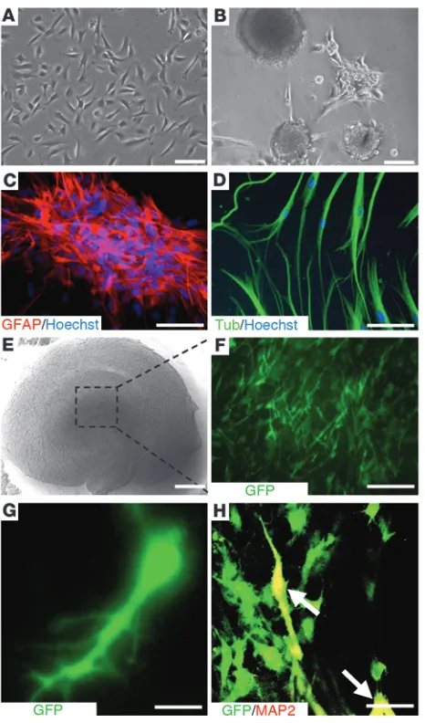
OE-MSCs are located within the olfactory lamina propria beneath the olfactory basal lamina. Transcript and membrane protein analyses have shown that OE-MSCs are closely related to bone marrow mesenchymal stem cells (BM-MSCs), but exhibit a high-level expression of genes involved in neurogenesis when compared with BM-MSCs (17). In vitro and under appropriate culture con-ditions, OE-MSCs are prone to give spheres expressing Nestin, glial fibrillary acid protein (GFAP), and polysialylated neural cell adhesion molecule (PSA-NCAM) markers (16, 17), which are also strongly expressed in neural stem cells (Figure 1, A–C). Moreover, OE-MSCs are found to express III–β-tubulin (Figure 1D) and can be differentiated into MAP2 neuronal cells (17).
In a proof-of-principle experiment, we determined whether, as previously described with mesenchymal stem cells (27), the hip-pocampus could provide a suitable environment for in vitro neural differentiation of human OE-MSCs. Dissociated sphere-derived OE-MSCs, constitutively expressing GFP, were laid on the top of freshly prepared mouse hippocampal slices cultivated on inserts (Figure 1, E and F). Three weeks after grafting, we observed that surviving GFP+ transplanted cells were still on the top of
organo-typic hippocampal cultures and some of them (nonquantified) displayed an interneuron-like morphology (Figure 1G) and/or expressed MAP2 mature neuronal markers (Figure 1H). Not a single GFAP-expressing cell was ever found within the exogenous cell population. Some grafted human OE-MSCs exhibited electro-physiological properties consistent with immature neurons (data not shown). With these encouraging in vitro results in hand, we chose to graft OE-MSCs into an in vivo mouse model of ibotenic acid–lesioned hippocampus.
Human OE-MSCs restore learning and memory after transplantation in injured hippocampus
For this first series of experiments, 3 major choices were made: (a) memory dysfunction was induced by provoking cell death within the hippocampal cornu ammonis (CA) and dentate gyrus (DG) layers using ibotenic acid, an excitotoxic NMDA agonist (28); (b) all GFP+ OE-MSCs were derived from a single stem cell originating
from a sphere and grafted within the lesion site; and (c) no immu-nosuppressant was delivered to mice transplanted with human stem cells, in order to avoid a putative confounding factor.
[image:3.585.49.282.81.477.2]Twenty-four hours after hippocampal lesion was induced, brains were screened using MRI (Figure 2, A and B) and animals with-out signs of bilateral inflammation within the hippocampus were
Figure 1
In vitro assessment of neurogenic characteristics of OE-MSCs. Olfac-tory stem cells (A) gave rise to spherical clusters (B), expressing the GFAP neural stem cell marker (C), when grown in appropriate medi-um. Cells from spheres were then allowed to differentiate into neu-rons expressing the III–β-tubulin neuron marker (D). Freshly prepared mouse hippocampal slices were cultivated on culture insert (E) and loaded with sphere-derived GFP+ OE-MSCs (F). 3 weeks after
cul-ture at the air-liquid interface, some GFP+ OE-MSCs (green) adopted
a neuron-like shape (G) and gave rise to MAP2-expressing neurons
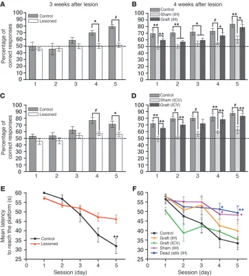
excluded from the study (n = 4). The extent of neurodegeneration, assessed by visualizing edema volume on each scan, was 2.5 mm (± 0.5) when the anteroposterior axis was considered. The pyramidal and granular layers (CA1-3, DG) were significantly damaged (Figure 2, C–F). To better appreciate cellular loss efficiency, we performed region-specific lesion (stratum pyramidale of the CA1 and upper part of the stratum granulosum layer DG) of a hippocampus and observed a reduction of the cell layer density when compared with neighboring intact layers (stratum pyramidale of the CA2/CA3 and lower part of the stratum granulosum layer DG) of the same hip-pocampus (Figure 2, G and H). Mnesic performances of each mouse were assessed 3 weeks after lesion and 4 weeks after transplantation, either in the olfactory tubing maze or the Morris water maze.
Hippocampal-dependent associative memory assessment in the olfac-tory tubing maze. We first used the olfactory tubing maze and an
associative memory paradigm that we previously devised (29). Three weeks after the lesion was induced, water-deprived mice (n = 24) were trained to associate a banana synthetic odor with a drop of water and a lemon synthet-ic odor with a nonaversive sound in a dedsynthet-icated maze (Supplemental Video 1; supplemental material available online with this article; doi:10.1172/JCI44489DS1). As shown in Figure 3A, lesioned mice (n = 16) were unable to learn the cue-reward associations across the 5 train-ing sessions and responded randomly in comparison with control mice (n = 8) (multiple ANOVA [MANOVA], F[1,22] = 8.68, P = 0.007). Animals exhibiting correct
lesions were selected for grafting of human GFP+
OE-MSCs at the initial sites of ibotenic acid delivery (225,000 cells per hemisphere; grafted intrahippocam-pal [IH] group, n = 8) or culture medium (sham-grafted IH group, n = 8). Four weeks after transplantation, asso-ciative memory was retested according to the same pro-cedure. Statistical analyses of the percentage of correct responses revealed a significant group effect (MANOVA, F[2,21] = 16.426; P < 0.001). The group effect was due to
significantly impaired performance in lesioned mice, whether sham-grafted or grafted (IH), overall percent-age of correct responses being significantly decreased in these 2 groups when compared with control mice (P < 0.001 and P = 0.037, respectively). However, as shown in Fig-ure 3B, transplanted mice significantly improved their ability to perform correct associations when compared with sham-grafted animals (P = 0.021). Interestingly, grafted animals dramatically improved their learning capabilities across the 5 training ses-sions and exhibited scores close to those of control animals, while sham-grafted animals continued to respond randomly. Addition-ally, intertrial interval analysis revealed no significant difference between sham-grafted (28.63 s SEM ± 1.44) and grafted (IH) (28.3 s SEM ± 1.75) groups, excluding any bias related to varia-tions in motor function. To confirm the validity of our findings, the olfactory cue–based test was backed up by a visual cue–based memory assessment using the Morris water maze.
[image:4.585.43.318.83.557.2]Hippocampal-dependent reference memory assessment in the Mor-ris water maze. To evaluate visuospatial learning and reference
Figure 2
Lesion assessment. 24 hours after IH ibotenic acid injec-tions, lesion extent was assessed in vivo by MRI (A and B). Examples of 4 axial contiguous T2-weighted images (slice thickness = 500 μm, TEeff = 60 ms, TR = 3000 ms,
memory, a second series of mice (n = 32) were trained to find an immersed platform in the Morris water maze (ref. 30 and Supple-mental Video 2). Lesions were found to induce dramatic deficits in spatial learning as shown by the significant difference in the mean escape latencies between control and lesioned groups (MANOVA, F[1,38] = 4.66; P = 0.037) (Figure 3E). As shown in Figure 3E, at day 5,
control mice reached the submerged platform significantly faster
(31.9 s SEM ± 3.9) when compared with lesioned mice (46.2 s ± 2.1) (ANOVA, F[1,38] = 9.307; P = 0.004),
while no difference was observed in the swimming speed between control (11.10 cm/s ± 0.87) and lesioned mice (10.98 cm/s ± 0.36). Twenty-four hours later, probe test showed a significantly longer time spent in the platform quad-rant (Q1) by control mice, where-as no difference wwhere-as observed in the lesioned group (Supplemen-tal Table 1). The latter data indi-cate a reference memory deficit, consecutive to the lesion. After confirming the lesion efficiency, a strictly similar protocol was applied for both grafted IH and sham-grafted IH groups. More-over, we used an additional con-trol group, which included mice grafted with dead OE-MSCs at the initial lesion sites (dead cells IH group, n = 8). Four weeks after transplantation, MANOVA of the latencies to reach the platform revealed a significant group effect (F[3,28] = 4.41; P = 0.012). The
group effect was due to impaired performance in sham-grafted IH (P = 0.023) and dead cells IH groups (P = 0.024): latencies were significantly increased in these 2 groups when compared with con-trol group, whereas no signifi-cant difference was observed for the grafted IH group (P = 0.406) (Figure 3F). Again, we observed a positive effect of OE-MSCs grafts, as indicated by the significant decrease in the latencies to reach the platform across the 5 training sessions, when compared with both sham-grafted IH and dead cell groups. Noticeably, swim-ming speed analysis revealed no intergroup difference (ANOVA, F[3,34] = 2.465; P = 0.081). Probe
test analyses revealed a signifi-cantly longer time spent in the platform quadrant (Q4) by both control and grafted IH groups, whereas no significant difference was observed in lesioned and dead cell groups (Supplemental Table 1).
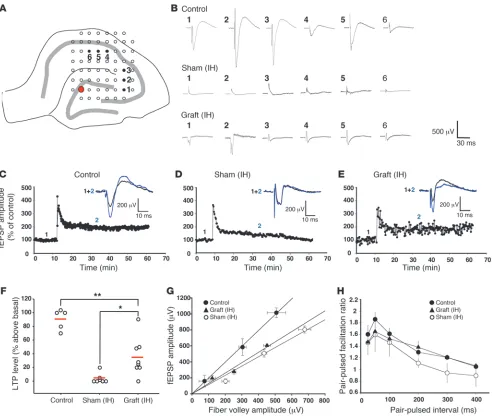
Transplantation of human OE-MSCs restores synaptic transmission and long-term synaptic plasticity
[image:5.585.43.405.83.483.2]Electrophysiological recordings were performed in order to sub-stantiate behavioral data. At the end of the cognitive tests, 20 mice
Figure 3
IH and ICV transplantation of human OE-MSCs improved hippocampus-dependent learning and memory. Cognitive capacities of mice were assessed in the olfactory tubing maze (A–D) and the Morris water maze (E and F). Associative or spatial memory in mice was assessed 3 weeks after lesion (A, C, and E) and 4 weeks after cell or culture medium transplantation (B, D, and F). (A–D) Mean percentage of correct responses was obtained during 5 training sessions of 20 trials per day. (A and C) Lesioned mice (n = 2 × 16) exhibited significant impairment in an associative memory task when compared with control mice (n = 2 × 8). 4 weeks after cell implantation in the lesioned sites (B) or in the lateral ventricles (D), grafted mice (grafted,
were sacrificed, acute hippocampal slices were prepared, and integ-rity of the trisynaptic hippocampal circuitry (loop DG-CA3-CA1) was tested using a multielectrode array including 60 extracellular electrodes. Electrical stimulation was delivered in the DG cell body layers and evoked responses (field excitatory postsynaptic poten-tials [fEPSPs]) were measured in CA3 and CA1 subfields (Figure 4A). Compared with control mice, almost no EPSP was elicited in CA3 and CA1 in slices from sham-grafted animals. In contrast,
although of smaller amplitude when compared with control group, fEPSPs were recorded in CA1 following DG stimulation in slices from grafted animals (Figure 4B).
[image:6.585.49.541.81.497.2]Then we investigated whether this improvement was associated with changes in LTP in the CA1 subfield, by applying a train of high-frequency stimulation (100 Hz, 1 s) to the Schaffer collateral pathway. Forty minutes after high-frequency stimulation, LTP magnitude was 90% ± 6% (n = 5) in control animals (Figure 4C),
Figure 4
while it was almost completely abolished in sham-grafted animals (4% ± 9%, n = 7) (t test; P < 0.001) when signals could be recorded in the CA1 area (Figure 4D). In contrast, a significant recovery of LTP (35% ± 10%, n = 8) was observed in grafted animals when compared with sham-grafted (t test; P = 0.0119) (Figure 4, E and F). How-ever, LTP in grafted animals remained significantly lower when compared with LTP recorded in control animals (Figure 4F) (t test;
P = 0.0019). Additionally, EPSPs zeroed after perfusion of NBQX, an AMPA receptor antagonist, confirming the synaptic nature of recorded EPSPs (data not shown).
In parallel, we investigated whether lesioning and cell grafting altered basal synaptic transmission and short-term synaptic plas-ticity. Basal synaptic transmission was studied by generating the input/output curve. The fEPSP amplitude obtained at various stimulus intensities was plotted against the corresponding fiber
volley amplitude (Figure 4G). In control animals, we observed a lin-ear relationship with a slope value of 1.92 (n = 4; r2 = 0.91).
Lesion-ing the animals led to a change in the synaptic signals, as the slope of the curve was flattened to 1.20 (n = 4; r2 = 0.94). In grafted
ani-mals, the slope was 1.13 (n = 4; r2 = 0.71), not significantly different
from values obtained in sham-grafted animals. Short-term synaptic plasticity was evaluated by pairing stimuli at intervals ranging from 25 to 400 ms. No significant change was detected in sham-grafted and grafted animals when compared with control group, although in lesioned animals, there was a lower paired-pulse ratio for the interstimulus intervals of 200, 300, and 400 ms (Figure 4H).
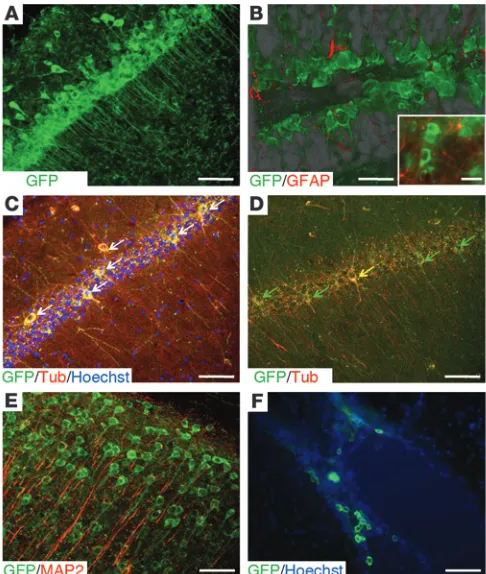
Human OE-MSCs survive, migrate, differentiate into neurons, and stimulate endogenous neurogenesis in mouse lesioned hippocampus
The functional recovery following transplantation was associated with an exogenous neurogenesis. Five weeks after transplantation, we found that 60,000 to 90,000 exogenous cells (survival rate rang-ing from 13% to 20%) had settled within the hippocampus. A large proportion of GFP+ cells were distributed along the different
dam-aged hippocampal fields, including CA1, CA3, and DG (Figure 5, A–C). While 69% ± 11% of the OE-MSCs were expressing III–β -tubu-lin, an immature neuron marker, and 0.9% ± 0.4% were positive for MAP2, a mature neuron marker (Figure 5, D–F), none of the exog-enous cells were immune-positive for the astrocytic marker GFAP (Figure 5G). Moreover, GFP+ cells were also observed in other
cere-bral areas, especially in cortices above the hippocampus (Supple-mental Video 3). Not a single exogenous cell was found in periph-eral structures such as kidneys, liver, or lung (data not shown). An additional experiment, based on transplantation of human OE-MSCs in unilaterally unlesioned hippocampus, demonstrated that stem cells migrated toward the contralateral lesioned hippocampus (Figure 5, H–K). Interestingly, transplantation of OE-MSCs in bilat-erally unlesioned hippocampi showed that cells do not migrate and stay within the tract generated by the injecting needle.
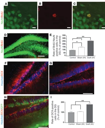
To quantify the impact of human OE-MSCs on endogenous hippocampal neurogenesis, newborn cells were labeled with the thymidine analog BrdU. In each group (n = 5 per group), the
per-Figure 5
Human OE-MSCs transplanted into lesioned mouse hippocampi sur-vived, migrated, and differentiated into neurons. (A) 5 weeks after transplantation, exogenous GFP+ human OE-MSCs were present
in the different fields (CA1, CA3, DG) of the lesioned hippocampus. (B and C) GFP+ human OE-MSCs were mostly found in pyramidal
(CA3, B) and granule cell layers (DG, C). (D) Within these layers, a high proportion (69%) of GFP+ human OE-MSCs (green) expressed
III–β-tubulin (red) (white arrows in D). (E) High magnification of the merged picture of human GFP+ OE-MSCs (green) expressing III–β
-tubulin (red). (F) A small proportion of human GFP+ OE-MSCs (green)
expressed MAP2 (white arrow), a marker for mature neurons (red). (G) No GFP+ human OE-MSC (green) was ever found to express the
astrocytic marker GFAP (red). (H) 4 weeks after lesioning the right hippocampus, GFP+ OE-MSCs were transplanted into the intact
hippo-campus (i.e., left hemisphere). At day 0 (D0) after transplantation, cells
formed clusters and were only observed within the injection site as a cell cluster (I). At D4 after transplantation, numerous GFP+ cells were
observed migrating outside the injection site toward the contralateral lesioned hippocampus (J). At D7 after transplantation, few GFP+
[image:7.585.50.271.81.555.2]centage of newly formed neurons expressing BrdU and NeuN was determined 5 weeks after the last BrdU injection (Figure 6, A–E). No GFP+ cell was found to be BrdU positive and no tumor
formation was ever observed. In confirmation, we found that not a single exogenous cell was positive for Ki67 (data not shown). Moreover, we performed a comparative CFSE proliferation assay showing that, in our culture conditions and prior transplantation, sphere-derived cells are not proliferative or are poorly proliferative (Supplemental Figure 1). Additionally, we quantified a significant increased neurogenesis within the subgranular zone and granule cell layers of the DG in both sham-grafted (P = 0.032) and grafted groups (P = 0.008) when compared with control group. The num-ber of BrdU+/NeuN+ cells was approximately 1.5-fold and 2.5-fold
higher in the sham-grafted group and in the grafted group, respec-tively. Moreover, newly generated neurons were significantly more numerous in the grafted group when compared with the sham-grafted group (P = 0.008). We also labeled immature neuronal cells with doublecortin (DCX) antibody, in order to assess the state of the endogenous neurogenesis just before sacrifice, 5 weeks after transplantation (Figure 6, F–H). Surprisingly, no GFP+ exogenous
cell was found to be DCX positive (Figure 6H). We observed a 1.8-fold increase in the number of endogenous DCX-positive cells in the grafted group when compared with either sham-grafted or control groups (P < 0.01) (Figure 6I).
Human OE-MSCs grafted within the lateral ventricles, survive, migrate, differentiate, and restore mnesic capacities
The positive results obtained during this first series of experiments led us to modify 1 of our 3 initial options. Using the same ani-mal model, we decided to graft GFP+ OE-MSCs, derived from a
single stem cell, within the cerebrospinal fluid of lateral ventricles. As previously described, we first confirmed the lesion efficiency in both tests (Figure 3, C and E). Four weeks after transplanta-tion, behavioral assessment using the olfactory tubing maze revealed a significant group effect (MANOVA, F[2,21] = 34.839; P < 0.001). The group effect was due to significantly impaired per-formance in lesioned mice (sham-grafted intracerebroventricular [ICV] group), with an overall percentage of correct responses sig-nificantly decreased in this group when compared with control (P < 0.001) and grafted ICV groups (P < 0.001). Across the 5
train-Figure 6
Human OE-MSCs stimulated endog-enous neurogenesis after transplantation in lesioned hippocampi. 5 weeks after grafting, brain sections of mice injected with BrdU twice a day during 3 days fol-lowing cell implantation (n = 5 for each group) were immunostained with anti-NeuN (green) and anti-BrdU antibodies (red) (A–D). Quantification of BrdU+/
NeuN+ cells in DG indicated an increased
number of mitotic cells in grafted (IH) mice when compared with sham-grafted (IH) (P < 0.05) and control (P < 0.01) mice (E). DCX immunohistochemistry revealed the presence of immature neurons in the DG of control (G) and grafted (IH) (F, H) mice. As shown in (H), no GFP+
[image:8.585.40.381.80.494.2]ing sessions, grafted animals improved their mnesic capacities significantly, while sham-grafted animals remained unable to per-form the task (Figure 3D). Intertrial interval analysis revealed no significant difference between sham-grafted (31.12 s SEM ± 2.16) and grafted (ICV) (28.42 s SEM ± 1.7) groups. In parallel, analysis of latencies in the Morris water maze test revealed a significant group effect (F[3,28] = 10.473; P < 0.001). The group effect was due to
sig-nificantly impaired performance in sham-grafted (P = 0.009) and dead cell groups (P = 0.009), with latencies significantly increased in these 2 groups, when compared with control and grafted ICV groups. No significant difference was observed between grafted ICV and control groups (P = 1) (Figure 3F). Whereas no significant difference was observed in the swimming speed between grafted ICV and both sham-grafted (P = 0.1) and control (P = 1) groups, similar analysis revealed that grafted mice with dead cells were significantly faster when compared with those in the grafted ICV group (P = 0.01). Probe test analyses revealed a significantly lon-ger time spent in the platform quadrant (Q4) by both control and grafted ICV groups, while no significant difference was observed in lesioned and dead cell groups (Supplemental Table 1).
Improved mnesic performances were also associated with the presence of GFP+ cells in the different layers of the lesioned
hippo-campi (Figure 7, A and B, and Supplemental Video 4). An extensive exogenous neurogenesis was observed, with stem cells differentiat-ing into cells expressdifferentiat-ing neuron-specific markers (Figure 7, C and D). No GFP+/GFAP+ cell was ever encountered. Exogenous cells
were found in cortical areas but remained always immunonegative for the mature neuronal marker MAP2 (Figure 7E), and no GFP+
cell was found in peripheral structures. Conversely, GFP+ cells
exhibiting an undifferentiated morphology and negative for neu-ral markers (III–β-tubulin, MAP2, GFAP) were found at the margin
of ventricular areas (Figure 7F). Regarding these data, we expected a migratory potential of OE-MSCs in response to specific signals generated by a lesion.
Lesioned hippocampi overexpress genes involved in cell chemoattraction
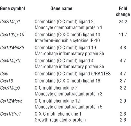
In order to understand OE-MSC homing, a comparative gene expression profile of lesioned and control hippocampi (3 vs. 3) was performed using mouse pangenomic DNA microarrays 4 weeks after surgery. In total, 114 transcripts with a fold change above 2.5 were found upregulated in lesioned hippocampi. The list of overexpressed genes indicates that 53% were involved in immune and inflammatory processes, 22% in cell metabolism, and 27% in various other processes (Supplemental Figure 2). Interestingly, among overexpressed genes, 8% code for chemo-kines and 2 of them, Ccl2 and Cxcl10, have a fold change above 10 (Table 1). Moreover, cytokines such as Spp1 and C3, known for their chemotactic properties, were also upregulated with a fold change of 11 and 162.5, respectively.
Discussion
[image:9.585.43.286.84.371.2]Stem cells are present in most adult tissues, where they contrib-ute to tissue regeneration. Such a feature is also observed in the olfactory mucosa, which has an extraordinary capacity to func-tionally self regenerate. Until recently, basal stem cells (hori-zontal and globose), located in the olfactory epithelium, were identified as the only 2 stem cells types in the nasal cavity (31). However, a new olfactory stem cell subtype, previously suspected to reside inside the lamina propria (15), has been confirmed and characterized by 2 distinctive groups at the same time (16, 17). Although the biological function of these ectomesenchymal stem cells remains to be unveiled, our present study and previ-ous data suggest that these cells could be useful for stem cell therapies (15, 32, 33). We report here what we believe is the first successful restoration of learning and memory after transplan-tation of adult human olfactory nasal stem cells, derived from the lamina propria, in a murine model that mimics effects of ischemic/hypoxic injury in the hippocampus exclusively (28). As a result, it is possible that this new stem cell type can be used for repairing the damaged or pathological brain.
Figure 7
Human OE-MSCs transplanted into the cerebrospinal fluid (CSF) of hippocampus-lesioned mice survived at least 5 weeks, migrated, and differentiated into neurons. (A) Increased density of GFP+ cells within
the pyramidal cell body layers demonstrated the ability of human OE-MSCs to migrate from the CSF toward the injury zone. (B) As dem-onstrated by confocal image reconstitution using projection transpar-ency (see also Supplemental Video 4), exogenous GFP+ cells were
also remarkably distributed within the granule cell body layers of the DG. The insert indicates that not a single human OE-MSC expressed the astrocytic marker GFAP (red). (C and D) Within the CA3 field, some pyramidal cell–like and interneuron-like GFP+ human OE-MSCs
(green) expressed III–β-tubulin (red) (white arrows in C, yellow arrow in D), but others were immunonegative for this immature neuronal marker (green arrows in D). (E) Exogenous GFP+ cells migrated as
well in other cerebral areas (cortical area in E), but remained immuno-negative for the mature neuronal marker MAP2 (red). (F) GFP+ cells
Although no immunosuppressant was delivered to the host animal, we found that a relatively large fraction (nearly 20%) of injected human cells invaded the hippocampal neuronal layers and that the vast majority (nearly 70%) differentiated into cells express-ing neuronal markers (III–β-tubulin, MAP2) and not glial marker (GFAP). In contrast to what has been observed in other transplan-tation studies (6, 34) and despite our previous experiments dem-onstrating that olfactory mucosa spheres can be driven to generate astrocytes and/or neurons under defined conditions in vitro (15), none of the grafted cells differentiated into astrocytes. Similarly, in our in vitro coculture experiment, no OE-MSC–derived astrocyte was ever observed. These surprising findings may be due to our initial choice of exclusively lesioning the hippocampus. A previ-ous in vitro study demonstrated that factors from this neurogenic brain area induced the differentiation of mesenchymal stem cells into neurons exclusively, and stem cell–derived astrocytes were found only when soluble factors were collected from the cortex or the cerebellum (27). Conversely, it has been shown by others that SVZ neural stem cells, dedicated to producing dopaminer-gic or GABAerdopaminer-gic neurons, differentiate only into glial lineages when transplanted ectopically (35). Accordingly, our data confirm the importance of the microenvironment in the differentiation process. It remains now to be seen whether similar outcomes can be achieved when olfactory stem cells are implanted in another lesioned cerebral area.
The central nervous system is known for being at least partially immunoprivileged (36), and we took advantage of this specificity to perform xenotransplantations without the use of any immunosup-pressant. Moreover, it has been demonstrated that mesenchymal stem cells display specific immune properties including hypoim-munogenicity and immunoregulatory and immunosuppressive activities (37). As a result, human mesenchymal stem cells are not fully rejected by the CNS of rodent recipients.
When compared with BM-MSCs, OE-MSCs in vitro exhibit a high proliferating profile under defined conditions and can be grown in large numbers. Here we show that specific culture condi-tions for sphere formation inhibit cell proliferation. This could explain why not a single dividing human cell has been found after brain implantation, as observed by BrdU and Ki67 stain-ing. This result indicates that, unlike embryonic stem cells (10), sphere-derived OE-MSCs are not prone to proliferation after brain transplantation. This property is obviously valuable and might be considered to represent a prerequisite for clinical trials based on cell therapy. In addition, as previously described in studies using mesenchymal and neural stem cell types (38), postmitotic OE-MSCs were able to boost the proliferation of resident stem cells. To date, the mechanisms by which OE-MSCs trigger endogenous neurogenesis remain largely unknown. However, in a parallel in vitro study, we found that OE-MSCs release trophic factors, trig-gering a significant increase in neurite length (data not shown). Together, these data support the hypothesis that OE-MSC trans-plantation creates a permissive microenvironment that stimulates endogenous neurogenesis and helps to restructure preexisting net-works acting on resident neural stem cells and neurons.
Although temporally simultaneous, the dual process of endoge-nous neurogenesis and exogeendoge-nous neuronal differentiation exhib-it dissimilarexhib-ities. Resident stem cells proliferate and give rise to functional neural stem cells (39) but display a limited migration. In contrast, exogenous stem cells stop multiplying and differen-tiate into neurons after a protracted migration to the damaged neuronal layers. We recently showed that OE-MSCs can be consid-ered as a mesenchymal stem cell subtype with neurogenic proper-ties (17). It is well established that mesenchymal stem cells are able to cross physical and metabolic frontiers such as the blood-brain barrier. Moreover, analysis of gene overexpression in lesioned hip-pocampus provides molecular signature evidence for the site-spe-cific migration process observed in this study. This site-spesite-spe-cific attraction could be similar to leukocyte trafficking and occurs via inflammation-associated factors, such as chemotactic cytokines or chemokines (40). Here, we demonstrate that many inflammatory molecules are still expressed in lesioned hippocampi 4 weeks after lesioning. We observed a robust overexpression of 9 genes coding for chemokines that could provide a chemoattractant microen-vironment supporting the migration of OE-MSCs. Ccl2 (Mcp-1), the most overexpressed chemokine in lesioned hippocampus, is known to have chemoattractive properties on mesenchymal stem cells (41) or neural stem cells (42, 43). Similarly, Cxcl10, another overexpressed chemokine, stimulates migration of mesenchymal stem cells (44). Two other strongly overexpressed genes coding for cytokines with chemoattractive properties, C3 and Spp1, could be involved in the OE-MSC–directed migration. SPP1 (osteopon-tin) exerts a chemoattractive effect on stem cells by recruiting mesenchymal stem cells via CD44 (45) or regulating the migra-tion of neuroblasts to injured cerebral structures (46). The protein encoded by C3, via the formation of C3a, also plays a chemoattrac-tive role for mesenchymal stem cells (47). In addition, we observed that OE-MSCs express several chemokine receptors and are responsive to overexpressed molecules that stimulate their migra-tion (data not shown). It is therefore not surprising that OE-MSCs can home to lesioned sites, and this selective migration is a critical step in stem cell regenerative therapies.
[image:10.585.54.275.132.345.2]The significant neurogenic effects observed in these experiments were associated with partial recovery of LTP and a remarkable
Table 1
Dysregulated transcripts coding for chemokines with a fold change above 2.5 in lesioned hippocampus in comparison with control
Gene symbol Gene name Fold change Ccl2/Mcp1 Chemokine (C-C motif) ligand 2 24.2
Monocyte chemoattractant protein 1
Cxcl10/Ip-10 Chemokine (C-X-C motif) ligand 10 11.7 Interferon-inducible cytokine IP-10
Ccl19/Mip3b Chemokine (C-C motif) ligand 19 4.8 Macrophage inflammatory protein 3b
Ccl4/Mip1b Chemokine (C-C motif) ligand 4 4.7 Macrophage inflammatory protein 3b
Ccl5 Chemokine (C-C motif) ligand 5/RANTES 4.7
Cxcl16 Chemokine (C-X-C motif) ligand 16 3.7
Ccl7/Mcp3 C-C motif chemokine 7 3.2 Monocyte chemoattractant protein 3
Ccl12/Mcp5 C-C motif chemokine 12 2.9 Monocyte chemoattractant protein 5
Cxcl1/Gro1 C-X-C motif chemokine 1 2.6 Growth-regulated α protein 2.6
recovery of learning and memory abilities. Surprisingly, such find-ings are correlated with the reestablishment of neuron networks mostly displaying the immature marker III–β-tubulin. However, previous studies reported that immature neurons express a more robust LTP than mature neurons (48, 49), supporting the hypoth-esis that immature neurons derived from a pool of endogenous stem cells (50) and/or exogenous stem cells in our study could contribute to the formation of a new mnesic trace. Combined with our data showing that OE-MSCs induce neuronal matura-tion/stimulation via trophic factors, it can thus be envisaged that exogenous cells participate in the reestablishment of a functional network. Otherwise, it has already been shown that embryonic (51) or neural (52) stem cells can form functional synapses and integrate into the host cortical circuitry after transplantation into the developing brain. Although previous studies have demon-strated hippocampal plasticity after cell transplantation (53–56), to the best of our knowledge, no study using human adult stem cells has ever demonstrated that cell therapy can induce the res-toration of functional neuronal networks within the adult hip-pocampus. Grafting neural stem cells, sampled from the brain, was previously shown to improve learning capacities of lesioned adult mice (6). While these results suggest that both types of stem cells could potentially be used for future treatments of memory loss, nasal olfactory stem cells present numerous advantages for a medical application.
Indeed, among all the cell types proposed for the future regen-erative medicine of the CNS, OE-MSCs have the enormous advan-tage of being easily accessible in living adults, obtainable under local anesthesia, and transplantable in an autologous manner, thus eliminating contentious ethical and technical considerations (57). By overcoming these 2 major hurdles, the use of OE-MSCs in clinical trials protocols should be facilitated, representing a first step for the translation to the clinic. In addition, the proof of prin-ciple that olfactory ensheathing cells, also derived from the nasal lamina propria, can be safely used in clinical protocols for spinal cord injury has been recently established. Moreover, and contrary to other cell types like BM-MSCs, nasal stem cells already reside in a neural environment, which represents a nonnegligible benefit for targeting brain disorders. Considering that the time window after injury is a critical point for using cell therapy, the short time scale necessary for the isolation and expansion of OE-MSCs is impor-tant to consider. As a consequence of their highly proliferative capacity in vitro under the control of defined factors, the number of cells is not a limiting factor.
Methods
Experimental design
We performed the ibotenate lesion on experimental day 1 followed by a control of the lesion efficiency, using MRI, at day 2. A functional control, using behavioral tests, was performed at week 3. Human OE-MSCs were transplanted at week 4, and animals were retested at week 8 before being sacrificed for either electrophysiological or immunohistological analy-ses. Anesthesia and surgical procedures were performed according to the French law on Animal Care Guidelines, and the Animal Care Committee of Aix-Marseille University approved our protocols. Human biopsies were obtained under a protocol that was approved by the local ethical committee (Comité de Protection des Personnes) of Marseille. Informed consent was given by each individual participating in the study, in accordance with the 1964 Helsinki convention and French law relating to biomedical research.
Animals
Adult male Balb/c mice (n = 77) and C57BL/6 (n = 40) (IFFA CREDO), 10 weeks old at the beginning of the experiment, were used. All animals were housed in individual cages and maintained on a 12-hour light/ 12-hour dark cycle at a constant temperature (22 ± 1°C). Food and water were provided ad libitum except during the habituation and training peri-ods in the olfactory tubing maze.
Experimental injury model: amnesic syndrome
We performed a bilateral lesion of the hippocampus in mice, by using an NMDA agonist (i.e., ibotenic acid; Sigma-Aldrich) leading to the loss of neuronal cells in the sites of injection by cellular excitotoxicity. Anesthe-tized Balb/c (n = 46) or C57BL/6 mice (n = 32) were inserted in a stereo-tactic frame, the skull surface was exposed, and holes were drilled at the appropriate sites to allow bilateral infusion of ibotenic acid (10 μg/μl) with a glass capillary (40-μm external tip) glued to a 1-μl Hamilton syringe con-nected to stereotactic syringe pump (KDS 310; KD scientific). Antero-pos-terior (AP), lateral (L), and vertical (V) coordinates for micro-infusions in the hippocampus, were taken relative to the bregma: (a) AP – 1.7 mm, L ± 1 mm, V – 1.5 mm; (b) AP – 2 mm, L ± 1.3 mm, V – 1.5 mm; (c) AP – 2.6 mm, L ± 1.5 mm, V – 1.5 mm. The infused volume of ibotenic acid or PBS was 30 nl per injection site. For migration kinetic experiments (n = 4), unilateral lesions were performed (right or left hemisphere, n = 2 + 2).
MRI
Mice were anesthetized using a mixture of air and isoflurane (3% during induction and 2% for maintenance) via the nose cone of a mouse head-holder device at a flow of 2 l/min. During data acquisition, body tempera-ture was maintained at 37°C using a heating blanket. All experiments were performed with a Bruker PharmaScan spectrometer (7 T magnet and 16-cm horizontal bore size). A dedicated mouse head coil (linear birdcage coil with 23-mm inner diameter) was used for signal transmission and reception.
Sixteen axial contiguous T2-weighted images (slice thickness = 0.5 mm) with fat suppression (band width = 900 Hz) were acquired 1 day after lesion with a turbo-RARE sequence (effective echo time [TEeff] = 60 ms, repetition time [TR] = 3000 ms, rare factor = 8, 8 averages), using a 21-mm field of view and 256 × 256 matrix. Mouse respiration was monitored and, to minimize motion artifacts, image acquisition was synchronized with the respiratory cycle.
Isolation and expansion of human olfactory mucosa cells
Human nasal olfactory mucosae were obtained by biopsy during routine nasal surgery under general anesthesia. Biopsies were immediately placed in growth medium containing DMEM/Ham F12 supplemented with 10% FBS and 1% penicillin/streptomycin (Invitrogen). OE-MSCs were purified from the lamina propria and cultivated as described before (17).
Generation of GFP+ clonal sphere-derived OE-MSCs for transplantation
Human OE-MSCs were infected with a GFP-lentiviral vector, and sphere for-mation was induced as previously described (17). Individual primary spheres were dissociated and clonally replated using the limit dilution method. On the day of transplantation, floating spheres were harvested, washed, and enzymatically (trypsin/EDTA) dissociated. Sphere-derived cells were centri-fuged and resuspended in serum-free DMEM-F12 at the respective concen-trations of 75,000 cells/μl and 225,000 cells/μl for IH and ICV injections.
Coculture experiment
21-day-old Balb/c mice and plated on culture inserts. Immediately after, a drop (1 μl) of freshly prepared cell suspension (10,000 cells/μl) was laid on top of the slice. Hippocampal slices were then cultured for 21 days at 37°C, 5% CO2, and medium was replaced 3 times per week.
Transplantation surgery
Thirty days after lesioning, mice with confirmed MRI and severe learn-ing deficits were blindly allocated to 4 groups. In the transplanted groups (grafted groups), mice received bilateral infusions of sphere-derived human OE-MSCs (450,000 cells in total), while in the untransplanted groups, ani-mals received an equal amount of culture medium without cells (sham-grafted groups) or dead OE-MSCs killed by 1 hour freezing step (dead cell groups). Cell suspensions or vehicle were injected with a 1 μl Hamilton syringe into the hippocampus (grafted [IH], n = 21; sham-grafted [IH],
n = 21; dead cells, n = 8) at the same coordinates as those used for ibotenate lesion or in the lateral ventricles (coordinates: AP – 0.58; L ± 1, V – 2.1) (grafted [ICV], n = 16; sham-grafted [ICV], n = 16). The infused volume was 1 μl per injection site; the rate of infusion was 0.5 μl/min.
Behavioral procedure
The procedures using the olfactory tubing maze (29) and Morris water maze (30) have been previously described (see Supplemental Data for a full description).
Electrophysiological analyses
Experiments were carried out on acute hippocampal slices (350 μm) obtained from mice of each of 3 groups (control n = 5, grafted [IH]
n = 8, and sham-grafted [IH] n = 7). Immediately after decapitation, brains were quickly dissected and placed in ice-cold buffer including 124 mM NaCl, 3.5 mM KCl, 25 mM NaHCO3, 1.25 mM NaH2PO4, 1 mM CaCl2, 2 mM MgSO4, and 10 mM glucose (bubbled with O2/CO2:95%/5%). Slices were then prepared with a vibratome (VT1000S; Leica) and maintained at room temperature in the same buffer supplemented with 1 mM CaCl2. Slices were transferred within the same buffer to a multi-electrode array (MEA-60; Multi Channel Systems) comprising 60 extracellular electrodes (S4). Slices were continually superfused with the medium at a flow rate of 2 ml/min–1 and maintained at 37°C. Monopolar stimulation was achieved with an external stimulator (MC Stimulus; Multi Channel Systems) by applying biphasic current pulses to 1 electrode of the array. Field EPSPs were then simultaneously recorded by the remaining electrodes in the dendritic subfields of hippocampal neurons. Basal synaptic transmission was monitored by delivering a 0.066 Hz stimulation. The input/output curve was generated by applying increasing stimulation intensities and by plotting the fEPSP amplitude as a function of the corresponding fiber volley amplitude. LTP was elicited by applying a 100-Hz stimulation train for 1 second. NBQX (10 μM), an AMPA receptor antagonist, was applied at the end of some experiments in order to verify that the signals recorded were effectively fEPSPs. Field EPSPs were further analyzed (MC Rack; Multi Channel Systems).
Cell quantification
The survival rate of OE-MSCs after transplantation and the percentage of transplanted OE-MSCs that expressed cell-specific marker were quanti-fied through fluorescent microscopy. Brains were perfused and coronal sections (35 μm thick) were serially collected. The number of OE-MSCs that survived inside the hippocampus 4 weeks after transplantation was estimated by counting the number of GFP+ cells in all sections containing hippocampus (n = 4 animals analyzed). The percentage of OE-MSCs that expressed cell-specific markers was estimated by counting the number of costained cells (GFP+ + marker X) and dividing this number by the total
number of GFP+ cells in the same section. For each animal (n = 4), the ros-trocaudal hippocampus was serially sectioned at a thickness of 35 μm, and each marker was analyzed every sixth section.
Assessment of endogenous neurogenesis
BrdU (Sigma-Aldrich) at a concentration of 6 mg/ml was injected intra-peritoneally (100 mg/kg) immediately after transplantation surgery and twice a day for the next 3 days, with a delay of 8 hours between each injection. Four weeks after the last injection, brain tissues (control, sham-grafted, grafted; n = 5 per group) were processed (see Supplemen-tal ExperimenSupplemen-tal Procedures). Note that no behavioral assessment was performed with these mice.
Comparative quantification of BrdU+/NeuN+ cells. Comparative analysis between each group was conducted in a blind manner. Fluorescent stain-ing of coded slides was visualized and quantified with a fluorescent micro-scope at ×40 and ×63. For each group, similar sections were stained and the number of BrdU+/NeuN+ cells was quantified in the subregions of the hippocampal DG, including granule cell body layer and subgranular zone. For each mouse, BrdU+/NeuN+ cell numbers were counted in 9 coronal sections (35-μm thick) covering the left DG in its rostrocaudal extension (3 dorsal sections, 175 μm apart, and 6 ventral sections, 175 μm apart), using an image analyzer (LUCIA; Laboratory Imaging). The number of BrdU+/NeuN+ cells in the control group was used as a baseline (index = 100) to evaluate changes in neurogenesis in other groups.
Comparative quantification of DCX+ cells. Using the method described above, the number of DCX+ cells was quantified in the granular cell body layer of the DG.
Immunostaining
Floating sections were incubated for 1 hour at room temperature (RT) with blocking buffer (3% BSA, 5% appropriate serum, 0.1% Triton X-100 in PBS) and for 90 minutes with the following primary antibodies dilut-ed in blocking solution: rabbit polyclonal anti-DCX (1/500; Abcam), rabbit polyclonal anti-GFAP (1/500; Dako), mouse monoclonal anti– III–β-tubulin (1/500; Sigma-Aldrich), rabbit polyclonal anti-MAP2 (1/500; Chemicon), fluorescently labeled mouse monoclonal anti-NeuN (1/1000; Chemicon), rabbit polyclonal anti-GFP (1/300; Chemicon), mouse anti-GFP (1/500; Roche), monoclonal anti-BrdU (1/100; Dako), and rabbit polyclonal anti-Ki67 (1/100; Abcam). Then slices were rinsed (3 × 5 min) in PBS and incubated for 90 minutes with cross-adsorbed Alexa Fluor 488– or 594–conjugated anti-rabbit or anti-mouse second-ary antibodies (1/500; Jackson Immunoresearch) in dark conditions. After several washes in PBS, slices were counterstained with 0.5 μg/ml Hoechst blue (33258; Molecular Probes) for 30 minutes and mounted with anti-fading medium (DAKO). For BrdU staining, DNA was dena-tured using a pretreatment in HCl [2N] and 0.2% Triton X-100 for 40 minutes at 37°C. Positive and negative controls were performed for all immunostaining of this study.
Microarray experiments
Briefly, for the Agilent microarray analysis, total RNA from freshly dis-sected hippocampi (3 control vs. 3 lesioned mice) were isolated 4 weeks after lesioning and then treated as described in Supplemental Methods. Genes with a fold change above 2.5 were clustered into functional groups using DAVID Functional Annotation Clustering Tool. Data are available on the ArrayExpress database (accession number E-MEXP-2682).
Statistics
when appropriate, by 2 × 2 comparisons based on the Bonferroni post-hoc, using the SPSS/PC + statistics 11.0 software (SPSS Inc.). For electrophysi-ological and endogenous neurogenesis data, statistical significance was assessed using unpaired Student’s t test and 2-tailed Mann-Whitney U test, respectively. The threshold for significance was set at P < 0.05.
Acknowledgments
We thank M. Baudry and I. Sancho-Martinez for comments during the preparation of this manuscript, Pierre Giraud and Laure Fron-zaroli-Molinieres (INSERM U641) for their help with hippocampal slice cultures, Catherine Cohen-Solal for the preparation of acute hippocampal slices, and Nicolas Boulanger and Béatrice Loriod for the microarray study (genomic platform TAGC, Ibisa, INSERM
U928). This study was supported by the French IRME and AFM Foundations, the ANR MALZ, and the FEDER PACA.
Received for publication July 24, 2010, and accepted in revised form April 27, 2011.
Address correspondence to: Emmanuel Nivet, Salk Institute for Biological Studies, 10010 North Torrey Pines Road, La Jolla, California 92037, USA. Phone: 858.453.4100, ext. 1324; Fax: 858.453.2573; E-mail: enivet@salk.edu.
Emmanuel Nivet’s present address is: Salk Institute for Biological Studies, La Jolla, California, USA.
1. Maguire EA, et al. Navigation-related structural change in the hippocampi of taxi drivers. Proc Natl Acad Sci U S A. 2000;97(8):4398–4403.
2. Wiltrout C, Lang B, Yan Y, Dempsey RJ, Vemu-ganti R. Repairing brain after stroke: a review on post–ischemic neurogenesis. Neurochem Int. 2007; 50(7–8):1028–1041.
3. Jin K, et al. Increased hippocampal neurogenesis in Alzheimer’s disease. Proc Natl Acad Sci U S A. 2004; 101(1):343–347.
4. Li B, et al. Failure of neuronal maturation in Alzheimer disease dentate gyrus. J Neuropathol Exp Neurol. 2008;67(1):78–84.
5. Lindvall O, Kokaia Z. Stem cells for the treat-ment of neurological disorders. Nature. 2006; 441(7097):1094–1096.
6. Yamasaki TR, Blurton-Jones M, Morrissette DA, Kitazawa M, Oddo S, LaFerla FM. Neural stem cells improve memory in an inducible mouse model of neuronal loss. J Neurosci. 2007;27(44):11925–11933. 7. Blurton-Jones M, et al. Neural stem cells improve
cognition via BDNF in a transgenic model of Alzheimer disease. Proc Natl Acad Sci U S A. 2009; 106(32):13594–13599.
8. Qu T, Brannen CL, Kim HM, Sugaya K. Human neural stem cells improve cognitive function of aged brain. Neuroreport. 2001;12(6):1127–1132. 9. Acharya MM, et al. Rescue of radiation-induced
cognitive impairment through cranial transplanta-tion of human embryonic stem cells. Proc Natl Acad Sci U S A. 2009;106(45):19150–19155.
10. Bjorklund LM, et al. Embryonic stem cells develop into functional dopaminergic neurons after trans-plantation in a Parkinson rat model. Proc Natl Acad Sci U S A. 2002;99(4):2344–2349.
11. Martinez-Cerdeno V, et al. Embryonic MGE precur-sor cells grafted into adult rat striatum integrate and ameliorate motor symptoms in 6-OHDA-lesioned rats. Cell Stem Cell. 2010;6(3):238–250. 12. Freed CR, et al. Transplantation of embryonic
dopamine neurons for severe Parkinson’s disease.
N Engl J Med. 2001;344(10):710–719.
13. Olanow CW, et al. A double-blind controlled trial of bilateral fetal nigral transplantation in Parkinson’s disease. Ann Neurol. 2003;54(3):403–414. 14. Bachoud-Levi AC, et al. Effect of fetal neural
trans-plants in patients with Huntington’s disease 6 years after surgery: a long-term follow-up study.
Lancet Neurol. 2006;5(4):303–309.
15. Murrell W, et al. Multipotent stem cells from adult olfactory mucosa. Dev Dyn. 2005;233(2):496–515. 16. Tome M, Lindsay SL, Riddell JS, Barnett SC.
Iden-tification of nonepithelial multipotent cells in the embryonic olfactory mucosa. Stem Cells. 2009; 27(9):2196–2208.
17. Delorme B, et al. The human nose harbors a niche of olfactory ectomesenchymal stem cells displaying neurogenic and osteogenic properties. Stem Cells Dev. 2010;19(6):853–866.
18. Lindsay SL, Riddell JS, Barnett SC. Olfactory
muco-sa for transplant-mediated repair: a complex tissue for a complex injury? Glia. 2010;58(2):125–134. 19. Shyu WC, et al. Implantation of olfactory
ensheath-ing cells promotes neuroplasticity in murine mod-els of stroke. J Clin Invest. 2008;118(7):2482–2495. 20. Kadar T, Dachir S, Shukitt-Hale B, Levy A.
Sub-regional hippocampal vulnerability in various animal models leading to cognitive dysfunction.
J Neural Transm. 1998;105(8–9):987–1004. 21. Grady MS, Charleston JS, Maris D, Witgen BM,
Lifshitz J. Neuronal and glial cell number in the hippocampus after experimental traumatic brain injury: analysis by stereological estimation. J Neuro-trauma. 2003;20(10):929–941.
22. Miller DB, O’Callaghan JP. Aging, stress and the hippocampus. Ageing Res Rev. 2005;4(2):123–140. 23. Bobinski M, et al. The histological validation of
post mortem magnetic resonance imaging-deter-mined hippocampal volume in Alzheimer’s disease.
Neuroscience. 2000;95(3):721–725.
24. Schmidt-Kastner R, Freund TF. Selective vulner-ability of the hippocampus in brain ischemia. Neu-roscience. 1991;40(3):599–636.
25. Feron F, et al. Autologous olfactory ensheathing cell transplantation in human spinal cord injury.
Brain. 2005;128(pt 12):2951–2960.
26. Mackay-Sim A, et al. Autologous olfactory ensheath-ing cell transplantation in human paraplegia: a 3-year clinical trial. Brain. 2008;131(pt 9):2376–2386. 27. Rivera FJ, Sierralta WD, Minguell JJ, Aigner L.
Adult hippocampus derived soluble factors induce a neuronal–like phenotype in mesenchymal stem cells. Neurosci Lett. 2006;406(1–2):49–54. 28. Jarrard LE. Use of excitotoxins to lesion the
hippo-campus: update. Hippocampus. 2002;12(3):405–414. 29. Roman FS, Marchetti E, Bouquerel A, Soumireu-Mourat B. The olfactory tubing maze: a new appa-ratus for studying learning and memory processes in mice. J Neurosci Methods. 2002;117(2):173–181. 30. Morris R. Developments of a water-maze procedure
for studying spatial learning in the rat. J Neurosci Methods. 1984;11(1):47–60.
31. Leung CT, Coulombe PA, Reed RR. Contribution of olfactory neural stem cells to tissue maintenance and regeneration. Nat Neurosci. 2007;10(6):720–726. 32. Murrell W, et al. Olfactory mucosa is a potential source for autologous stem cell therapy for Parkin-son’s disease. Stem Cells. 2008;26(8):2183–2192. 33. Pandit SR, Sullivan JM, Egger V, Borecki AA,
Oleskevich S. Functional effects of adult human olfactory stem cells on early–onset sensorineural hearing loss. Stem Cells. 2011;29(4):670–677. 34. Shetty AK, Rao MS, Hattiangady B. Behavior of
hippocampal stem/progenitor cells following graft-ing into the injured aged hippocampus. J Neurosci Res. 2008;86(14):3062–3074.
35. Seidenfaden R, Desoeuvre A, Bosio A, Virard I, Cre-mer H. Glial conversion of SVZ–derived committed neuronal precursors after ectopic grafting into the adult brain. Mol Cell Neurosci. 2006;32(1–2):187–198.
36. Galea I, Bechmann I, Perry VH. What is immune privilege (not)? Trends Immunol. 2007;28(1):12–18. 37. Uccelli A, Moretta L, Pistoia V. Mesenchymal
stem cells in health and disease. Nat Rev Immunol. 2008;8(9):726–736.
38. Munoz JR, Stoutenger BR, Robinson AP, Spees JL, Prockop DJ. Human stem/progenitor cells from bone marrow promote neurogenesis of endogenous neural stem cells in the hippocampus of mice. Proc Natl Acad Sci U S A. 2005;102(50):18171–18176. 39. Toni N, et al. Synapse formation on neurons born
in the adult hippocampus. Nat Neurosci. 2007; 10(6):727–734.
40. Fox JM, Chamberlain G, Ashton BA, Middleton J. Recent advances into the understanding of mesenchymal stem cell trafficking. Br J Haematol. 2007;137(6):491–502.
41. Belema-Bedada F, Uchida S, Martire A, Kostin S, Braun T. Efficient homing of multipotent adult mesenchymal stem cells depends on FROUNT-mediated clustering of CCR2. Cell Stem Cell. 2008; 2(6):566–575.
42. Belmadani A, Tran PB, Ren D, Miller RJ. Chemo-kines regulate the migration of neural progenitors to sites of neuroinflammation. J Neurosci. 2006; 26(12):3182–3191.
43. Magge SN, et al. Role of monocyte chemoattractant protein-1 (MCP-1/CCL2) in migration of neural pro-genitor cells toward glial tumors. J Neurosci Res. 2009; 87(7):1547–1555.
44. Croitoru-Lamoury J, Lamoury FM, Zaunders JJ, Veas LA, Brew BJ. Human mesenchymal stem cells constitutively express chemokines and chemokine receptors that can be upregulated by cytokines, IFN-beta, and Copaxone. J Interferon Cytokine Res. 2007; 27(1):53–64.
45. Raheja LF, Genetos DC, Yellowley CE. Hypoxic osteocytes recruit human MSCs through an OPN/ CD44-mediated pathway. Biochem Biophys Res Com-mun. 2008;366(4):1061–1066.
46. Yan YP, Lang BT, Vemuganti R, Dempsey RJ. Per-sistent migration of neuroblasts from the subven-tricular zone to the injured striatum mediated by osteopontin following intracerebral hemorrhage.
J Neurochem. 2009;109(6):1624–1635.
47. Schraufstatter IU, Discipio RG, Zhao M, Khaldoy-anidi SK. C3a and C5a are chemotactic factors for human mesenchymal stem cells, which cause pro-longed ERK1/2 phosphorylation. J Immunol. 2009; 182(6):3827–3836.
48. Schmidt-Hieber C, Jonas P, Bischofberger J. Enhanced synaptic plasticity in newly generated granule cells of the adult hippocampus. Nature. 2004; 429(6988):184–187.
49. Wang S, Scott BW, Wojtowicz JM. Heterogenous properties of dentate granule neurons in the adult rat. J Neurobiol. 2000;42(2):248–257.
18(2):93–114.
51. Wernig M, et al. Functional integration of embry-onic stem cell-derived neurons in vivo. J Neurosci. 2004;24(22):5258–5268.
52. Englund U, Bjorklund A, Wictorin K, Lindvall O, Kokaia M. Grafted neural stem cells develop into functional pyramidal neurons and integrate into host cortical circuitry. Proc Natl Acad Sci U S A. 2002; 99(26):17089–17094.
53. Hattiangady B, Rao MS, Zaman V, Shetty AK. Incor-poration of embryonic CA3 cell grafts into the adult hippocampus at 4-months after injury: effects of
com-bined neurotrophic supplementation and caspase inhibition. Neuroscience. 2006;139(4):1369–1383. 54. Hattiangady B, Shuai B, Cai J, Coksaygan T, Rao
MS, Shetty AK. Increased dentate neurogenesis after grafting of glial restricted progenitors or neu-ral stem cells in the aging hippocampus. Stem Cells. 2007;25(8):2104–2117.
55. Shetty AK, Zaman V, Hattiangady B. Repair of the injured adult hippocampus through graft-mediat-ed modulation of the plasticity of the dentate gyrus in a rat model of temporal lobe epilepsy. J Neurosci. 2005;25(37):8391–8401.
56. Zaman V, Shetty AK. Fetal hippocampal CA3 cell grafts transplanted to lesioned CA3 region of the adult hippocampus exhibit long-term survival in a rat model of temporal lobe epilepsy. Neurobiol Dis. 2001;8(6):942–952.
57. Feron F, Perry C, McGrath JJ, Mackay-Sim A. New techniques for biopsy and culture of human olfac-tory epithelial neurons. Arch Otolaryngol Head Neck Surg. 1998;124(8):861–866.