BMC Musculoskeletal Disorders (2001) 2:3
Research article
MCMV induces neointima in IFN-
γ
R-/- mice: Intimal cell apoptosis
and persistent proliferation of myofibroblasts
Damir Hamamdzic*
1
, Russell A Harley
2
, Debra Hazen-Martin
2
and
E Carwile LeRoy
1
Address: 1Departments of Microbiology and Immunology and and 2Pathology and Laboratory Medicine Medical University of South Carolina, Charleston, South Carolina, 29425, U.S.A
E-mail: Damir Hamamdzic* - [email protected]; Russell A Harley - [email protected]; Debra Hazen-Martin - [email protected]; E Carwile LeRoy - [email protected]
*Corresponding author
Abstract
Background: CMV infections have been linked to vasculopathies like atherosclerosis and Scleroderma. CMV infects vascular endothelium with intermittent shedding of the virus and the development of latency.
Methods: We adopted a model of arteritis, developed by Presti et al. (1998), triggered by murine cytomegalovirus (MCMV) infection. Our studies focused on neointima formation. Groups of mice include: 1) immunocompetent 129S, 2) immunocompetent 129S receiving whole body irradiation and MCMV, 3) IFN-γR-/- receiving MCMV, and 4) IFN-γR-/- receiving MCMV and whole body irradiation.
Results: Mice were inoculated with MCMV (5 x 10⁴ or 1 x 10⁵ PFU's) by i.p. injection; hearts and abdominal aortas were collected and histopathology evaluated. Infected immunocompetent animals exhibited widespread perivascular inflammation, which subsided by 8 weeks. Intimal pathology was not observed in any control group. Immunocompetent animals receiving MCMV and irradiation developed mild to moderate intimal lesions associated with medial and adventitial inflammation. IFN-γR-/- mice infected for 4 months and receiving whole body irradiation 2 months after infection developed pathology characterized by extensive adventitial and medial infiltrate and significant neointima, suggesting that infection and immunosuppression were co-requisites of neointima formation. Immunohistochemical analysis revealed myofibroblasts as a major component of neointima. The disease is characterized by up-regulation of growth factors (TGF-β1, PDGF-A and B). Apoptosis was detected in the intimal layer of affected aortas. Active proliferation of myofibroblasts and infiltrating cells was also detected.
Conclusion: These results indicate that CMV infections may lead to intimal injury that results in the formation of neointima characteristic of autoimmune vasculopathies.
Editorial note
A commentary on this article can be found at [http://
www.biomedcentral.com/1471-8219/2/5]
Published: 31 July 2001
BMC Musculoskeletal Disorders 2001, 2:3
Received: 20 June 2001 Accepted: 31 July 2001 This article is available from: http://www.biomedcentral.com/1471-2474/2/3
Introduction
Cardiovascular diseases, a major health concern in in-dustrialized countries [1–4], include vasculopathies such as atherosclerosis [5] and autoimmune vascular diseases such as lupus [6], graft versus host disease [7], and sys-temic sclerosis [8]. Many factors are involved in the de-velopment and progression of these diseases including lifestyle (diet, smoking, and lack of physical exercise); genes, and environment [1–4]. Also, infectious agents, including both bacterial (Chlamydia) [9,10], and viral (CMV) [11,12], have been implicated. CMV, a herpes vi-rus, causes chronic asymptomatic infections in immuno-competent individuals, termed latency [13]. However, in situations of immunocompromise, CMV is reactivated, which in many cases leads to organ failure and death [13]. Epidemiological reports indicate that chronic CMV infections in humans may play an important role in pathogenesis of vascular diseases such as atherosclerosis [14] and systemic sclerosis [11,12]. In addition, a recent report [15] revealed that SSc autoantibodies bind to CMV late protein UL94 and induce apoptosis in endothelial cells therefore implicating molecular mimicry as a po-tential mechanism accounting for the link between SSc and CMV.
Here we report that MCMV infections of gene-targeted mice lacking IFN-γR and subjected to whole body irradi-ation develop vascular lesions that over 4 months progress to severe vasculopathy characterized by signifi-cant neointima formation, a prominent feature of au-toimmune vasculopathies in humans. Furthermore, imunohistochemical analyses indicate the presence of significant lymphohistiocytic infiltrate in the adventitia of affected arteries containing both T and B-lym-phocytes. Neointima stained positive for both -smooth muscle actin and PCNA indicating proliferation of smooth muscle cells possibly mediated by growth factors
TGF-β1, PDGF-A and PDGF-B, while TUNEL indicated
apoptosis in the intimal layer in affected arteries.
Materials and methods
Mice
All experiments described in this study confirm with "The guide for the Care and Use of Laboratory Animals" published by the US National Institutes of Health (NIH Publication No. 85-23, revised 1996). CD-1 mice used for generating the MCMV stock were purchased from Charles River Laboratories (Wilmington, MA). Experi-mental groups included: 1) adult, immunocompetent
129S mice, and 2) adult B6,129S IFN-γR-/- mice, both
obtained from Jackson Laboratories (Bar Harbor, MN). Food and water were provided ad libitum. All mice were housed in hepa filtered cages in the approved animal fa-cility and monitored daily for the development of clinical manifestations of infection.
Preparation of MCMV stock and infection protocol MCMV strain Smith stock was purchased from American Type Tissue Collection (Rockville, MD) This virus stock had been propagated in SC-1 cells (mouse embryo fi-broblast cell line). To enhance pathogenicity, the virus was passed three times in adult immunocompetent CD-1 mice, which were infected with MCMV by i.p. injection with 5 x 10⁵ plaque forming units (pfu). Two weeks after infection mice were sacrificed, salivary glands were col-lected, and virus stock was prepared as a 10% weight/ volume homogenate. Concentration of virus in these ho-mogenates was determined by a standard plaque assay on an infected 3T-12 fibroblast cell line. Final MCMV stock contained 4.2 x 10⁶ PFUs/ml of salivary gland
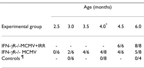
ho-Table 1: Overview of the time course and incidence of vascular pathology in IFN-γR-/- mice
Age (months)
Experimental group 2.5 3.0 3.5 4.0* 4.5 6.0
IFN-γR-/-MCMV+IRR - - - - 6/6 8/8
IFN-γR-/- MCMV 0/6 2/6 4/6 4/8 4/6 5/8
Controls ¶ - 0/6 - 0/8 - 0/4
[image:2.612.314.553.109.238.2]Mice were infected at the age of 2 months. * = Irradiation. Results are presented as number of animals affected / total number of animals in the group. ¶ = 129S, immunocompetent mice infected with MCMV.
Table 2: Overview of experimental groups of mice and seveverity of vascular disease.
Lesions*
Genetic MCMV Intima Media Adventitia
IFN-γR-/- + Genetic ++++ ++++
IFN-γR-/- + - ++ ++ ++
Wild type + + + + +
Wild type + - 0 0 0
Controls¶ - -/+ 0 0 0
[image:2.612.56.292.577.689.2]mogenate. Control animals were infected with the same concentration of salivary gland homogenate obtained from control uninfected CD-1 mice.
Experimental protocol
Two months old mice were injected i.p. with either MCMV or control salivary gland homogenate. Starting at day 15 after infection and for as long as 6 months of age, mice were sacrificed on a biweekly basis by i.p. injection of sodium nembutal. Hearts and abdominal aortas were collected and histopathological and immunohistochemi-cal analyses were performed. To both accelerate the de-velopment of vascular pathology and to deepen the immunocompromised state, these mice were irradiated with a sublethal dose of gamma radiation (300 Rads) at 4 months of age. Tissue sections were coded and scoring was performed in a blinded fashion.
Histology
H&E, Movat trichrome, and Verhoeff van Gieson, stain-ing was performed on 4 µm sections of aortas fixed for 16 hours in neutral buffered formalin (10%), and embedded in paraffin.
Antibodies
Polyclonal antibodies specific for CD4, CD8, γ/δ-TCR,
TGF-β1, PDGF-A, PDGF-B, MCP-1 and polyclonal
anti-body to PCNA were purchased from Santa Cruz Biotech-nology Inc., (Santa Cruz, CA). Polyclonal antibody to B-lymphocyte marker (B-220) was purchased from Caltag Laboratories (Burlingame, CA). Monoclonal antibody specific for α-smooth muscle actin (clone 1A4) was pur-chased from Sigma (St. Louis, MO).
Immunohistochemical analysis
Tissues were collected, fixed in formalin, and embedded in paraffin. After deparaffinizing and two 5-minute washes in PBS, the sections were blocked for 20 minutes with a solution of 2% unconjugated goat anti-mouse IgG in 1 x PBS. Indicated dilutions of the primary antibodies were added to the slides and incubated for 2 hours at room temperature followed by two 5-minute washes with PBS. Biotinylated secondary IgG diluted 1:100 was added to the slides and incubated for 30 minutes. After two additional 5-minute PBS washes, the tissue sections were treated with a 3% solution of hydrogen peroxide to quench endogenous peroxidase activity. After two addi-tional 5-minute washes, the slides were incubated for 30 minutes in the solution containing the
avidin-biotin-per-oxidase complex. Diaminobenzidine/H2O2 substrate
was allowed to react with the peroxidase labeled tissue sections for 20 minutes. After two 1-minute rinses in dis-tilled water, the tissue sections were counterstained with hematoxilin, cleared and dehydrated by successive gra-dations through 70%, 95%, and 100% ethanol followed
by a final passage in xylene. The slides were mounted and analyzed by bright field microscopy.
Tunel
Animals were sacrificed and tissues collected and proc-essed as described for immunohistochemistry. TUNEL assay was performed utilizing a kit for in situ detection of apoptosis, Oncogene, (Cambridge, MA), according to
manufacturer's instructions. Briefly, 5 µm sequential
sections were deparaffinized and fixed with 4% parafor-maldehyde for 20 minutes at room temperature followed by 30 minute wash in PBS. After a 5 minute rinse with PBS, 50 µl of TUNEL reaction mixture was added to tis-sue sections and incubated in humidified chamber for 60 minutes at 37°C followed by three 5 minute rinses in PBS and directly analyzed under the microscope.
Results
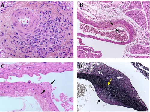
ani-mals revealed severe vasculitis characterized by signifi-cant inflammatory infiltrate in the adventitia and media (Table 2), (Fig. 2). At the same time hearts showed no signs of disease (data not shown). Verhoeff-van Gieson (Fig. 2C) and Movat pentachrome (Fig. 2D) staining in-dicated the focal destruction of elastic tissue, as well as potential trafficking of the infiltrating cells through these breaks in the elastica. The extent of vascular damage in all mice from this group was similar. The most striking feature of the MCMV induced vascular disease was the formation of neointima in aortas of MCMV infected im-munodeficient mice after irradiation (Table 2), (Fig 2).
At the same time there was no vascular pathology in any of the control groups (Table 2) (Fig 2A). Verhoeff-van Gieson staining of aortas collected from IFN-γR-/- mice
30 days after irradiation (not shown) and IFN-γ
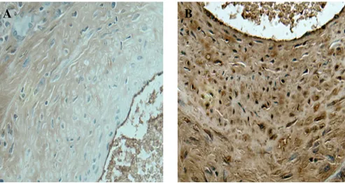
[image:4.612.56.557.238.613.2]R-/-mice 60 days after irradiation (Fig. 2) indicated that the intimal vascular lesion induced by MCMV infection was characterized by collagen deposition and smooth muscle proliferation. We utilized monoclonal antibody specific for α-SMA, a marker for myofibroblast cells, and found that neointima seen in the aortas of MCMV infected ex-perimental animals contained myofibroblasts. (Fig. 3).
Figure 1
Beginning of arterial wall thickening induced by infecting IFN-γR-/- mice with MCMV. A. H&E staining of small aortic branch 21 days after MCMV infection showing focal lymphohistiocytic adventitial infiltrate. B and C. Beginning of intimal thickening 2 months after MCMV infection (H&E staining) Spaces between black arrows indicates intimal thickening. D. Verhoeff-van Gie-son staining showing small breaks in the elastic tissue of aorta 2 months after MCMV infection. White arrows indicate focal adventitial inflammation and fibrosis, black arrow indicates intimal thickening, yellow arrow points to small breaks in the elastic tissue. Objective magnification x 20
A
C
B
Immunohistochemical characterization of immune re-sponse to MCMV infection
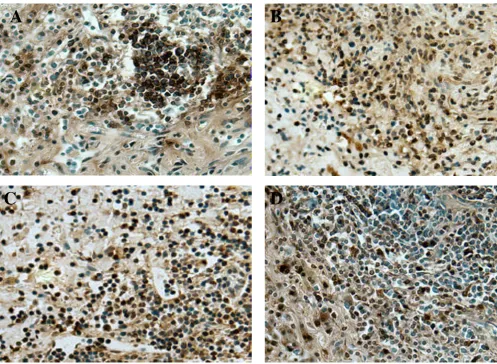
Immunohistochemical characterization of the inflam-matory infiltrate seen in the adventitia and media of the affected aortas revealed both helper T-lymphocytes
(CD4+) (Fig. 4B) and cytotoxic T-lymphocytes (CD8+)
(Fig. 4C) throughout the lesion. Since one of the charac-teristics of autoimmune vasculopathies is the appear-ance of high numbers of γ/δ-TCR [16] bearing cells, we studied this cell type in our model of vascular disease.
[image:5.612.56.555.86.455.2]Results from these experiments revealed a large number of γ/δ-TCR bearing lymphocytes throughout the lesion (Fig. 4D). We also investigated the presence of B-lym-phocytes in the vascular lesion of MCMV infected mice. While T-lymphocytes were identified throughout the le-sion these cells were mostly found in small follicles (Fig. 4A).
Figure 2
Neointima formation in the aortas of MCMV infected IFN-γR-/- mice 2 months after whole body irradiation. A. H&E staining of abdominal aorta collected from uninfected IFN-γR-/- mouse 2 months after whole body irradiation. Arrow points to the intima. B. H&E staining of a large abdominal vessel collected from MCMV infected IFN-γR-/- mouse 4 months after infection and 2 months after whole body irradiation. Space between arrows indicates the thickness of the neointima C.
Verhoeff-van Gieson stain of a large abdominal vessel collected from MCMV infected IFN-γR-/- mouse 4 months after infection and 2 months after whole body irradiation. Verhoeff-van Gieson stain: muscle (brown-yellow), collagen (red), cell cytoplasm (yellow), nuclei (blue-black). Focal transmural necrotizing vasculitis is seen. . Space between black arrows indicate intimal thick-ening and white arrows indicate focal adventitial inflammation and fibrosis. D. Movat staining of a large abdominal vessel col-lected from MCMV infected IFN-γ R-/- mouse 4 months after infection and 2 months after whole body irradiation. Movat stain: elastica (black), collagen and reticular fibers (yellow), mucosubstance (blue-green), red blood cells (red). Space between black arrows indicates the thickness of the neointima. Yellow arrows indicate normal external and internal elastica. Objective magni-fication x 20.
A
B
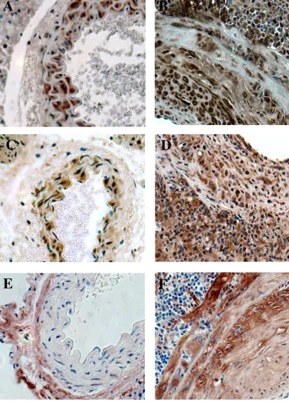
Investigation of the role of cytokines and growth factors Growth factors possibly responsible for the neointimal proliferation were studied based on their putative impor-tance in autoimmune vasculopathies in humans. Immu-nohistochemical analysis indicated a significant
overproduction of TGF-β1, PDGF-A, and PDGF-B in the
MCMV induced intimal lesion (Fig. 5). While TGF-β 1
and PDGF-A immunoreactivity in affected arteries is lo-calized to all three layers (intima, media, adventitia) (Fig. 5B and 5D), PDGF-B staining is localized to the me-dia and neointima (Fig. 5F). MCP-1, a chemokine that has been implicated in pathogenesis of several autoim-mune vascular diseases including SSc, was noted in the adventitia and media of affected arteries (Fig. 6). We did not detect MCP-1 positive staining in neointima (data not shown).
Role of apoptosis and proliferation
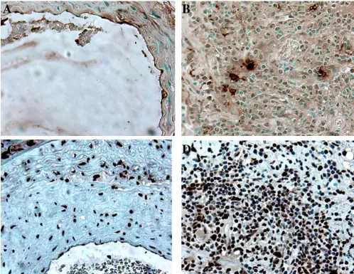
Further, we investigated the role of apoptosis and prolif-eration in vascular pathology triggered by MCMV infec-tion of immunosuppressed mice. In situ TUNEL revealed positive staining in the intimal layer of affected aortas (Fig 7A). In addition, a small portion of infiltrating in-flammatory cells stained positive by TUNEL (Fig. 7B). Smooth muscle/myofibroblast cells forming the neointi-ma did not reveal any cells positive by TUNEL (Fig. 7A). Immunohistochemical staining for PCNA was used to
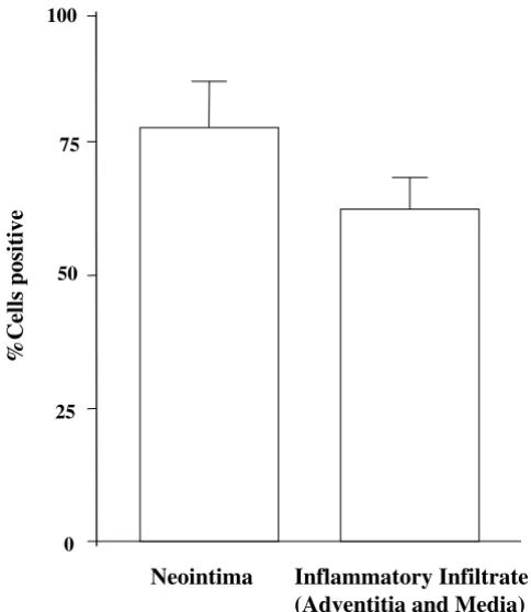
identify cell proliferation. The majority of cells in the ne-ointima (Fig. 7C and Fig. 8), as well as infiltrating inflam-matory cells (Fig. 7D and FIG. 8), exhibit immunoreactivity for PCNA, indicating active prolifera-tion.
Discussion
The major finding of this study is the formation of ne-ointima associated with MCMV infection in immuno-compromised mice. The disease described in our study resembles pathological processes seen in other autoim-mune vasculopathies. However, it is important to note that while there are undoubtedly autoimmune aspects of atherosclerosis, this issue is still under debate [17]. The formation of neointima characteristic of the vasculopa-thy in autoimmune diseases by this approach has, to our knowledge, not been previously reported. While MCMV vasculitis in IFN-γR-/- mice has been previously report-ed [18], that study does not focus on intimal prolifera-tion. In our experimental conditions we were able to
increase yield from 62% affected animals in IFN-γ
[image:6.612.61.555.86.348.2]R-/-mice infected with MCMV to 100% by including whole body irradiation of chronically infected IFN-γR-/- mice (Table 1). In addition, a striking increase in the severity of intimal lesions was noted in irradiated mice (Table 2). We propose that there are two possible explanations for the increase in both the number of affected animals, as Figure 3
Immunohistochemistry of neointima. A. Negative control for α-SMA staining (primary antibody omitted). B. Section of the aorta collected from MCMV infected IFN-γR-/- mouse 4 months after infection and 2 months after whole body irradiation were stained with monoclonal anti α-SMA. Objective magnification x 40
well as increase in the severity of vascular disease in
MCMV infected IFN-γR-/- mice receiving whole body
ir-radiation treatment. First, sublethal irir-radiation addi-tionally immunosuppresses the animals allowing unlimited replication of MCMV which may lead to direct viral damage. Second, the immune system after recover-ing from effects of radiation (reconstitution) contains large number of MCMV specific CTLs that may be capa-ble of inducing intimal pathology while attempting to clear the infection.
CMV infections have been implicated in a number of vas-cular diseases. CMV invades the vasvas-cular endothelium with the development of both latency and intermittent shedding of the virus to distal tissues, often by detach-ment of infected endothelial cells. In immunocompetent individuals CMV infections are of chronic or latent type
[image:7.612.57.555.85.449.2]and are usually not associated with clinically detectable disease. In immunocompromised patients such as ne-onates, transplant recipients and patients with AIDS, CMV infections are a leading cause of organ failure and death. Nonetheless, the mechanisms of CMV reactiva-tion and inducreactiva-tion of vascular lesions are not well under-stood. Clinical and epidemiological studies strongly suggest the association of CMV infection in the progres-sion of numerous vascular diseases, including athero-sclerosis [16–20], rapidly progressive coronary artery disease, endothelialitis in heart transplant recipients [21–24], coronary restenosis [25,26], and inflammatory aortic disease [27,28] as well as SSc [11,12]. We attempt-ed to reproduce the endpoint of such diseases by using immunosupression and CMV infection, allowing us to investigate cellular and molecular events leading to this pathology. While SSc is an autoimmune disease affecting Figure 4
Characterization of inflammatory infiltrate. Aortas collected from MCMV infected IFN-γR-/- mice 4 months after infec-tion and 2 months after whole body irradiainfec-tion. A. Immunostaining specific for B-lymphocytes (B220+). B. Staining specific for helper T-lymphocytes (CD4+). C. Staining specific for cytotoxic T-lymphocytes (CD8+). D. Immunostaining specific for γ/δ T-cell receptor bearing T-lymphocytes (γ/δ-TCR+). Objective magnification x 40
A
B
Figure 5
Investigation of the role of growth factors in the pathogenesis of MCMV induced neointima formation. A,C,E
abdominal aortas collected from control uninfected IFN-γR-/- mice 2 months after whole body irradiation. B,D,F aortas col-lected from MCMV infected IFN-γR-/- mice 4 months after infection and 2 months after whole body irradiation.A&B staining specific for TGF-β1. C & D staining specific for PDGF-A. E & F staining specific for PDGF-B. Objective magnification x 40
A
B
D
F
C
microvasculature, some recent studies also described in-creased incidence of vascular pathology in the large arte-rial vessels of SSc patients [29–31]. However, it is still unclear if macro vascular pathology is a primary feature of SSc or an indirect effect of some of the downstream events related to this disease.
Inoculation of immunocompetent mice with MCMV causes persistent productive infection in salivary glands followed by latency [32,33]. This latency is characterized by the presence of the viral genome whereas infectious viral particles could not be detected. In addition it was possible to reactivate virus either in vivo or in vitro from tissue explants. While there are several lines of evidence to suggest that the immune system, or its absence, plays a role in MCMV reactivation from latency [34,35] the mechanism is not known. Latent CMV in many organs can be reactivated from its dormant state by several means of immunosuppression, including transmission from donors to immunosuppressed recipients of trans-plants, and blood transfusion.
Prior studies describe development of MCMV associated vascular lesions in large arterial vessels of mice. The i.p. injection of suckling mice induces vascular lesions in the aorta and pulmonary artery, which are characterized by cellular infiltrates containing T-lymphocytes [36]. A more recent study describes the development of MCMV induced vascular lesions in the aorta of both
immuno-competent mice and those lacking IFN-γ responsiveness
where IFN-γ is determined to be a major antiviral factor preventing the long term persistence of aortic vascular lesions [18]. The present study focused on neointima for-mation, which was seen only in immunocompromised mice. Our results indicated presence of both CD4+ help-er, and CD8+cytotoxic T-lymphocytes in the adventitia of the affected arteries. Significant portion of these
T-lym-phocytes are γ/δ-TCR+ This subpopulation of
lym-phocytes has been identified as part of the pathogenesis of autoimmune diseases. For example, it has been shown that γ/δ-TCR+ lymphocytes from SSc patients recognize and lyse endothelial cells at a significantly higher rate then γ/δ cells isolated from control subjects [16]. Also, a
recent study indicates that γ/δ-TCR+ bearing
lym-phocytes play an important role in immune response of immunosuppressed transplant recipients to CMV infec-tion [37]. NK cells, natural antibodies, and complement, potentially important in controlling CMV infection, await further study.
Growth factors TGF-β, and PDGF are contributing
fac-tors in vascular diseases. TGF-β upregulates α-smooth muscle actin gene transcription [38]. Since we identified
significant α-smooth muscle actin expression in our
model and this coincided with the expression of TGF-β, it is possible that TGF-β mediates α-smooth muscle actin expression in our model. The elevated expression of PDGF in our model is also significant since the expres-Figure 6
Immunohistochemical investigation of the expression of MCP-1. A. Negative control for MCP-1 (primary antibody omitted). B. Section of the aorta collected from MCMV infected IFN-γR-/- mouse 4 months after infection and 2 months after whole body irradiation were stained with polyclonal antibody specific for MCP-1. Objective magnification x 40
[image:9.612.56.555.85.319.2]sion of these growth factors by endothelial cells can be induced by direct immune cell – endothelial cell interac-tion, promoting smooth muscle cell migration and pro-liferation in atherosclerosis [39]. CMV, as well as other viruses, induces expression of a number of cytokines and growth factors in cells in vitro, including TGF-β, a profi-brogenic cytokine [40,41]. MCP-1 is a member of the β -chemokine (C-C) family [42] and this -chemokine has been implicated in pathogenesis of SSc [43]. Our results (Fig. 6) indicate that this chemokine may play a role in recruitment of inflammatory cells to CMV infected vas-culature.
We investigated the role of apoptosis in pathogenesis of MCMV induced intimal lesion since apoptosis of virally
infected cells is one of the mechanisms by which host may limit the virus spread. The presence of apoptotic T-lymphocytes has been reported to be a feature of MCMV infection [44]. Apoptosis of vascular endothelium in-duced by immune effectors has been described in both SSc patient vessels as well as those of UCD chickens (one of the animal models for SSc) [45]. Recently, it has been reported that IgG autoantibodies from SSc patients bind CMV late protein UL94 and this induces apoptosis in hu-man endothelial cells [15]. We detected the presence of apoptotic cells in the intimal layer as well as in inflam-matory infiltrate of the affected aortas (Fig. 7A & 7B). In the future we will ask if apoptosis in our model is induced by anti-endothelial autoantibodies, MCMV infection of endothelium, or is induced and/or perpetuated by in-Figure 7
Investigation of apoptosis and proliferation in the pathogenesis of MCMV induced neointima formation. A and
B. TUNEL performed on the aorta collected from MCMV infected IFN-γR-/- mouse 4 months after infection and 2 months after whole body irradiation. C. and D. Immunostaining specific for PCNA. A. C. Neointima.B. D. inflammatory infiltrate. Objective magnification x 40
A
B
[image:10.612.55.554.87.472.2]flammatory cells. Apoptosis of vascular endothelium may be the trigger that initiates the uncontrolled repair mechanism characterized by the proliferation of smooth muscle cells/myofibroblasts, leading to the development of neointima in MCMV induced vascular disease.
The development of CMV induced vasculopathies is a major cause of morbidity and mortality in immunosup-pressed patients. CMV infections have been identified as important mediator of organ failure and death in trans-plant recipients and AIDS patients. Although the role of CMV infection in pathogenesis of vascular diseases in immunocompetent patients is less clear, epidemiological studies strongly suggest that chronic or latent infections may accelerate the development of diseases such as atherosclerosis and SSc. In fact, it is fair to hypothesize that several diseases thought to be of "idiopathic" origin may be triggered by chronic infections with CMV and other infectious agents in the genetically susceptible host. Animal models, such as those described in this pa-per, will allow us to better understand the role that viral infections play in the pathogenesis of vascular diseases.
It is crucial to identify factors responsible for contribut-ing to the development of vascular pathology in order to develop better preventive as well as treatment modali-ties.
Competing interests
None declared
Abbreviations
CMV = cytomegalovirus, MCMV = murine cytomegalovi-rus, MCP-1 = monocyte chemoattractant protein-1, PCNA = proliferating cell nuclear antigen, PDGF = plate-let derived growth factor, SSc = systemic sclerosis, TGF-β1 = transforming growth factor-β1, TUNEL = terminal
TdT-mediated dUTP nick end labeling, α-SMA = α
-smooth muscle actin
Acknowledgements
The authors wish to thank Dr. Jean-Michel Goust for helpful discussions, and Margaret Romano for the preparation of histological sections. Work supported by:Medical University of South Carolina Institutional Re-search Grant(DH),RGK Foundation gift(ECL);Kate and Claiborne Johnson gift (ECL).
References
1. Bouchard C: Physical inactivity. Can J Cardiol 1999, 15 Suppl G:89G-92G
2. Paradis G, Fodor JG: Diet and the prevention of cardiovascular diseases.Can J Cardiol 1999, 15 Suppl G:81G-88G
3. Reeder B, Bouchard C: Obesity and cardiovascular diseases.Can J Cardiol 1999, 15 Suppl G:69G-72G
4. Pickering T: Cardiovascular pathways: socioeconomic status and stress effects on hypertension and cardiovascular func-tion.Ann N Y Acad Sci 1999, 896:262-277
5. Fuster V: Human lesion studies.Ann N Y Acad Sci 1997, 811:207-24 6. Moder KG, Miller TD, Tazelaar HD: Cardiac involvement in
sys-temic lupus erythematosus.Mayo Clin Proc 1999, 74:275-284 7. Cook NS, Zerwes HG, Rudin M, Beckmann N, Schuurman HJ:
Chronic graft loss: dealing with the vascular alterations in solid organ transplantation. Transplant Proc 1998, 30(5) :2413-2418
8. Kahaleh MB, LeRoy EC: Autimmunity and vascular involvement in systemic sclerosis (SSc).Autoimmunity 1999, 31:195-214 9. Yamashita K, Ouchi K, Shirai M, Gondo T, Nakazawa T, Ito H:
Dis-tribution of Chlamydia pneumoniae infection in the athero-sclerotic carotid artery.Stroke 1998, 29(4):773-778
10. Muhlestein JB: Bacterial infections and atherosclerosisJ Investig Med 1998, 46(8):396-402
11. Pandey JP, LeRoy EC: Human cytomegalovirus and the vascu-lopathies of autoimmune diseases (especially scleroderma), allograft rejection, and coronary restenosis. Arthritis Rheum
1998, 41:10-15
12. Neidhart M, Kuchen S, Distler O, Bruhlman P, Michel BA, Gay RE, Gay S: Increased serum levels of antibodies against human cy-tomegalovirus and prevalence of autoantibodies in systemic sclerosis.Arthritis Rheum 1999, 42:389-392
13. Mocarski ES: Cytomegalo viruses and their Replication.Fields Virology. Edited by Fields BN et al. Pennsylvania, Lippincott-Raven,
19962447-2491
14. Nieto FJ, Adam E, Sorlie P, Farzadegan H, Melnick JL, Constock GW, Szklo M: Cohort study of Cytomegalovirus infection as a risk factor for carotid intimal-medial thickening, a measure of subclinical atherosclerosis.Circulation 1996, 94:922-927 15. Lunardi C, Bason C, Navone R, Millo E, Damonte G, Corrocher R,
[image:11.612.54.296.89.368.2]Puccetti A: Systemic sclerosis immunoglobulin G antibodies bind the human cytomegalovims late protein UL94 and in-duce apoptosis in human endothelial cells. Nat Med 2000, 6(10):1183-1186
Figure 8
Morphometric analysis of PCNA positive staining.
Both neointima and adventitia/media were analyzed by aver-aging cell numbers from three selected high power view fields (objective magnification 40X). Data were collected from three individual animals and are expressed as % positive cells ± SD.
0 25 50 75 100 %Cells positive
16. Kahaleh MB, Fan PS, Otsuka T: Gammadelta receptor bearing T cells in scleroderma: enhanced interaction with vascular en-dothelial cells in vitro.Clin Immunol 1999, 1(2):188-195 17. Shoenfeld Y, Sherer Y, Harats D: Atherosclerosis as an
infec-tious, inflammatory and autoimmune disease.Trends Immunol
2001, 22(6):293-295
18. Presti RM, Pollock JL, Dal Canto AJ, O'Guin AK, Virgin IV HW: In-terferon γ regulates acute and latent murine cytomegalovi-rus infection and chronic disease of the great vessels.J Exp Med 1998, 188:577-588
19. Sorlie PD, Adam E, Melnick SL, Folsom A, Skelton T, Chambless LE, Barnes R, Melnick JL: Cytomegalovirus/herpesvirus and carotid atherosclerosis: the ARIC study.J Med Virol 1994, 42:33-37 20. Melnick JL, Adam E, DeBakey ME: Cytomegalovirus and
athero-sclerosis.Bioessays 1995, 17:899-903
21. Koskinen P, Lemstrom K, Bruggeman C, Lautenschlager I, Hayry P: Acute cytomegalovirus infection induces subendothelial in-flammation (endothelialitis) in the allograft vascular wall. A possible linkage with enhanced allograft atherosclerosis.Am J Pathol 1994, 144:41-50
22. Dummer S, Lee A, Breinig MK, Kormos Ho RM, Griffth B: Investiga-tion of cytomegalovirus infecInvestiga-tion as a risk factor for coro-nary atherosclerosis in the explanted hearts of patients undergoing heart transplantation.J Med Virol 1994, 44:305-309 23. McDonald K, Rector TS, Braunlin EA, Kubo SH, Olivari NT: Associ-ation of coronary artery disease in cardiac transplant recipi-ents with cytomegalovirus infection.Am J Cardiol 1989, 64 :359-362
24. Grattan MT, Moreno-Cabral CE, Stames VA, Oyer PE, Stinson EB, Shumway NE: Cytomegalovirus infection is associated with cardiac allograft rejection and atherosclerosis. JAMA 1989, 261:3561-3566
25. Speir E, Modali R, Huang ES, Leon MB, Shawl F, Finkel T, Epstein SE: Potential role of human cytomegalovirus and p53 interac-tion in coronary restenosis.Science 1994, 265:391-394 26. Zhou YF, Leon MB, Waclawiw MA, Popma JJ, Yu ZX, Finkel T, Epstein
SE: Association between prior cytomegalovirus infection and the risk ofrestenosis after coronary atherectomy.N Engl J Med
1996, 335:624-630
27. Yonemicu Y, Nakagawa K, Tanaka S, Mori R, Sugimachi K, Sueishi K: In situ detection of frequent and active infection of human cytomegalovirus in inflammatory abdominal aortic aneu-rysms: possible pathogenic role in sustained chronic inflam-matory reaction.Lab Invest 1996, 74:723-736
28. Tanaka S, Komori K, Okadome K, Sugimachi K, Mori R: Detection of active cytomegalovirus infection in inflammatory aortic aneurysms with RNA polymerase chain reaction.J Vasc Surg
1994, 20:235-243
29. Hafner J, Schneider E, Burg G, Cassina PC: Management of leg ul-cers in patients with rheumatoid arthritis or systemic sclero-sis: the importance of concomitant arterial and venous disease.J Vasc Surg. 2000, 32(2):322-329
30. Ho M, Veale D, Eastmond C, Nuki G, Belch J: Macrovascular dis-ease and systemic sclerosis.Ann Rheum Dis. 2000, 59(1):39-43 31. Stafford L, Englert H, Gover J, Bertouch J: Distribution of macro
vascular disease in scleroderma.Ann Rheum Dis 1998, 57(8) :476-479
32. Pollock JL, Virgin HW: Latency, without persistence, of murine cytomegalovirus in spleen and kidney.J Virol 1995, 69 :1762-1768
33. Kurz S, Steffens HP, Mayer A, Harris JR, Reddehase MJ: Latency ver-sus persistence of intermittent recurrences: evidence for la-tent state of murine cytomegalovirus in the lungs.J Virol 1997, 71:2980-2987
34. Mayo D, Armstrong JA, Ho M: Activation of latent murine cy-tomegalovirus infection: cocultivation, cell transfer, and the effect of immunosuppression.J Infect Dis 1978, 138:890-896 35. Balthesen M, Messerle M, Reddehase MJ: Lungs are a major organ
site of cytomegalovirus latency and recurrence.J Virol 1993, 67:5360-5366
36. Dangler CA, Baker SE, Kariuki Njenga M, Chia SH: Murine cytome-galovirus associated arteritis.Vet Pathol 1995, 32:127-133 37. Dechanet J, Merville P, Lim A, Retiere C, Pitard V, Lafarge X,
Michel-son S, Meric C, Hallet MM, Kourilsky P, Potaux L, Bonneville M, Moreau JF: Implication of gammadelta T cells in the human
immune response to cytomegalovirus. J Clin Invest 1999, 103(10):1437-1449
38. Hautmann MB, Madsen CS, Owens GK: A transforming growth factor beta (TGFbeta) control element drives TGFbeta-in-duced stimulation of smooth muscle alpha-actin gene ex-pression in concert with two CArG elements.J Biol Chem 1997, 272(16):10948-56
39. Funayama H, Ikeda U, Takahashi M, Sakata Y, Kitagawa S, Takahashi Y, Masuyama J, Furukawa Y, Miura Y, Kano S, Matsuda M, Shimada K: Human monocyte-endothelial cell interaction induces plate-let-derived growth factor expression. Cardiovasc Res 1998, 37(1):216-224
40. Michelson S, Alcami J, Kim SJ, Danielpour D, Bachelerie F, Picard L, Bessia C, Paya C, Virelizier JL: Human cytomegalovirus infection induces transcription and secretion of transforming growth factor beta 1.J Virol 1994, 68(9):5730-5737
41. Taichman RS, Nassiri MR, Reilly MJ, Ptak RG, Emerson SG, Drach JC: Infection and replication of human cytomegalovirus in bone marrow stromal cells: effects on the production of IL-6, MIP-1alpha, and TGF-beta1.Bone Marrow Transplant 1997, 19(5) :471-480
42. Van Coillie E, Van Damme J, Opdenakker G: The MCP/eotaxin subfamily of CC chemokines.Cytokine Growth Factor Rev 1999, 10:61-86
43. Hasegawa M, Sato S, Takehara K: Augmented production of chemokines (monocyte chemotactic protein-1 (MCP-1), macrophage inflammatory protein-1 alpha (MIP-lalpha) and MIP-lbeta) in patients with systemic sclerosis: MCP-1 and MIP-lalpha may be involved in the development of pulmo-nary fibrosis.Clin Exp Immunol 1999, 117(1):159-165
44. Yoshida H, Sumichika H, Hamano S, He X, Minamishima Y, Kimura G, Nomoto K: Induction of apoptosis of T cells by infecting mice with murine cytomegalo virus.J. Virol. 1995, 69:4769-4775 45. Sgonc R, Gruschwitz MS, Dietrich H, Recheis H, Gershwin ME, Wick
G: Endothelial cell apoptosis is a primary pathogenic event underlying skin lesions in avian and human scleroderma.J Clin Invest 1996, 98:785-792
Pre-publication history
The pre-publication history for this paper can be ac-cessed here:
http://www.biomedcentral.com/content/backmatter/ 1471-2474-2-3-b1.pdf
Publish with BioMed Central and every scientist can read your work free of charge
"BioMedcentral will be the most significant development for disseminating the results of biomedical research in our lifetime."
Paul Nurse, Director-General, Imperial Cancer Research Fund
Publish with BMCand your research papers will be:
available free of charge to the entire biomedical community
peer reviewed and published immediately upon acceptance
cited in PubMed and archived on PubMed Central
yours - you keep the copyright
[email protected] Submit your manuscript here:
http://www.biomedcentral.com/manuscript/