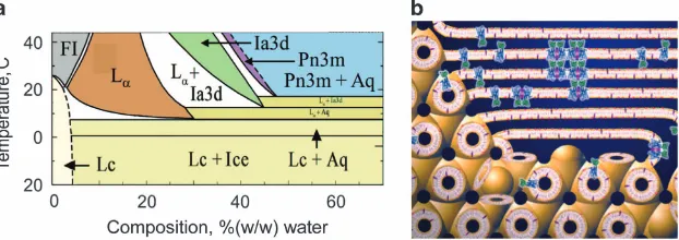
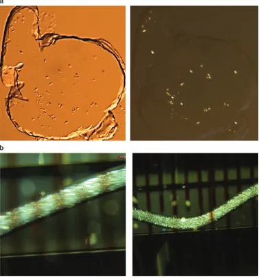
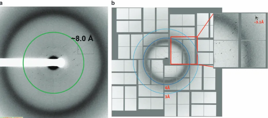
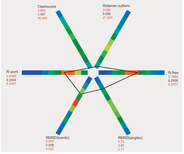
X ray laser diffraction for structure determination of the rhodopsin arrestin complex
Full text
Figure


Related documents
The International Study of Unruptured Intracranial Aneurysms (ISUIA) grouped patients according to the presence or absence of prior aneurysmal subarachnoid hemorrhage and the
A new upper bound is presented for the computational complexity of the parsing problem for TAGs, under the constraint that input is read from left to right in such
malignant tissues, that epigenetic subtypes exist within human breast cancers, and in particular, that a unique epigenetic gene signature has stronger prognostic value than
The Statistical Office publishes three series of monthly energy bulletins: Coal - Hydrocarbons - Electrical energy.. Each of these bulletins
The Journal of the New Zealand Institute of Architects and New Zealand Building Progress, in particular, are mines of information concerning the American presence within
On the basis of our experience during the past 6 years performing MR imaging examinations in patients with non-MR imaging– conditional CIEDs, including those who are pacemaker
The authors reported gray matter volume loss in the cerebellar hemispheres, vermis, and whole brain stem and white matter loss in the whole brain stem, midbrain, pons, middle
Aslan & Alpaslan (1999) reported that Turkish version of CTQ has three factor that physical abuse (e.g., “I was punished with a belt, a board, a cord, or some other hard
