Thesis by Jeffrey A. Frelinger
In Partial Fulfillment of the Requirements for the Degree of
Doctor of Philosophy
California Institute of Technology Pasadena, California
1973
ACKl~OWLEDGMENTS
I thank Ray Owen for his help and counsel during my stay at Caltech. His insight and knowledge have provided a continuing model for me. I thank Lee Hood and Paul Hargrave for helping me learn protein chemistry. I thank the other members of my committee
Ed Lewis, Herschel Mitchell, and Dick Russell for their interest in my work.
I am grateful to Ron Acton who showed me Science can be a business, and Dave Dusenbeiy who showed me it was fun. I thank my fellow
students, Tom Douglas, Sue Melvin, and Su Rosenberg for tolerating me, especially during the composition of this thesis. I also thank all
the other people who made this work possible, Frank, Richard and Dale in the machine shop, Ed, Bill and Jane in the stockroom, Milton and the animal room crew.
I thank the National Institutes of Health and Caltech for financial support.
ABSTRACT
Transf errin polymorphism, detected by differences in electro-phoretic mobility among allelic forms, is widespread in vertebrates. In pigeons, two alleles have been reported. In a developmental study, I have shown that for a period following hatching the young bird.'s transferrin phenotype reflects the maternal rather than its own genotype, although the squab is actively synthesizing transferrin in its liver. This probably reflects transfer of maternally derived protein through the egg. As the period of maternal transfer
corresponds to the period of immunoincompetence on the part of the squab, the transferrin is known to be bacteria-and fungistatic, I investigated the possibility of differences in the funistatic effects of the three transferrin phenotypes, using yeast as an assay organism. I found that transferrin derived from heterozygotes is much more
effective in the inhibition of yeast growth ·than that from homozygotes. Under the breeding conditions in our flock·, embryonic mortality
was significantly lower among the progeny of heterozygous females. This suggests that transferrin polymorphism is maintained by
selection against the progeny of homozygous females. An algebraic consideration of this form of selection leads to the predictiori that a population at equilibrium for allele frequencies would be in
populations are in Hardy-Weinberg equilibrium.
Transferrin was purified (>
95%
pure) from all three phenotypes;all had similar amino acid compositions and molecular weights.
Peptide mapping of the allele products revealed a difference in one
peptide, which on analysis was consistent with a single amino acid
substitution (Ser -)Asp). Mixtures of homozygous type transferrins,
while appearing electrophoretically identical to the heterozygous
type, do not equal its behavior in yeast inhibition. This suggests
the presence of "hybrid molecules" synthesized in heterozygotes.
I found that pigeon transferrins can be dissociated to 40,000 M.W.
fragments by heating in sodium dodecyl sulfate (SDS). Removal of
SDS results in reassociation into 80,000 M.W. dimers. All
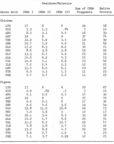
prepara-tions show only a single amino terminal (alanine). CNBr cleavage
produces only four fragments in spite of the fact that there are
eight methionines. Sequence data may indicate multiple amino acid
sequences. Quantitative hydrozanalysis yields only a single carboxyl
terminal (serine). These data suggest that pigeon transferrin may
TABLE OF CONTENTS
CHAPTER TITLE PAGE
Dedication--- ii
Acknowledgments---~--- iii
Abstract--- iv
GENERAL INTRODUCTION--- 1
I Maternally Derived Transferrin in Pigeon Squabs---
17
Additional Data--- 20
Reference--- 21
II Maintenance of Transferrin Polymorphism in Pigeons--- 24
III Transf errin Polymorphism and the Hardy.Weinberg
29
Ratios---IV Chemical Basis of Transf errin Polymorphism in Pigeons--- 39V The Covalent Structure of Transferrins---
52
INTRODUCTION
Transferrin is a large, non-heme, iron binding protein, which binds two moles of iron per mole of protein. Since its initial description in 1951 (1,2), it has been investigated by physical biochemists interested in its iron binding properties (3-17); by protein chemists interested in its covalent structure and the in the num~er of polypeptides per mole of intact protein
(18-50); by physiologists to discover its role in iron transport and control of erythropoesis (51-66); and by geneticists who have been intrigued by its widespread polymorphism (57-116). There have been few attempts to correlate the studies conducted in different fields. Most of the experiments were performed using human serum transferrin, chicken egg white or rabbit serum trans-ferrin; little work has been done on protein derived from other species.
The chemical studies of transferrin have centered on investigation of the iron binding site, carried out principally by spectroscopy. It has now been firrnaly established that the iron binding occurs to three ionized tyrosine residues and a single bicarbonate ion, with some involvement of nitrogen ligands (3,7,12,13,16,17), in
is precisely what would be expected when charge density and size are considered (117).
Earlier work (4) had suggested that the two iron binding sites were equivalent in each molecule. Recently Luk (11) has proposed, on the basis of fluorescence spectroscopy, that the iron binding sites are far enough apart to be non-interacting, but are not equivalent, as only one mole of Pr3+ or Nd3+ is bound per mole of protein. These data, based on lanthanide binding, may be interpreted differently, as due to severe allosteric effects caused by the
large size of the Pr3+ or Nd3+ ions which might' destroy the conformation of the second metal binding site. The distinction between identical and equivalent must be very carefully made. The two sites are not equivalent for lanthanide binding, but they could
3+
easily be equivalent for binding Fe , a much smaller ion. At present, therefore, the equivalence of the iron binding sites must be considered open to doubt.
not subsequently confirmed by others (30,34). A single polypeptide chain model has been reported by Feeney (30) for chicken transferrin. Mann et al. (34) have proposed a similar model for htnnan serum
transferrin. It has recently been suggested that the transferrin structural gene was formed by a fused tandem duplication (41). This is based on the analysis of cyanogen bromide fragments of
chicken ovotransferrin (41), in which molecular weight and charge similarities of the pairs of the fragments suggested some duplicate structure.
In two other species, turtles (36) and cattle (26), evidence for subunit structure has been found by ultracentrifugation. Hagfish transferrin has been reported to have a molecular weight of only 40,000, approximately one-half the counnon weight (36). However, Aisen et al. (18) attempting to repeat this work, reported a molecular weight.of 80,000 for hagfish transferrin.
Marrnnalian milk also contains an iron binding protein (118). This protein, lactoferrin, was originally thought to be related to
transferrin. The issue was confused by proof of identity of a serum and a milk iron binding protein in rabbits (21,53). The current belief is that there are at least two distinct iron binding proteins in milk, transferrin and lactoferrin, whose ratios may vary among species. In rabbits the milk iron binding protein is almost
(118, 122). In pigeon milk, the only iron binding protein which has been identified i.s transferrin (this thesis).
The role of transferrin in erythropoeisis is well established. Iron-saturated transferrin functions as an iron donor for hemoglobin synthesizing reticulocytes (51,52,61,63). In vitro this process
requires citrate or other iron chelates as cofactors (5). Transferrin preferentially binds to reticulocytes rather than to erythrocytes. Iron-saturated transferrin is more likely than iron free transferrin to bind to the reticulocytes.
Transferrin is synthesized primarily in the liver of mannnals (123), although its synthesis can also be detected in leukocytes (139). In birds, the oviduct also synthesizes ovotransferrin (104).
Susceptibility to systemic fungal infection, particularly candidasis, has been associated with decreased serum transferrin levels. This is particularly important in patients who are immuno-suppressed, either intentionally, as in the case of kidney transplant recipients, or unintentionally as in the case of leukemic patients (126). The mechanism of the antifungal activity of serum transferrin bas been of interest for many years. The first obervation of the
(132), and can be effectively inhibited by this mechanism. This appears to be the mode of action of transferrin on microbes in vitro.
Iron is known to have contraceptive properties (133). Transferrin has been shown to be present in seminal fluid (134, 135), and a related protein, lactoferrin, is found in vaginal secretions (136). The obvious mechanism seems to be that iron saturates the transferrin and allows local infection to take place. This provides an environment not healthy for either the sperm or the eggs, and their decreased viability and shorter functional life decreases the probability of fertilization as well as survival of the embryos.
Transferrin has been found to be polymorphic in most vertebrate species examined. The only exceptions seem to be those which have recently gone through a population bottleneck, such as the American Bison (137). Polymorphic transferrins are usually differentiated by differences in electrophoretic mobility on either starch or
acrylamide gel. Usually, one particular all·ele is frequent and there are many low-frequency variants. At least eighteen alleles are
known in man (138), many represented only by a single family. Some alleles, however, are common in some populations but rare in others. C transferrin is the most common allele but transferrin D is common in negro populations and Dchi in oriental and some American Indian populations (67,138).
first reported by Mueller et al. (101). The allelic transferrins differ in electrophoretic mobility on starch gels. The two alleles have been reported at nearly equal frequency in such diverse places as California, Wisconsin, Missouri, and North Ireland (78, 81, 101). In pigeons as in other species studied, transferrin appears to be inherited as an autosomal codominant trait. This is based on the observation that artificial mixtures of homozygous transf errins
appear identical to heterozygous material on electrophoresis. Ashton has suggested that transferrin polymorphism in cattle may be maintained by heterozygote superiority. He reported excess numbers of hetero-zygotes in some populations, and in the progeny of some mating
combinations (68, 69, 72). Subsequent study of other breeds of cattle (70) has failed to confirm Ashton's conclusions. In mice, conflicting reports on the effects of transferrin type on fertility have appeared (71, 102). Some workers have found no relationship (102), while others have shown small but statistically significant effects among some combinations of genotypes (71). These data have been used to explain the absence of transferrin A in wild mouse populations. In vole populations, the disadvantage of one of the alleles has been indicated by field studies, but no evidence of heterozygote
REFERENCES
1. Koechiin, B.: J. Am. Chem. Soc.: 74:2649 (1952)
2. Warner, R. C.; Weber, I.: J. Biol. Chem. 191:173 (1951)
3. Aasa, R.; Aisen, P.: J. Biol. Chem. 243:2399 (1968)
4. Aasa, R.; Malmstrom, B. G.; Saltman, P.; Vanngard, T.: Biochim.
Biophys. Acta 12:203 (1963)
5. Aisen, P.; Leibman, A.: Biochem. Biophys. Res. Corrnn. 32:220 (1968)
6. Aisen, P.: Biochem. Biophys. Res. Corrnn. 30:407 (1968)
7. Azari, P.; Phillips, J. L.; Arch. Biochem. Biophys. 138:32 (1970)
8. Bezkorovainy, A.: Biochim. Biophys. Acta. 127:535 (1966)
9. Davis, P.; Saltman, P.; Benson, S.: Biochim. Biophys. Res. Comm.
~:56 (1962)
10. Lehrer, S. S.: J. Biol. Chem. 244:3613 (1969)
11. Luk, C. K.: Biochem. 10:2838 (1971)
12. Komatsu, S. K.; Feeney, R. E.: Biochem. &_:1136 (1967)
13. Phillips, J. L.; Azari, P.: Arch. Biochem. Biophys. 151:445 (1972)
14. Warner, R. C.; Weber, I.: J. Am. Chem. Soc. 12:5094 (1953) 15. Williams, J.; Phelps, C. F.; Lowe, J.M.: Nature 226:858 (1970)
16. Zschocke, R.H.; Chiao, M. T.; Bezkorovainy, A.: Eur. J.
Biochem. lZ_:l45 (1972)
17. Warner, R.C.: Trans. N. Y. Acad. Sci. 16:182 (1953)
18. Aisen, P.; Liebman A.; Sia, C.: Biochem. 11:3461 (1972)
19. Aisen, P.; Koenig, S. H.; Schillinger, W. E.; Scheinberg, I. H.;
20. Allison, R. G.; Feeney, R. E.: Arch. Biochern. Biophys. 124:548
(1968)
21. Baker, E.; Shaw, D. C.; Morgan, E. H.: Biochern. l.:1371 (1968)
22. Bezkorovainy, A.; Grohlich, D.; Ge-rbeck, C.: Biochern. J. 110:
765 (1968)
23. Bezkorovainy, A.; Rafelson, M.; Likttite, A.: Arch. Biochern.
Biophys. 103:371 (1963)
24. Boffa, G. A.; Drilhou, A.; Favre, A.; Zakin, M. M.; Fine, J.M.;
C. R. Acad. Sci. Serus D 267:1067 (1968)
25. Clark, J.; Osuga, D.; Feeney, R. E.: J. Biol. Chern. 238:3621
(1963)
26. Efrernov, G. D.; Smith, L. L.; Barton, B. P.; Huisman, T. H.J.:
Animal Blood Groups, Biochern. Genet. 1:159 (1971)
27. Elleman, T. C.; Williams, J.: Biochern. J. 116:515 (1970)
28. Ezekiel, E.; Lai, L. Y. C.; Kalder, E.: Comp. Biochern. Physiol.
10: 69 (196 3)
29. Frenoy, N.; Groussault, Y.; Bourrillon, R.: Biochirnie 53:
1207 (1971)
30. Greene, F. C.; Feeney, R. E.: Biochern. l.:1366 (1968)
31. Jamieson, G. A.; Jett, M.; DeBernardo, S. L.: J. Biol. Chern.
246: 3686 ( 19 71)
32. Jep·psson, J. O.: Acta Chim. Scand,. 21:1686(1967)
33. Jordan, S. M.; Morgan, E. H.: Comp. Biochem. Physiol. 29:
34. Mann, K. G.; Fish, W.; Cox, A. C. ;. Tanford, C.: Biochem. 9:1348
(1970)
35. Osuga, D.; Feeney, R. E.: Arch. Biochem. Biophys. 124:560 (1968)
36. Palmour, R. M.; Sulton, H. E.: Bioch~m. 10:4026 (1971)
37. Parker, W. C.; Bearn, A. G.: Science 133:1014 (1961)
38. Parker, W. C.; Bearn, A.G.: Jo Exp. Med. 115:83 (1962)
39. Parker, W. C.; Hagstrom, J. W. C.; Bearn, A.G.: J. Exp. Med.
118:975 (1963)
40. Patras, B.; Stone, W. H.: Proc. Soc. Exp. Biol. Med. 107:118
(1961)
41. Phillips, J. L.; Azari, P.: Biochem. 110:1160 (1971)
42. Robinson, J. C.; Pierce, J. E.: Arch. Biochem. Biophys. 106:
348 (1964)
43. Rosseneu-Motreff, M. Y.; Soetewey, F.; Lamote, R.; Peters, H.:
Biopolymers 10:1039 (1971)
44. Stratil, A.; Spooner, R. L.: Biochem. Genet. 2_:347 (1971)
45. van Eijk, H.G.; van Dijk, J.P.; van Noort, W. L.; Leijnse, B.;
Monfoort, C. H.: Scand. J. Haem. ~:267 (1972)
46. Webster, R. O.; Pollara, B.: Comp. Biochem. Physiol. 30:509
(1969)
47. Williams, J.: Biochem. J. 83:355 (1962)
48. Williams, J.: Biochem. J. 108:57 (1968)
50. Zschoke, R.H.; Bezkorovainy, A.: Biochem. Biophys. Acta.
200:241 (1970)
51. Baker, E.; Morgan, E. H.: Biochem. 8:2954 (1969)
52. Baker, E.; Morgan, E. H.: Biochem. 8:1133 (1969)
53. Baker, E.; Jordan, S.; Toffery, A.; Morgan, E. H.: Life Sci.
~: 89 (1969)
54. Baker, E.; Morgan, E. H.: Quart. J. Exp. Physiol. 54:173
(1969)
55. Douglas, T. A.; Renton, J.P.; Watts, C.: Brit. J. Haematology
20:185 (1971)
56. Douglas, T. A.; Renton, J.P.; Wright, R.: Am. J. Obst. Gyn.
102 :1169 (1968)
57. Fielding, J.: Edwards, S.; Ryall, R.: J. Clin. Path. 22:677
(1969)
58. Fletcher, J.: Clin. Sci. 11.:273 (1968)
59. Fletcher, J.; Huehns, E. R.: Nature 218:1211 (1968)
60. Kornfeild, S.: Biochim. Biophys. Acta. 194:25 (1969)
61. Martinez-Medellin, J.; Schulman, H. M.: Biochim. Biophys.
Acta. 264:272 (1972)
62. Morgan, E. H.; Baker, E.: Biochim. Biophys. Acta. 184:442 (1969)
63. Morgan, E. H.: Biochim. Biophys. Acta 244:103 (1971)
64. Schade, S.; Bernier, G.; Conrad, M.: Brit. J. Haematol. 17:
65. Tarvydas, H.; Jordan, S.; Morgan, E. H.: Brit. J. Nutrit.
22:565 (1968)
66. Tojvanen, P.; Ross, T.; Hirvonen, T.: Scand. J. Haematol.
6 :113 (1969)
67. Arends, T.; Gallango, M. L.: Science 143:367 (1964)
68. Ashton, G. C.: Nature 183:404 (1958)
69. Ashton, G. C.: Genetics 52:983 (1965)
70. Ashton, G. C.; Lampkin, G. H.: Genet. Res. £:209
(1965)
71. Ashton, G. C.; Dennis, M. N.: Genetics 67:253 (1971)
72. Ashton, G. C.; Hewetson, R. W.: Animal Prod~ 11:533 (1969)
73. Baker, C.M.A.; Croizier, G.; Stratil, A.; Manwell, C.: Advances
in Genetics 15:147 (1970)
74. Baker, C. M. A.: Genetics 58:211 (1968)
75. Baker, C. M. A.: Comp. Biochem. Physiol. 20:949 (1967)
76. Baker, C. M. A. ; Manwell, C. ; Labisky, R. F. ; Harper, J. A. :
Comp. Biochem. Physic! . .!..Z.:467 (1966)
77. Baker, C. M.A.; Hanson, H. C.: Comp. Biochem. Physic!. 17:
997 (1969)
78. Brown, R. V.; Sharp, H.B. Jr.: Animal Blood Groups, Biochem.
Genet • .!_113 (1970)
79. Brush, A.H.: Comp. Biochem. Physiol. 25:159 (1968)
80. Chen, S. H.; Sutton, H. E.: Genetics 56:425 (1967)
82. Cohen, B. C.: Genet. Res. _!_:431 (i960)
83. Fesus, L.; Rasmussen, B. A.: Animal Blood Groups, Biochem.
Genet. 257 (1971)
84. Fowle, K. E.; Cline, J. H.; Klosterman, E.W.; Parker, C. F.:
J. Animal Sci. ~:1226 (1967)
85. Gaines, M. S.; Krebs, C. J.: Evolution _?2:702 (1951)
86. Gaines, M. S.; Myers, J.; Krebs, C. J.: Evolution 25:443 (1971)
87. Giblett, E. R.; Hickman, C. G.; Smithies, 0.: Nature 183:1589
(1959)
88. Gilmour, D. G.; Morton, J. R.: Theor. Appl. Genet. 41:47 (1971)
89. Hershberger, W. K.: Trans. Am. Fish Soc. 99:207 (1970) 90. Jamieson, A.: Heredity 20:419 (1965)
91. Jeppsson, J. O.: Biochim. Biophys. Acta 140:465 (1967)
92. Klein, P.A.; Roop, B. L.; Roop, W. E.: Nature 212:1376 (1966)
93. Kristjansson, F. K.: J. Reprod. Fertil. 8:311 (1964)
94. Kuzmenko, L. G.; Tsitol, Genet. 1:469 (1968)
95. Lai, L. Y. C.: Folia Primetologica 11.:193 (1972)
96. Lush, I.E.: Nature 189:981 (1961)
97. Mao, S. H.; Dessquer, H. C.: Comp. Biochem. Physiol. 40A:
669 (1971)
98. McDermid, E. M.; Vos. G. H.: S. Afr. J. Med. Sci. 36:7 (1971)
99. McDougal, E. I.; Lowe, V. P. W.: J. Zool. Soc. Lond. 155:133
(1968)
101. Mueller, J.; Smithies, O.; Irwin, M. R.: Genetics 47:1385
(1962)
102. Nagai, J.; Kristjansson, F. K.: Can. J. Genet. Cytol. 12:307
(1970)
103. Nadler, C.: Comp. Biochem. Physiol. 2:]_:487 (1968)
104. Ogden, A. L.; Morton, J. R.; Gilmour, O. G.; McDermid, E. M.
Nature 195:1026 (1962)
105. Roop, W.; Putnam, F.: J. Biol. Chem. 242:2507 (1967)
106. Shreffler, D. C.: Proc. Nat. Acad. Sci. (US) 46:1378 (1960)
107. Shreffler, D. C.: J. Heridity 54:127 (1961)
108. Spooner, R. L. and Baxter, G.: Biochem. Genet. 2:371 (1969)
109. Stratil, A.: Comp. Biochem. Physiol. 24:113 (1968)
110. Tamarin, R.H.; Krebs, C. J.: Evolution 23:183 (1969)
111. Vohs, P.A.; Carr, L. R.: Condor 2!_:413 (1969)
112. Wang, A. C.; Shustes, J.; Epstein, A.; Fudenberg, H. H.:
Biochem Genet _!.:347 (1968)
113. Wang, A. C.; Sutton, H. E.; Riggs, A.: Am. J. Hum. Genet.
18:454 (19b6)
114. Watanabe, S.; Nozawa, K.; Suzuki, S.: Proc. Jap. Acad. 41:
326 (1965)
115. Watanabe, S.; Suzuki, S.: Proc. Jap. Acad. 42:178 (1966)
116. Wright, J.E.; Atherton, L. M.: Trans. Am. Fish Soc. 99:
179 (1970)
117. Feeney, R. E.; Komatsu, S. K.: Struct. Bonding _!.:146 (1966)
119. Blanc, B.; Isliker, H.: Bull. Soc. Chim. Biol. 43:929 (1961)
120. Masson, P. L.; Heremans, J. F.: Comp. Biochem. Physiol. 39B:
119 (1971)
121. Jorden, S. M.; Morgan,
E.
H.: Nature 215:76 (1967)122. Groves, M. L.: Biochim. Biophys. Acta 100:154 (1965)
123. Goldsworthy, P.
D.;
Mccarter, H. R.; McGuigan, J.E.; Peppers,G. F.; Volwiler, W.: Arn. J. Physiol. 218:1428 (1970)
124. McFa.rlane, H.; Reddy, S.; Adcock, K. J.; Adeshina, H.; Cooke,
A. R.; Akene, J.: Brit. Med. J.
i:
268 (1970)125. McFarlane, H.; Okubadejo, M.; Reddy, S.: Am. J. Clin. Path.
~:587 (1972)
126. Caroline, L.; Rosner, F.; Kozinn, P. J.: Blood 34:441 (1969)
127. Schade, A. L.; Caroline, L.: Science 100:14 (1944)
128. Feeney, R. E.: Arch. Biochem. Biophys. 34:196 (1951)
129. Feeney, R. E.; Nagy, D. A.: J. Bacteriol. 64:629 (1952)
130. Fraenkel-Conrat, H.; Feeney, R. E.: Arch. Biochem. 29:101
(1950)
131. Oran, J.
D.;
Reittler, B.: Biochim. Biophys. Acta 170:351(1968)
132. Kochan, I.; Golden, C. A.; Bukovic, J. A.: J. Bacteriol. 100:
64 (1968)
133. Loewit, K.; Fodisch, H.J.; Zambelis, N.; Egg, D.: Contraception
134. Roberts, T. K.; Boettcher, B.: J. Reprod. Fertil. 18:347 (1969) 135. Stratil, A.: Proc. 11th European Congr. Blood Groups and
Protein Polymorphisms in Animals P. 417 (1968)
136. Masson, P. L.; Heremans, J. F.; Ferin, J.: FertiL and Steril.
19:679 (1968)
137. Braend, M.; Stormont, C.: Nature 197:910 (1963)
138. Giblett, E. R.: Genetic Markers in Human Blood, F. A. Davis
Company, Philadelphia (1969)
CHAPTER 1
Maternally Derived Transferrin in Pigeon Squabs
The publication contained in this chapter is from Science
Reprinted from
26 March 1971, Volume 171, Pages 1260-1261
SCIENCE
Maternally Derived Transferrin in Pigeon Squabs
Abstract. With the use of genetically marked trans/ errin, a major portion of circulating trans/ errin from a newly hatched squab was found to be derived from the mother through the egg. The transfer ls not through the parental crop milk. The squab does not accumulate enough trans/ errin of its own making to be detectable until it is about 8 days old. The maternally derived protein remains detectable until 14 days after hatchi,1g. The squab actively synthesizes a portion its own trans/ errin from hatching onward.
Transferrin is a nonheme iron-bind-ing protein (I) that is widely distrib-uted throughout the chordates from lamprey to man (2). One of its prin-cipal functions is the transport of iron to reticulocytes for incorporation into hemoglobin (3). It may be important in nonspecific resistance to disease, as well as for its iron transport function. Transferrin has been found to be poly-morphic in most species, the products of different alleles usually differing in electrophoretic mobility ( 4).
Although the development of trans-ferrin has been studied in several mam-malian species, including mouse, rabbit, and man (5), I am not aware of any reports on developmental studies of transf errin in birds. In mammals, there is very little transport of transferrin to the developing fetus ( 6) ; in this, trans-ferrin differs from certain immunoglob-ulins, which are known to be- trans-ported intact to the embryo.
Pigeons have been shown to be poly-morphic at a transferrin locus, and have only two reported variants, T/A and
TfB (7). The alleles are inherited as autosomal codominants, and ·in popu-lations so far examined they are present in nearly equal frequency. Transferrin is found in the pigeon blood, egg white,
e~g yolk, crop milk, and probably in the semen. In all these fluids it is under the cont.rol of the same gene. In pigeons,
which differ in this respect from chick-ens ( 8), the electrophoretic mobility of transferrin iin all these fluids is identi-cal. In this report, therefore, transferrin derived from egg white will not be dis-tinguished by the specific term ovo-transferrin, which is often used.
Pigeons (Columba livia) were main-tained in the laboratory in mating cages ( 1 pair per cage) or in large fly-pens. Eggs were checked daily and the day of hatching was designated as day 0.
For adults and squabs over 4 days old, when more than 0.5 ml of blood was to be removed, the birds were bled from the brachial vein by syringe and needle. The blood was allowed to clot and the serum was collected. For younger birds, blood was collected di-rectly onto filter paper fo~ electropho-resis. In order to determine whether this would give consistent serum patterns, several squabs were killed by exsangui-nation and their serums were collected. The results for collection by filter paper and by syringe were identical.
Transferrin typing was carried out on horimntal starch gels by the use of tris-citric acid buffer (pH 7.5) in the gel and borate buffer (pH 8.7) as elec-trolyte (9). An ice pack was placed on top of the gel to prevent excessive heating; the gels were stained with Coo-massie blue.
prepared· in rabbits. Approximately 5
mg of purified pigeon transf errin in Freund's complete adjuvant (Difeo) was injected into toe pads. Four weeks later, 2 mg of this transferrin in.1 ml of saline was injected intraperitoneally; and 3 days after that, 3 mg in 1 ml of saline was injected intravenously. The rabbits were bled 1 week after the last injection. When assayed by immuno-electrophoresis against pigeon normal serum, the rabbit antiserum gave a sin-gle band in the transferrin position.
For determination of the synthesis of transferrin by newly hatched squabs, the squabs were injected intraperitone-ally with [3H]leucine (specific activity, 38.5 c/mrnole; New England Nuclear) and allowed 20 minutes to incorporate the isotope. The squabs were killed; their livers were washed iri ice-cold phosphate buffered saline (PBS) (JO)
and homogenized in a Waring Blendor for 2 minutes in 15 ml of PBS. The homogenate was centrifuged 1 hour at 30,000g and the pellet was discarded. A portion of the supernatant was pre-cipitated with an equal volume of 10 percent trichloroacetic acid (TCA) and counted in a toluene omnifiuor scintil-lation mixture. One-half milliliter of supernatant was reacted with 0.5 ml of antiserum for 1 hour at room tempera-ture, and then for 2 days at 4 °C. The pellets were washed four times in cold PBS, precipitated with TCA, filtered, and counted as above.
The following matings were made to test for maternally derived transf errin:
T/A/TJA ~ x TfB!TfB ~; the recipro-cal TJ8/TfB ~ x TJA/TJA cS; and
T/A/TJB ~ x Tf"ITJA g. In the AA ~ X BB ~ mating the squab is genotypi-cally AB. However, until 8 days after hatching it appears to have only A type transf errin in its serum, as assayed on starch gels. On the eighth day, tho squab's own type becomes detectable. A similar result is obtained in BB ~ X
AA ~ matings, the offspring showing only B type until 8 days. In AB ~ X
BB ~ matings, although half the squabs are homozygous BB, all show the heterozygous phenotype until they reach 14 to 16 days of age.
Because transferrin is found in crop
milk produced by both the male and the female, protein transfer by this route could affect the results. One pair of squabs of AB genotype was separated at hatching; one squab was put with the A parent, the other with the B parent. Both squabs followed the same pattern; that is, t.he squabs showed the maternal
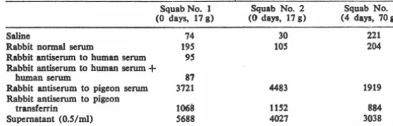
Table 1. Active synthesis of transferrin by squab liver. Phosphate buffered saline (0.S ml) and
O.S ml of liver homogenate were centrifuged at 30,000g. The supernatant and O.S ml of tho indicated serums were reacted, pellets were washed and precipitated with TCA as described in
the text. The values are given in counts per minute per filter and represent the average of duplicate portions of the same homogenate, subjected to the same treatments.
Squab No. 1 Squab No. 2 Squab No. 3 (0 days, 17 g) (0 days, 17 g) (4 days, 70a)
Saline 74
Rabbit normal scrum 19S
Rabbit antiserum to human scrum 9S
Rabbit antiserum to human scrum + human scrum 87 Rabbit antiserum to pigeon serum 3721
Rabbit antiserum to pigeon
transferrin 1068
Supernatant (0.S/ml) S688
phenotype regardless of the source of crop milk.
These experiments demonstrate the transfer of a maternally derived intact protein through the egg, or as a formal possibility, a portion of the maternal protein-synthesizing machinery. Because the homozygous offspring of matings of AB ~ x AA ~ show the heterozygous type even though they do not possess the Tf8 gene, the protein's source can. only be maternal.
These observations necessitated a study of active synthesis of transferrin by squabs. We tested squabs at various times after hatching for their ability to incorporate [BH]leucine into protein re-acting with mooospecific antiserums to transferrin. The results are given in Table 1. Antiserum to human serum, alone and wJth added human serum, served as coprecipitation controls; these controls are not different from the rab-bit normal serum or saline controls. The antiserum to transferrin precipi-tates about 20 percent of the total labeled protein in the supernatant, while the antiserum to pigeon whole serum precipitates almost all of the counts.
With the· use of genetically marked transferrin, this study has shown that, in pigeons, newly hatched squabs derive part of their plasma proteins from their mothers. The transferred protein repre-sents the major portion of the young squab's serum transf errin, even though the young bird is actively synthesizing transferrin during this period. This con-clusion is reasonable when the growth rate of a young pigeon is considered. The squab, weighing about 16 g at hatching, doubles its weight in the first 2 days, and by 2 weeks it weighs 150 g ( 11). This rapid rate of growth involves a large increase in mass, and extensive synthesis is required even to maintain a constant concentration of transf errin. All the observations are consistent
30 221
lOS 204
4483 1919
1152 884
4027 3038
with the following model. A large amount of transferrin is absorbed intact by the developing embryo, through the egg white or the yolk, or both. Some time prior to the day of hatching, the squab initiates its own synthesis of transferrin. The failure to detect the squab's own type in genotypically heterozygous squabs is a result of in-ability to detect low levels of
transfer-rin. Unlike mammals, therefore, pigeons proviide the major part of their off-spring's transferrin supply by maternal transfer, even though the squab is also engaged in active transferrin synthesis.
JEFFREY A. FRELINOER
Division of Biology,
California Institute of Technology, Pasadena 9 J 109
Reference1 aad Notel
t. R. Feeney and S. Komatsu, Struct. Bonding
t, 146 (1966).
2. R. Webster and B. Pollara, Comp. Blochem.
Phynol. 39, 509 (1969); E. Giblctt, Progr. Med. G•Mt. 2, ~ (1962).
3. E. Baker and E. Mor11an, Blochnnbtry I,
1133 (1969); J. Pletcher, Clln. Sci. 37, 273 (1969); S. Kornfeld, Blochlm. Blophy1. Acta
1'4, 2S (1969).
4. C. Baker, Comp. Blochem. Phynol. 21, 949
(1967); J. Clark, D. 01u11a, R. Feeney, I.
Biol. Chem. 213, 3621 (1963); E. E7.eldel, L.
Lal, E. Kaldor, Comp. Btochem. Ph;ynol. 10,
69 (1963); E. Glblett, C. Hickman, 0.
Smlth-le1, Naturt1 113, 1'89 (19,9).
5. D. Shreftler, Proc. Nat. Acad. Sci. U.S. 46,
1378 (1960); E. Morpn, Amt. I. E1q1. Biol. Med. Sci. 47, 361 (1969); D. Gllten and A. Bluucci, I. Clln. Invest. 48, 1433 (1969); P. Tojvanen, T. Roni, T. Hirvonen, Scand. I.
Hoematol. 6, 113 (1969).
6. E. Baker and E. Moraan, Quart. I. Exp.
Physlol. Co,. Med. Sci. 54, 173 (1969).
7. J. Mueller, O. Smithies, M. Irwin, Genetlc:t 47, 138' (1962); R. Brown and H. Sharp,
Anlm. Blood Group1 Blochem. Gmet. 1, 113 (1970).
8. A. 011den, J. Morton, D. Gilmour, E.
Mc-Dermid, Natvre 195, 1026 (1962); A. Stratil,
Comp. Btoch•m. Ph;ynol. 24, 13 (1968); C. Baker, Genetics !I, 211 (1968).
9. F. Kristjanason, Gen•tlc1 48, 1059 (1963). 10. 0.01,M phosphate and 0.07,M NaCl (pH 7.2). 11. W. Levi, Th• Pl,eon (Levi, Sumter, S.C., ed. 3, 19,7), pp. 269-271; and personal observa-tion.
12. I thank Dr. Ray Owen for valuable
dlscua-1ion, aupport, and criticism. Supported by ABC contract AT 0076706 FP and PHS arant GM 00086.
26 October 1970
[image:25.618.229.508.142.232.2]ADDITIONAL DATA
In order to test further the identity of egg white and sermn transferrin in the pigeon, antisera were prepared in rabbits to pigeon serum- and egg-white derived transferrins. The anti-egg white
transf errin was prepared in a manner similar to that described for the antiserum to serum. transferrin in the preceding publication. Egg white and sermn transferrins were assayed by Ouchterlony double diffusion with both antisera. They gave lines of identity with both reagents (fig. 1). These sera, therefore, detect no difference between pigeon egg white and serum transferrins.
Ferguson, h~wever, reported a difference inelectrophoretic
mobility between egg white and serum transferrin from feral pigeons (1). Samples of Pasadena pigeon egg white were sent to Dr. Ferguson. He found that Irish pigeons differ from U.S. pigeons in the characteristic of egg white transferrin mobility (personal communication). U.S.
REFERENCE
Figure Legend
Immunodiffusion test of the identity of servm.and egg white
transf errin. Top and bottom wells of each pattern contain purified
serum transferrin; left and right wells purified egg white transferrin.
The center well, A, contains anti-serum transferrin; the center well,
-24-CHAPTER 2
Maintenance of Transferrin Polymorphism in Pigeons
Reprinted from
Proc. Nat. Acad. Sci. USA
Vol. 69, No. 2, pp. 326-329, February 1972
The Maintenance of Transferrin Polymorphism in Pigeons
(Saccharomyces cerevisiae / eggw h ite)
JEFFREY A. FRELINGER
Division of Biology, California Institute of Technology, Pasadena, Calif. 91109
Communicated by Ray D. Owen, November 26, 1971
ABSTRACT Tran11ferrin, a nonheme iron-binding
protein, is polymorphic in most vertebrate specie!! that
have been examined. In pigeon!!, it i11 controlled hy an
autosomal gene, with two known rodon1inant alleles,
TJA and Tf11• The two allele!! arc found in nearly equal
frequencies and the three genotypes are at Hardy-Wein-berg equilibrium in all population!! studied. This report shows that ovotransferrins from heterozygous females inhibit microbial growth, by use of yeast as an as11ay organism, better than ovotransferrins from either of the homozygous types, or those from a mixture of homozygous
types. Heterozygous females hatch. a larger percentage of
their eggs than homozygous females. This difference is
probably accounted for by the transferrin effect. The
fail-ure of the mixtfail-ure of ·the homozygous type11 to act like
the heterozygous type calls into question the currently accepted structure of transferrin a11 a monomeric protein. The greater fecundity of heterozygous females can acrount
for the maintenance of transferrin polymorphism in
pigeons.
How genetic polymorphism is maintained is one of the major
unanswered questions in genetics today. The belief that it
depends upon a selective advantage of heterozygotes is widely held but not well supported by data. This paper presents evi-dence for maintenance of transferrin polymorphism in pigeons by the differential inhibition of microbial growth hy
different transferrin phenotypes.
Transferrin is a nonheme iron-binding protein found in the plasma of vertebrates and the eggs of birds. It is found to he polymorphic in most species examined. In each case appro-priately studied, transferrin is found to be controlled by co-dominant alleles of an autosomal gene (I, 2). There have been many attempts to find mechanisms for the main-tenance of this polymorphism. Ashton and his as:;ociates (3,
4) have reported evidence in some cuttle for excess of het-erozygotes, and postulated superior heterozygote fitness. This is not confirmed in other groups of cattle (5, 6), although
lowered milk production of one homozygous class has been reported (6). In other domestic animals (pigs, mice), no effect
of transferrin type on fertility has been found (7, 8). In Mic-rotus, lowered fitness of both one homozygous and one het-erozygous type has been suggested by field studies, but nose-lective mechanism has been proposed (9, 10). Recently, Mor-ton and Giltnour have reported an association between ma-ternal transferrin type and hatchability in chickens (11 ).
Mueller et al. (12) reported that there are two alleles, which act as Mendelian codominants, present in pigeon
popula-tions. These observations have been confirmed in population
studies by others (13, 14) and in this laboratory by family studies. Starch gel electrophoresis used in our laboratory
dis-326
plays no difference in mobility between transferrins of
differ-ent ti:-;sue sources i11 the same bird, so the term transferrin
will be used t.o describe the rnolrcule r<'ganlless of the tissue source. In all populations of pigeons that have been studierl, frequenri<'s of the two :dl<'lc:-; have hef'n dose to 0.5 for each allele (13, 14). The fact t.hat only two alleles exist, a11d that they exist in similar frequencies with the genotypes at Harrly-Weinberg equilibrium in such diverse places as
Ire-land, Wi:-;consin, l\Iissouri, and California, suggests that pow-erful selective pressures may be operating to mai11tain this
polymorphism.
The data reported here sug11,est that. in pigeons, the poly-morphism is maintai11erl by fertility differe11ces among fe-males, due to great.er rrsistance of th<' offspring of
h<'terozy-gous females to embryo11ic and early posthatching i11fection. I have reported (I Ii) that young squahs express, for a con-siderable period, not their own tra11:-;ferri11 type but the
ma-ternal type. This i11terrnl corresponds dosPly to tl1e period of immu110-incompetcnr<', thus giving the squabs of het-erozygous females an ext<'ndecl period of e11ha11ecd protection from microbial infection. Consideration of this kind of
se-lection leads to a modt>l that predicts rnai11tc11a11c·<' of poly-morphism in the ahse11c·e of a discernible exC'es:-; of
het-nozygotes in the population (Frelinger and Crow, rna11uscript
in preparation).
MATEHIALS AND METllOl>S
Pigeons. Two breeds of pigeo11 (Columba livia) were usrd
i11 these studies, White Kings and Tumblers. The birds were
maintained in pairs in mating rages or in flypcns. All 11rsts
were checked for eggs three times a week. Eggwhites were collected from infertile eggs and stored frozen at -20°C'
un-til use. Eggwhites and scra were typed by starch gel electro-phoresis as described (15).
Transferriri Purification. Pigeon tram,;ferrin was purified
by dialyzing eggwhite against 5 m.1\1 sodium acetate (pll 5),
centrifuging, and discarding the precipitatr. The supernatant was chromatographed on carboxymethyl 8ephadex C-25 in the same buffer. The breakthrough was pooled, 1 ml of 1 mM
FeCla in 0.5 ~[ sodium carbonate waH added, and the solu-tion was dialyzed against 0.01 M Tris-0.06 M NaCl, and
chromatographed on DEAE 8ephadex A-25. This gave a
product about 803 pure as judged hy .12so/.\410 ratios and
by immuno-electrophoresis.
Yeast Growth. Wild-type diploid yeast, Saccharomyces
-26-Proc. Nat. Acad. Sci. USA 69 (19n)
yeast nitrogen base (Difeo), 2% dextrose, 2 mg of tryptophan per ml, 0.1 mg of biotin per ml, and 100 µg of streptomycin per ml. For the eggwhite experiments, this me-dium was made 10% eggwhite from a single typed eggwhite, filtered through a 0.3 µm Millipore filter, and 10 ml of this solution was inoculated with about 106 colony-forming units of yeast per ml. The yeast was grown in suspension at 30°C, in a shaking water bath. When purified transferrin was used, the protein was first dialyzed against 1000-fold excess of 0.01 M citric acid (pH 4.7) to remove the iron, followed by di-alysis against 0.05 M morpholinoprane sulfonic acid buff er (pH 7.0). The medium was also made up in 0.05 M morpho-linopropane sulfonic acid bUffer and sterilized by filtration as before. The inoculum used for this experiment gave a final concentration of about 7 X 101 colony-forming units/ml.
The final transferrin concentration was 2 mg/ml, approxi-mately the same as a 10% eggwhite solution •. Yeast growth was monitored by serial dilution and duplicate plating on 2% agar plates (I% yeast extract, 2% dextrose, and 2% pep-tone) at int~rvals during the growth period. Tests of statisti-cal significance were "two-tailed" t tests.
RE~ULTS
Following the current convention for naming electrophoretic variants, the alleles described by Mueller as T/Ll, 'fJL2 (12) have been renamed as TJA and TJB, TJA controlling the faster-migrating transferrin. For the pigeons in this laboratory the allele frequencies are A = 0.52 and B = 0.48 (n = 97). These genotypes do not differ significantly from Hardy-W einberg equilibrium in their distribution in the flock, or in either breed. The allele frequencies reported by Ferguson (14) in Northern lreland were A = 0.592 and B = 0.408. Brown and Sharp (13) in Missouri reported A = 0.381 and B = 0.619. It appears that this polymorphism is both rela-tively st.able a.nd widespread.
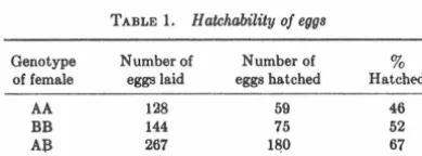
Hatchability
The hatching records of the female birds in our colony were correlated with transferrin type of the females. The values for Tumblers are shown in Table 1. Kings have poorer per-formance in our flock, but show !limilar differences. among fe-male genotypes. A significantly greater portion of the eggs of AB females hatch than do the eggs of the two homozygous types.
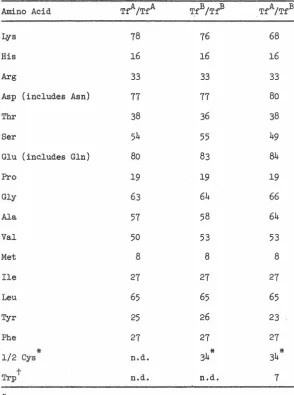
[image:32.618.348.527.95.402.2]Inhibition of yeast growth
Fig. 1 shows typical curves for the growth of yeast in medium containing individual eggwhites, all at the same eggwhite concentration. Variability of the AB inhibition was common among individual AB eggwhites, but in no case did AB in-hibit growth less than the homozygous types. Table 2 shows
TABLE 1. H atchability of eggs
Genotype Number of Number of 3 of female eggs laid eggs hatched Hatched
AA 128 59 46
BB 144 75 52
A:B 267 1&0 67
Chi square = 7.99; P <0.02.
Transferrin Polymorphism in Pigeons
107 ~- AB - AA+BB ~AA - BB
10~
A 104 o.___.__,,2 __ ..__4..__.___.6 __ _,__e..__.___.10---'--'12
-o- AA .__ AB
hr in culture
hr in culture
327
Frn. 1. Growth of yeast in meqium containing eggwhite. (A) shows one of the experiments iµ which AB transferrin did not inhibit yeast growth greatly. (B) shows an experiment in which the AB transferrin totally inhibited the yeast growth, while the yeast grown in A tranferrin grew at its characteristic rate.
a composite of growth rates of these growth curve experi-ments. These rates are calculated by computation of the number of yeast doublings during the culture period divided by the elapsed time in hours minus the 2-hr lag phase. Addi-tion of FeCb at a concentraAddi-tion of 0.01 mg/ml abolished the differences among transferrin genotypes. The two homozy-gous types and, most interestingly, mixtures of the two types, give similar yeast growth rates. This finding suggests an in-teraction in the heterozygote, absent from the simple mix-ture of the homo~ygous eggwhites. However, mixtures of the two homozygous types are indistinguishable from the heterozygous type on starch gels.
[image:32.618.118.313.602.674.2]328 Genetics: Frelinger
DISCUSSION
These experiments show for the first time in vitro a differ-ential inhibition of microbial growth by transferrin from dif
-ferent phenotypes. The bacteriostatic and fungistatic prop-erties of transferrin are well documented, predating, iu fact, the modern description of transferrin (16). Transferrin has been shown to inhibit a wide variety of iron-dependent mi cro-organisms inch~ding Saccharomcye8 cerevi8iae, Staphlococcus aureus, Candida albicans, Shigella dysenteria, Pasture/la sep-tica P8eudomona8 Clostridium welchii, Lysteria moncytogens, and
1
Salmonella t;phimurium (i7). In chickens, embryonic death is frequently associated with microbial contamination of the egg (18). Salmonella can penetrate through the egg shell to contaminate the egg (19). Levi (20) has reported that while Salmonella mfection may be asymptomatic in adults, it is a frequent cause of death in young squabs .. He also cites Candida albicans, Pasturella, and Mycobacterium tuberculosis as common in pigeon~. Thus, it appears that the opportunities for the infection of the egg and young squab are substantial, and the bacteriostatic and f ungistatic prop-erties of transferrin constitute, presumably, an important early line of defense. BecauHe the transferrin both in the egg and the young bird is of maternal origin, this critical selective period is based on the maternal genotype rather than the embryo's own constitution. This leads to the survival of a greater proportion of the progeny of heterozygous mothers; but produces distributions of surviving genotypes indis-tinguishable from th~ Hardy-Weinberg equilibrium (Fre-linger and Crow, manuscript in preparation).
TABLF. 2. Yeast growth in the presence of eggwhite and of purified transf errin of three types•
Number of egg
-Transferrin type Material Growth rate whitest
A (TJA/TJA) Eggwhite 0.495 ± 0.0637 6
Purified 0.34 5 (pool)
transferrin
B (Tf8/Tf8 ) Eggwhite 0. 393 ± 0. 037 4 13
Purified 0.30 5 (pool)
transferrin
Mixture of A and B Eggwhite 0.42!i ± 0.0509 4 (TJA/TJA +
TJB/TJB)
AB (TJA/Tf8 ) Eggwhite 0.162 ± 0.067 10
Purified 0.14 6 (pool)
transferrin
• Gosset's t test for significance revealed no significant differ
-ence (O.!i > P > 0.3) between A and B. These data were therefore pooled and tested against the mixtures of A and B. This com
-parison also yielded no significant difference (0.7 > P > 0.5).
Therefore, these data (A, B, mixture of A and B) were pooled and compared to AB. The difference is highly significant (P <
0.0003).
t For purified transferrin, number of eggwhites is the number of different eggwhites in the pool from which the transferrin was purified. In the case of eggwhites, it represent.s the number of individual egg whites that were separately tested. The results are given as the mean growth rate ± the standard error.
Proc. Nat. Acad. Sci. USA 69 (1972)
Several workers have reported that transferrin is a mono-meric protein (1, 2). The failure of the mixture of the two homozygous types to display the functional characteristic of the heterozygous type suggests the presence of &.n inter-action product in the heterozygote. This is difficult to explain in terms of a monomeric protein, if the alleles represent a structural locus controlling the primary structure of the tein. The difference in the action of the heterozygous pro-tein suggests the presence of dimeric propro-tein, so that "hybrid" molecules can be produced in the heterozygote. In cattle, a second gene affecting the carbohydrate portion of the tra.nsferrin molecule has recently been reported by Spooner (21). If the difference between the allelic transferrins of pigeons is in their carbohydrate rather than in their pri-mary protein structures, it then becomes possible to resolve the difference between heterozygous transferrin and the mix-ture of the two homozygous types. Experiments are cur-rently in progress to determine if the inherited difference is in the amino acid or in the carbohydrate portions of the mol e-cules.
E
~ c:
:::l g' 106
E
0
I >.
c:
0 0
u
+ control
a A tronsferrin o B tronsferrin • AB tronsferrin
2 4 6 8 10 12 14 16 18 20 22 24
hr in culture
/
0
a
Proc. Nat. Acad. Sci. USA 69 (1972)
The results report for the first time a direct in vitro test of
a proposed selective mechanism for the maintenance of a polymorphism. The data are consistent with the hypothesis that the polymorphism is maintained by selection against the offspring of homozygous females because of their greater susceptibility to infection both as embryos and as young birds. This interpretation is suggested by both the in vitro
data on inhibition of yeast growth and the in vivo results
rep-resented by the hatchahility data.
A preliminary report was pre~;ented at the Genetics Society of America meeting in August 1971. I thank Dr. Ray D. Owen for guidance and support during t.his work and Dr. H. K. Mitchell for suggesting morpholinopropane ~mlfonic acid buffer. This work was supported by Atomic Energy Commission contract AT(04-3)-767 and U.S. Public Health Service training grant GM 00086.
I. Feeney, R. & Komatsu, 8. (1966)Struct. Bonding (Br.rlin) 1, 146-207.
2. Baker, C. M. A., Crozier, G., Stratil, A. & Manwell, C. (1970) Advan. Genet. 15, 147-174.
3. Ashton, G. C., Francis, J. & IW,i;on, J. B. (1966) Aust. J. Biol. Sci. 19, 821-829.
4. Ashton, G. C. & Fallon, G. R. (1962) J. Reprod. Fert. 3,
93-104.
5. Ashton, G. C. & Lampkin, G. H. (1965) Genet. Res. 6,
209-215.
Transferrin Polymorphism in Pigeon:,; 329
6. Fowle, K. E., Cline, J. H., Klosterman, E. W. & Parker,
Q. F. (1J)67) J. Anim. Sci. 26, 1226-1231.
7. Nagai, J. & Ktistjensson, K. (1970) Can. J. Gcnrl. Cytol. 12,
307-3lfi.
8. Fesus, L. & Hasmw;en, B. A. (1971) A nim. Blood Groups,
Biochem. Gcnrt. 2, fi7-58.
. 9. Tamerin, H. JI. & Krebs, C. J. (1969) Evolution 23, 183-211.
10. Gaine;;, M. 8., Myers, J. H. & Kreh;;, C. J. (1971) Evolution 25, 443-4fi0.
11. Gilmour, D. G. & Morton, J. H. (1971) Theor. Appl. Genet. 41, 57--66.
12. Mueller, J. 0., Smithies, 0. & Irwin, J\1. H. (1962) Genetics
47, 1381)-1392.
13. Brown, IL V. & 8harp, H.B., Jr. (1970) Anim. RloodGroups, Hiochrm. Genet. 1, 1 rn-11 fi.
l 4. Ferguson, A. (1971) Comp. /Jiochem. Physiol. 38B, 4 77--486. lfi. Frelinger, J. A. (1971) Scirnce 171, 1260--1261.
16. 8r.ha<le, A. L. & Caroline, L. (1944) Scimcr 100, 14-lfi.
17. Kochan, I., Uol<le11, C. A. & Bukovic, J. A. (1!!68) J. Hac-terZ:ol. 100, 64-70.
18. Quarle~, C. L., (:entry, IL F. & Bre-;sler, G. 0. (1970)
Poultry Sci., 49, 60--66.
19. Williams, J.E. & Whittemore, A. D. (1967) Avian Dis. 11,
Studying Microbial Penetration through the Outer Htruc-467-490.
20. Levi, W. (1957) in The Pigeon (Levi Publiishing Co., Sumter, S.C.), :{rd ed., pp. :181-448.
21. Spooner, R. L. & Baxter, G. (1969) Biochem. Genet. 2,
CHAPTER 3
Transferrin Polymorphism and the Hardy-Weinberg Ratios
-30-TRANSFERRIN POLYMORPHISM AND THE HARDY-WEINBERG RATIOS
The iron binding protein, transferrin, is polymorphic in almost all
vertebrate species where it has been studied. In pigeons the two alleles,
TfA and TfB, are present in approximately equal frequency in populations
widely scattered throughout the world and the three genotypes are in
Hardy-Weinberg (H.W.) proportions in each population (Ferguson 1971).
A mechanism to account for the gene frequency stability has recently been
proposed (Frelinger 1972). Eggs from heterozygous females inhibit microbial
growth more strongly than those from either homozygote, and it is suggested
that this probably accounts for the observed differences in egg-hatch from
the three maternal genotypes. Because a larger fraction of their eggs
hatch, heterozygous females are effectively more fertile than homozygotes,
in that they contribute more offspring to the population, and this is
sufficient to maintain the two alleles in balanced polymorphism.
It is known that strong selection can cause wide departures from H.W.
ratios in a randomly mating population if the enumeration is made after
selection, but not if this is done before. In this case selection acts
through embryonic mortality so it might appear that enumeration of adults
would show departures from H.W. ratios, especially in view of the strong
selection that is implied by the large differences in egg hatch. But, the
selection is based on the maternal genotype, not that of the embryo. We
shall show that in this case the equilibrium population is in H.W. proportions,
as observed. However, if the population has not reached gene frequency
equilibrium there will be a slight excess of heterozygotes.
described and desi~ate the various Pa!"~-~ters in a randomly matin~ population
as follows:
Genotype
Adult freauency
Relative contribution of fe~ales
Relative contribution of males
p
1-s
1
W
=
P(l-s) + 2Q + P.(1-t)=
1 - sP - tR~
=
P + Q, the frequency of allele Aq
=
Q + R, the frequency of allele BA?.
2Q
1
1
::rn
n
1-t
1
The frequencies of the three ~enotY}"les next generation will be
(1) '?' = (P + Q - sP) (P + Q)/1-! = {p 2 spP)/H
(2) 2Q'
=
( (P t Q - sP) (Q + n) + (Q + R - tn) (P + Q)]/W =(3) R'
=
(Q + R - tR) (Q + R)/H=
(q 2 tqR)/\!.?otal
1
w
1
(2pq sa)' -tp:q) r~
Adding (1) to half of (2) ve obtain the A eene frequency next ~er.eration,
which after sone algebraic rearranger'lent is
(4) p'
=
p + (tpR - sqP)/2W.There will be an equilibriwn when tpR
=
sqP.We use the quantity H = Q2 /PR as a measure of the departure fron E. ~l.
(5)
-32-~·2
(2no - sgP - tpR)2
H'
=
P'R' ::: 2 24(p - spP) (q - tqR)
= p2q2 - po(saP + tnR) + (sqP + tpR)2/4
p2q2 - pq(sqP + tpR) + stpqPR
!:otice that the n'llI:'!erator and denon.inator a.re the sa!'ie except for the last
term. If we subtract one from the other we have
(6) (sqP + tpR)2/~ - stpqPR
=
(sqP - tpR)2/4 ~ 0Comparison of {6) and (4) shows that i f the population is at gene frequency equilibriun it is in H.W. proportions, but until this stage is reached there
will be an excess of heterozygotes.
near equilibrium, P ~ p2 and R ~ q2, and (4) becoJYles
(7) p' - p ~ (tpq - sqp 2 2 )/2•(;
= pq(s+t) [~ _ ]
2W s+t p
The equilibrium value of p (when p' - p
=
0) is t/(s+t), as in simpleover-dominance for fitness. The equilibrium is locally stable, as can be seen
from (7) because eene frequency change is always in the direction of the equilibrium value. This can be interpreted as a special case of fertility
The excess of heterozygotes in non-equilibrium populations implied QY
(6) is caused, not by differential mortality per !!:,, but by the resulting unequal gene frequencies in the contribution of male and female parents.
This corresponds to the standard principle that when the parental gene
:frequencies are unequal in the two sexes random mating leads to an excess
of heterozygotes (Wright 1969, p. 4; Crow and Kimura 1970, p. 44). On the
other hand, if the population sampled is actually made up of several randomly
mating subpopulations with differing gene frequencies, there may well be an
overall deficiency of heterozygotes.
The heterozygote excess shown by (6) is very slight in most cases.
Figure 1 shows the situation graphically, where the population genotype
frequencies are shown by a point in the triangle. The numerical values of
s and t (.3l3 and .224) are based on the egg hatch data of Frellnger (1972).
From any starting point the population moves in a single generation of random
mating to a point to the left and below the H.W. parabola. Actually in this
example the points are so close to the curve as to be indistinguishable in
the graph. The population then follows the parabola to the point of gene
:frequency equilibrium. The final value is determined by the ratio of s to
t, whereas the rate of approach is determined by their absolute values.
The fact that the departure fran H.W. proportions is so slight during
this process explains the regular finding of H.W. ratios, which then does not
-34-References
Bod.mer,
w.
F. 1965. Differential Fertility in Population Genetic Models. Genetics 51: 411-424.Crow, J. F., and M. Kimura. 1970. An Introduction to Population Genetics Theory. Harper.
Ferguson, A. 1971. Geographic and Species Variation in Transferrin and Ovotransferrin Polymorphism in Columbidae. Comp. Biochem. Physiol.
38B: 477-486.
Frelinger, J. A. 1972. The Maintenance of Transferrin Polymorphism in Pigeons. Proc. Nat. Acad. Sci. USA 69: 326-329.
Wright, s. 1969. Evolution and the Genetics of Populations. Vol. 2. The
Acknowledgments
Vle thank Dr. ?.ay D. Owen for advice and encouragement. JF was
Division of Biology
California Institute of Technology
Pasadena, California 91109
Laboratory of Genetics
University of Wisconsin
Madison, Wisconsin 53706
-36-Jeffrey A. Frelinger
1.0
0.8
0.6
R
0.4
0.2
0 0.2
-37-0.4 0.6 0.8 1.0
-38-Legend for Figure l.
The approach to equilibrium for selection caused by maternally determined
embryonic mortality. The population is represented by a point in the triangle.
The proportion of AA is given by the horizontal distance from the R axis,
the proportion of BB by the vertical distance from the P axis, and the
proportion of AB by the horizontal or vertical distance to the diagonal.
Populations in Hardy-Weinberg ratios lie on the parabola. The arrows represent
the behavior of populations during the first generation of random mating.
A~erward the population moves along the parabola to the equilibrium point.
Although the departures are too small to show on the graph, the actual points
are slightly below and to the left of the parabola, indicating a slight excess
CHAPTER 4
Chemical Basis of Transferrin Polymorphism in Pigeons
-40-CHEMICAL BASIS OF TRANSFERRIN POLYMORPHISM IN PIGEONS
Jeffrey A. Frelinger
Division of Biology, California Institute of Technology
Transferrin is a non-heme iron-binding protein, polymorphic in most
vertebrate species examined. Mueller et al. (1962) first reported
electro-phoretic variants of pigeon (Columba livia) transferrin. Frelinger (1972)
has recently proposed that the gene frequency stability observed in pigeons
is accounted for by superior inhibition of microbial growth by transferrins
from heterozygotes. However, mixtures of homozygous type transferrins do not
mimic transferrins from heterozygotes in this effect. For this reason we
have investigated the chemical basis for the observed allelic variation. To
our knowledge, the only species in which a chemical difference has been
identified is in humans (Wang and Sutton, 1965; Wang et al., 1966; Jeppson
and Sj8quist, 1966) although there have been other attempts (Roop and Putnam,
1967; Herschberger, 1970). We present data here suggesting a single amino
acid difference between alleles in the two forms of pigeon transferrin.
MATERIALS AND METHODS
~· Egg whites were collected, stored and typed as previously described
(Frelinger, 1971; Frelinger, 1972). Five to ten whites of the same phenotype
were pooled and purified by ion exchange and gel filtration chromatography
(Frelinger, 1972). This preparation was> 99% pure as judged by SDS gel,
cellulose acetate, starch gel, and immunoelectrophoresis.
Carbohydrate analysis. Purified transferrins were freed of carbohydrate
contamination by gel filtration on Biegel P-100 in 0.06 M NaCl, 0.01 M Tris,
pH 8.0. The peak was precipitated two times with 50% saturated ammonium
sulfate, and two times with cold 50% ethanol. Carbohydrate was analyzed by
gas chromatography as previously reported (Niedermeier, 1971). Sialic acid
/
was determined by the method of Warren (Warren, 1959). The results of two
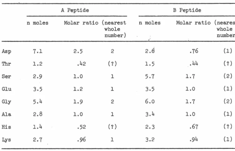
HCl, 0.2 M 2-mercaptoethanol at 37°C for 4 hours and carboxymethylated by 10% excess of iodoacetamide, then extensively dialyzed against 0.2 M ammonium
bi-carbonate, and lyophilized.
Peptide mapping. TPCK trypsin (Worthin~on) was incubated with 6-8 mg reduced carboxymethylated protein suspended in 100 µl ammonium bicarbonate; 1 µl of
M
trypsin (100 mg/ml in O.OOl~HCl) was added and the mixture was incubated for 1 hour at 37°C followed by the addition of 1 µl trypsin solution and a second hour of incubation. The mixture was lyophilized and suspended in 50 µl water, spotted and chromatographed in n-butanol/glacial acetic/water (540:160:800),
followed by electrophoresis at pH 3.5 in pyridine-acetate buffer (Bennet, 1967). The maps were stained with ninhybrin-collidine stain. For elution the peptides were stained with 1% ninhydrin in ethanol. Peptides were located and eluted with water, or 50% (v/v) NH
40H.
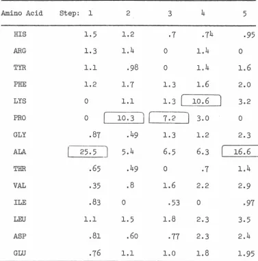
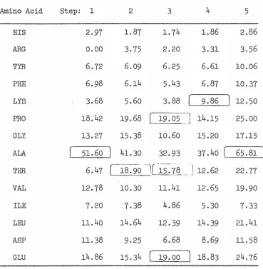
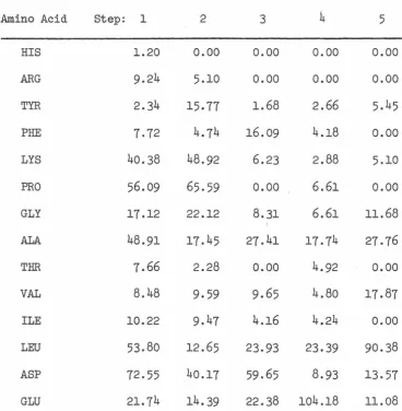
Amino acid analysis. Peptide eluates were lyophilized and hydrolyzed 18 hours in 6 N HCl at 110°C. The hydrolysates were analyzed on a Beck.man 121C or Durrum D-500 amino acid analyzer. Total amino acid compositions were carried out similarly except that 24 and 48 hour hydrolysis times were used to enable the determination of serine and threonine content by extrapolation to zero time.
Performic acid oxidized material (Hirs, 1967) was used to determine 1/2 cystines as cysteic acid. Tryptophan was analyzed by oxidation with N-bromosuccinimide
(Spande and Witkop, 1967).
RESULTS AND DISCUSSION