APC = antigen presenting cell; CIA = collagen-induced arthritis; FLS = fibroblast-like synovial cells; IKK = IκB kinase; MMP = matrix metallo-proteinases; NFκB = nuclear factor kappa B; PDGF = platelet-derived growth factor; PI(3)K = phosphatidylinositol 3-kinase; RA = rheumatoid arthritis; SCW = streptococcal cell wall; Th cells = T helper cells; TNF = tumor necrosis factor.
Introduction
Chronic inflammation perpetuates and amplifies itself through the numerous autocrine and paracrine loops of cytokines, acting on the cells within the lesion. The vicious circle can be broken either by neutralizing the biological activities of extracellular inflammatory mediators or by inhibiting cytokine production. The pattern of gene expres-sion is controlled by transcription factors, which relay into the nucleus signals emanating from the cytoplasmic mem-brane. In the nucleus, transcription factors selectively bind their cognate sites in the regulatory elements of targeted genes and activate or repress transcription. It appears that the complexity of inflammatory pathways is significantly reduced on the level of transcription factors. Whereas the cell within the inflammatory lesion is subjected to many dozens, perhaps hundreds, of extracellular stimuli, only a handful of inducible transcription factors, including AP-1,
C/EBPs, STATs, NF-AT, and NF-κB, appear to play a major role in the regulation of inflammatory genes. This suggests that neutralization of these transcription factors may provide an efficacious therapeutic strategy. A pivotal role for the transcription factor NF-κB in regulation of inflammation has been well recognized [1,2]. I attempted, in a recent general article, to summarize the latest devel-opments in the field and to discuss the feasibility of anti-NF-κB therapy for chronic inflammation [3]. The present review focuses on the role of NF-κB in the particular fea-tures of RA pathology.
The NF-
κκ
B signaling pathway
NF-κB is a collective name for dimeric transcription factors comprised of the Rel family of proteins that include RelA (p65), c-Rel, RelB, NF-κB1 (p50), and NF-κB2 (p52) [4]. The most abundant form found in stimulated cells is
Commentary
NF-
κκ
B in rheumatoid arthritis: a pivotal regulator of
inflammation, hyperplasia, and tissue destruction
Sergei S Makarov
Center for Inflammatory Disorders, Thurston Arthritis Research Center, and Department of Endodontics, University of North Carolina at Chapel Hill, Chapel Hill, North Carolina, USA
Correspondence:Sergei S Makarov, 4109 Thurston, CB 7280, University of North Carolina at Chapel Hill, Chapel Hill, NC 27599-7280, USA. Tel: +1 919 966 9565; fax: +1 919 966 1739; e-mail: smak@med.unc.edu
Abstract
The transcription factor NF-κB has been well recognized as a pivotal regulator of inflammation in rheumatoid arthritis (RA), but recent developments revealed a broad involvement of NF-κB in other aspects of RA pathology, including development of T helper 1 responses, activation, abnormal apoptosis and proliferation of RA fibroblast-like synovial cells, and differentiation and activation of bone resorbing activity of osteoclasts. In agreement with this, studies in animal models of RA have demonstrated the high therapeutic efficacy of specific inhibitors of NF-κB pathway, indicating the feasibility of anti-NF-κB therapy for human disease.
Keywords:apoptosis, hyperplasia, inflammation, inhibitors, nuclear factor NFκB, rheumatoid arthritis, therapy Received: 2 January 2001
Revisions requested: 26 February 2001 Revisions received: 6 March 2001 Accepted: 8 March 2001 Published: 26 March 2001
Arthritis Res2001, 3:200–206
This article may contain supplementary data which can only be found online at http://arthritis-research.com/content/3/4/200
© 2001 BioMed Central Ltd
commentary
review
reports
primary research
the RelA/NF-κB1 (p65/p50) heterodimer, often referred to as a ‘classic’ NF-κB. In unstimulated cells, NF-κB resides in the cytoplasm in a latent form, and must translocate to the nucleus to function. The cytoplasmic retention of NF-κB is provided by its interaction with inhibitory proteins known as IκB. Stimulation leads to a phosphorylation-targeted protea-somal degradation of IκB, allowing the ‘active’ NF-κB to enter the nucleus and initiate transcription.
The signal-induced IκB processing involves the consecu-tive steps of IκB phosphorylation, ubiquitination, and pro-teasomal degradation, which are controlled by three large multiprotein complexes; namely, IκB kinase (IKK), or signal-some, IκB ubiquitin ligase, and 26S proteasome [5]. The IκB ubiquitin ligase and 26S proteasome are considered mostly constitutively active, while IKK activity is rapidly induced in response to stimulation. The core components of the signalsome include the catalytic subunits IKKα/IKK-1 and IKKβ/IKK-2, and a scaffold protein IKKκ/NEMO that links the catalytic kinase subunits with the upstream activa-tors. Both IKKα and IKKβ can phosphorylate IκB in vitro, but knockout studies indicate that IKKβhas a pivotal role in cytokine-inducible activation of NF-κB [5].
In addition to the spatial control of NF-κB function, the ability of nuclear NF-κB to initiate transcription is regulated by interactions with numerous transcriptional coactivators and basal transcriptional machinery. The pathways con-trolling NF-κB nuclear translocation and its transcription function are regulated independently, but act in synergy in the activation of NF-κB-dependent gene expression [6].
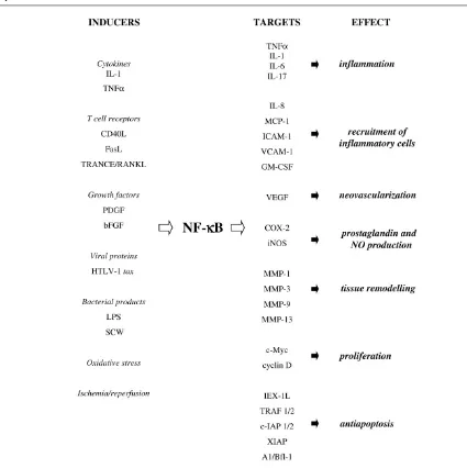
NF-κB can be activated by a variety of pathogenic stimuli, including bacterial products and viral proteins, cytokines, growth factors, radiation, ischemia/reperfusion, and oxida-tive stress. The coordinated activation of NF-κB that occurs in almost every cell type involved in inflammatory response, including neutrophils, macrophages, lympho-cytes, and endothelial, epithelial, and mesenchymal cells, is an integral part of the defensive response to pathogens and stress. The activation of NF-κB is required to induce expression of diverse inflammatory and immune response mediators. More than 150 NF-κB responsive genes have been identified, among them cytokines, chemokines, cell adhesion molecules, and growth factors [7]. A few exam-ples of inducers and targets of NF-κB are presented in Figure 1. It is worth noting that the list of inducers and targets of NF-κB almost perfectly matches the list of pivotal mediators of RA pathology.
NF-
κκ
B is activated in RA
Activated NF-κB has been detected in human synovial tissue on the early stage of joint inflammation [8], as well as in specimens obtained at the late stages of the disease. Analyses of nuclear extracts from synovial explants revealed the presence of increased NF-κB DNA binding
activity in RA patients, but not in osteoarthritis patients [9]. Immunohistochemical studies detected nuclear RelA (p65) and NF-κB1 (p50) mostly in RA endothelium and synovial lining, particularly in CD14-positive cells, and no staining in the normal synovium [10]. Immunostaining with antibodies against the ‘active’ (dissociated from IκB) NF-κB similarly revealed the presence of active NF-κB in the nuclei of macrophage-like synoviocytes in synovial lining and in vascular endothelium. The active NF-κB was found in both RA and osteoarthritis samples, although the pattern was different: patients with acute RA more com-monly showed vessel staining and, conversely, showed less frequent staining of the synovial lining as compared with osteoarthritis patients [11]. Miyazawa et alused an alternative approach with a NF-κB reporter gene construct to analyze NF-κB-dependent transcription in single clones of primary RA fibroblast-like synovial cells (FLS) in vitro. Constitutively active NF-κB was detected in some of the clones and, noteworthy, these clones spontaneously pro-duced large amounts of IL-6 [12]. These findings combine to provide compelling evidence that NF-κB activation is a common feature of human RA synovium. Activation of NF-κB has also been detected in different animal models of RA, including adjuvant arthritis in rats [13], pristane-induced [14] and streptococcal cell wall (SCW)-pristane-induced arthritis in rats [14,15], and collagen-induced arthritis (CIA) in mice [16].
NF-
κκ
B and synovial inflammation
Initiation of chronic inflammation in RA is associated with development of an autoimmune response that progresses to a sustained, self-perpetuated inflammation. Experimen-tal evidence suggests that NF-κB activation plays a pivotal role both at the stage of initiation and the stage of perpet-uation of chronic inflammation in RA.
NF-
κκ
B in the initiation of chronic
inflammation
upregulating the expression of NF-κB-dependent mole-cules MHC class II, CD80, and CD86 [19–22]. The importance of NF-κB activation in immune response was supported by the observations in knockout mice. Inactiva-tion of RelB and NF-κB2 in relb–/–and nfkb2–/–caused an impairment of APC function. The loss of NF-κB1 and c-Rel in nfkb1–/– and rel–/– mice resulted in multiple defects in the activation of T and B cells, and weakened responses to pathogens [23].
NF-
κκ
B facilitates T helper 1 subset development
After activation, CD4+T helper (Th) cells can differentiate into Th1 or Th2 effector subsets. These two types of cells produce distinct profiles of cytokines and regulate [image:3.612.81.506.94.520.2]differ-ent immune responses: Th1 (IFNγ and IL-12 dominant) cells mediate cellular immunity and activate macrophages, and they are considered proinflammatory; Th2 (IL-4 and IL-5 dominant) cells, which potentiate antiparasite and humoral immunity, and inhibit macrophage activation, are considered anti-inflammatory. As NF-κB controls the expression of Th1 cytokines IL-2, IFNγ, and IL-12, activa-tion of NF-κB should facilitate Th1 subset development. Indeed, transgenic mice expressing, in the T lineage, an inhibitor of NF-κB (a nondegradable mutant of IκBα, also called ‘super-repressor’ IκBα [srIκBα]), had weakened Th1 and enhanced Th2 responses [24,25]. T cells from transgenic mice expressing Rac2, an upstream NF-κB activator, accordingly developed Th1 type responses [26]. Figure 1
commentary
review
reports
primary research
NF-
κκ
B and the perpetuation of chronic
inflammation
Secreted products of activated T cells and direct cell–cell contacts induce activation of macrophages, the major pro-ducers of inflammatory cytokines in RA synovium. NF-κB controls the expression of cytokines IL-1βand TNFα, the essential mediators of inflammation in RA. TNFαand IL-1 are potent inducers of NF-κB activation, suggesting an interdependence of persistent NF-κB activation and sus-tained levels of IL-1 and TNFα. Indeed, expression of srIκBα abrogated the induction of IL-1β and TNFα in human macrophages and primary FLS [14,27]. A recent study by Aupperle et al indicates that, in RA FLS, IKKβ/IKK-2 is the principal kinase in activation of NF-κB in response to IL-1 and TNFα. Expression of a DN mutant form of IKK-2 inhibited cytokine-inducible activation of NF-κB and abrogated synthesis of IL-6 and IL-8, as well as expression of ICAM-1 and collagenase-1. In contrast, the DN IKKα/IKK-1 had no effect [28]. The notion that IKKβ/IKK-2 is the key convergence pathway for cytokine-induced NF-κB activation is consistent with results of genetic studies in IKK knockout mice [5].
It is worthy of note that suppression of NF-κB inhibited expression of many proinflammatory molecules, including IL-1, TNFα, IL-6, IL-8, ICAM-1 and VCAM-1, but had little, if any, effect on the expression of anti-inflammatory cytokines IL-10 and IL-1 receptor antagonist [14,29–31]. This suggests that NF-κB activation facilitates the impaired balance of proinflammatory and anti-inflammatory molecules in the arthritic joint.
NF-
κκ
B and hyperplasia
Normal synovium is a delicate tissue lining the joint capsule but, in RA, the synovium transforms into an aggressive, tumor-like structure called pannus, which invades and erodes the joint. Experimental evidence sug-gests that NF-κB activation may facilitate synovial hyper-plasia by promoting proliferation and inhibiting apoptosis of RA FLS.
Proliferation
NF-κB serves as a positive regulator of cell growth in myoblasts and fibroblasts by inducing the expression of c-Myc and cyclin D1, proteins required for cell cycle pro-gression [32–34]. Our studies in primary rat FLS have shown that stimulation with platelet-derived growth factor (PDGF) and basic fibroblast growth factor induced NF-κB activation, which was required for induction of c-Myc and DNA synthesis [32] (J Romashkova, S Makarov, unpub-lished observations). In contrast, the mitogenic activity of insulin-like growth factor-1, which did not activate NF-κB, was not influenced by NF-κB inhibitors (J Romashkova, S Makarov, unpublished observations). Another function of NF-κB in mitogenic signaling in FLS is to protect cells against cytotoxicity of c-Myc. Although c-Myc is required
for proliferation, it causes cell death unless certain survival factors are provided. PDGF is one such factor that over-comes the pro-apoptotic proclivity of c-Myc. We found that blocking NF-κB activation abrogated the protective effect of PDGF, indicating that, in PDGF signaling, NF-κB transmits two signals: one is required for the induction of c-Myc; and the second is an anti-apoptotic signal that neu-tralizes c-Myc cytotoxicity, conceivably by inducing the expression of a protective gene (or multiple genes) [32]. As c-Myc is heavily overexpressed in RA synovium, NF-κB activation may contribute to synovial hyperplasia by inhibit-ing c-Myc-induced apoptosis and promotinhibit-ing proliferation. A point of interest is that the pathway via which PDGF induced NF-κB activation involved phosphatidylinositol 3-kinase (PI(3)K) and protein 3-kinase B/Akt (see later). As the PI(3)K/Akt pathway has been implicated in the pathogene-sis of numerous human malignancies, this suggests that similar mechanisms may operate in the promotion of hyperplasia in RA and cancer.
Apoptosis
Many pro-apoptotic stimuli, including TNFα, radiation, and chemotherapy, induce NF-κB activation. NF-κB activation delivers, in most cell types, an anti-apoptotic signal that counteracts cell death. NF-κB suppression of apoptosis appears to be a transcriptional event since it activates expression of anti-apoptotic genes TRAF1 and TRAF2, c-IAP1 and c-IAP2, the 2 homologs A1/Bfl-1 and Bcl-xL, IEX-1, and XIAP (reviewed in [35]). In our studies, blocking NF-κB activation in primary rat SCW FLS strongly potentiated the cytotoxicity of TNFα and FasL. Consistent with this, administration of distinct inhibitors of NF-κB (proteasomal inhibitors and adenoviral gene trans-fer of srIκBα) in vivo resulted in accelerated apoptosis in joints of rats with pristane-induced and SCW-induced arthritis [14]. These studies are in agreement with that published by Zhang et al, who observed that NF-κB acti-vation was required to protect RA FLS against the cytotox-icity of TNFα in a human RA/SCID mouse model, presumably through induction of anti-apoptotic protein XIAP [36]. These findings indicate an important role for NF-κB in protecting FLS against apoptosis in RA syn-ovium, conceivably by inhibiting the cytotoxicity of TNFα and FasL. Because TNFαis a potent mitogen in RA FLS, NF-κB appears as a master switch determining whether TNFαexerts mitogenic or pro-apoptotic effects.
and Her-2/neu [41], and to inhibit cell death induced by deregulated c-myc [32], H-Ras [40], and TNFα[41]. Pre-cisely how Akt activates NF-κB-dependent transcription is a matter of debate. Several groups, including that of the present author, demonstrated an involvement of IKK, thus implicating IκB-dependent mechanisms [32,37,39,41,42], while results by other workers demonstrate Iκ B-indepen-dent mechanisms, in which Akt potentiates the transcrip-tional function of NF-κB [38,40]. It is possible that the mechanisms connecting Akt with NF-κB, as well as the contribution of NF-κB pathway in the anti-apoptotic func-tion of Akt, are cell type and stimulus specific. A role for Akt in the pathogenesis of RA and the contribution of Akt to NF-κB activation in RA synovium are not known, but there is circumstantial evidence that this link may exist. The activity of the PI(3)K/Akt pathway is negatively regu-lated by the phosphatase PTEN, a tumor suppressor fre-quently inactivated in human malignancies [43] and in some autoimmune diseases, such as Cowden disease and Sjögren’s syndrome [44]. The importance of the PTEN-PI(3)K/Akt connection in autoimmune disease was demonstrated by observations in PTEN+/– heterozygous mice that exhibited defects in Fas-induced apoptosis and developed autoimmune disease [45], and in PI(3)K trans-genic mice that developed strikingly similar pathology [46]. Although PTEN mutations were not found in RA, the expression of PTEN is strikingly reduced in RA synovial lining [47], suggesting that deregulated PTEN may result in activation of the PI(3)K/Akt pathway and contribute to activation of NF-κB. The role of Akt and the Akt–NF-κB connection in RA clearly merits further investigation.
NF-
κκ
B and tissue remodeling
FLS are considered the major effectors of RA joint destruction. Inflammatory milieu in the RA joint results in a highly invasive phenotype of RA FLS. Several lines of evi-dence suggest that NF-κB activation contributes to the destructive potential of RA FLS. First, NF-κB mediates transcriptional activation of several matrix metallopro-teinases (MMP). The promoter of MMP-1 has a NF-κB binding site that is required for the induction of MMP-1 by IL-1β[48]. Furthermore, NF-κB activation was found nec-essary for the induction of MMP-3, MMP-9, and MMP-13 expression [49–51]. Second, NF-κB activation is neces-sary for the induction of cyclooxygenase-2 and inducible nitric oxide synthetase, the enzymes that catalyze synthe-sis of proinflammatory prostaglandins and nitric oxide metabolites [7]. Finally, NF-κB activation is required for the induction of vascular endothelial growth factor, an endothelial cell-specific mitogen and a pivotal regulator of angiogenesis in RA [52].
Osteoclast is another cell type in which NF-κB activation may have a prominent role in RA joint destruction. Bone marrow-derived osteoclast precursors are recruited to RA synovium by elevated levels of NF-κB-dependent
cytokines IL-1αand IL-1β, TNFα, IL-6, and IL-17 [53,54]. The contacts with activated T cells and osteoblast/stromal cells expressing high levels of TRANCE/RANKL/ODF promote osteoclast maturation and induce bone resorbing activity, while administration of osteoprotegrin, a soluble decoy receptor for RANKL, prevents osteoclast differenti-ation and precludes bone loss in animal arthritis [20,55]. The critical importance of NF-κB in bone turnover was underscored by the observations in double-knockout
nfkb1–/–nfkb2–/–mice that develop osteopetrosis owing to accumulation of immature osteoclasts [56].
The NF-
κκ
B pathway as a therapeutic target
Many conventional anti-inflammatory and anti-rheumatic drugs, including glucocorticoids, aspirin, sodium salicy-late, sulfosalazine, and gold compounds, are inhibitors of NF-κB activation. The list of therapeutics that inhibit NF-κB also includes numerous natural and synthetic antioxidants, immunosuppressants, and natural plant com-pounds, suggesting that the ability to suppress NF-κB activation at least partially accounts for their therapeutic effects [57]. This interpretation, however, is complicated by the fact that most of the conventional drugs are not very potent inhibitors of NF-κB, and that they can also affect other signaling pathways. More clear answers were obtained using animals with genetically inactivated NF-κB signaling. Inactivation of c-Rel and NF-κB1 in rel–/– andnfkb1–/–knockout mice rendered the animals refractory to development of CIA [58]. Transgenic mice expressing srIκBα in the T lineage were similarly refractory to CIA [25]. These genetic studies are in a good agreement with the experiments that utilized highly specific inhibitors of NF-κB. In our studies, liposomal delivery of NF-κB decoys (double-stranded oligonucleotides containing NF-κB binding sites) effectively prevented the recurrence of SCW arthritis in rats. Intriguingly, the suppression of arthritis was evident not only in the ipsilateral, treated joints, but also in the contralateral, untreated joints, indi-cating systemic effects of local anti-NF-κB therapy [14]. Administration of NF-κB decoys in a similar study signifi-cantly reduced the severity of CIA in rats, and inhibited the production of IL-1 and TNFα within the joints [59]. In a study by Palombella et al, a proteasomal inhibitor of IκB degradation afforded protection against SCW-induced arthritis in rat [15]. Together, these animal studies strongly support the feasibility of using NF-κB inhibitors in RA.
of liver against apoptosis, and that inactivation of the
NF-κB pathway may be associated with detrimental side effects (reviewed in [23]). One way to circumvent this problem is to target the components of NF-κB signaling that are required for cytokine-inducible activation of NF-κB, without interfering with basal NF-κB activity. One such approach has been recently described by May et al
[60]. The authors designed a peptide derived from IKKγ/NEMO to block the assembly of IKK signalsome. The peptide strongly suppressed cytokine-inducible NF-κB activation, but spared basal NF-κB activity. Using the cell-permeable inhibitory peptide in vivoafforded the suppres-sion of inflammatory responses in animal models of peritonitis and ear edema.
Another concern is that systemic suppression of NF-κB may impair defensive responses to pathogens. The unwanted effects of anti-NF-κB therapy can be diminished by targeting NF-κB inhibitors to certain tissues or cell types. In this regard, gene delivery of NF-κB inhibitors may have distinct advantages (reviewed in [61]). Local delivery should alleviate the possible side effects associated with systemic exposure and minimize the risk of general immunosuppression.
Acknowledgements
This work was supported by National Institutes of Health Grants AR/AI 44564, 5-P60-AR30701-14 and AR/AI 44030, and by an Investigator Award from the Arthritis Foundation.
References
1. Barnes PJ, Karin M: Nuclear factor-kappa B: a pivotal transcrip-tion factor in chronic inflammatory diseases. N Engl J Med 1997, 336:1066–1071.
2. Tak PP, Firestein GS: NF-kappa B: a key role in inflammatory diseases. J Clin Invest2001, 107:7–11.
3. Makarov SS: NF-kappa B as a therapeutic target in chronic inflammation: recent advances. Mol Med Today 2000, 6: 441–448.
4. Baldwin AS Jr: The NF-kappa B and I kappa B proteins: new discoveries and insights. Annu Rev Immunol 1996, 14: 649–683.
5. Karin M, Ben Neriah Y: Phosphorylation meets ubiquitination: the control of NF-kappa B activity. Annu Rev Immunol2000, 18:621–663.
6. Schulze-Osthoff K, Ferrari D, Riehemann K, Wesselborg S: Regu-lation of NF-kappa B activation by MAP kinase cascades. Immunobiology1997, 198:35–49.
7. Pahl HL: Activators and target genes of Rel/NF-kappa B tran-scription factors. Oncogene1999, 18:6853–6866.
8. Gilston V, Jones HW, Soo CC, Coumbe A, Blades S, Kaltschmidt C, Baeuerle PA, Morris CJ, Blake DR, Winyard PG: NF-kappa B activation in human knee-joint synovial tissue during the early stage of joint inflammation [abstract]. Biochem Soc Trans 1997, 25:518S.
9. Asahara H, Asanuma M, Ogawa N, Nishibayashi S, Inoue H: High DNA-binding activity of transcription factor NF-kappa B in syn-ovial membranes of patients with rheumatoid arthritis. Biochem Mol Biol Int1995, 37:827–832.
10. Handel ML, McMorrow LB, Gravallese EM: Nuclear factor-kappa B in rheumatoid synovium.Localization of p50 and p65. Arthri-tis Rheum1995, 38:1762–1770.
11. Marok R, Winyard PG, Coumbe A, Kus ML, Gaffney K, Blades S, Mapp PI, Morris CJ, Blake DR, Kaltschmidt C, Baeuerle PA: Acti-vation of the transcription factor nuclear factor-kappa B in human inflamed synovial tissue. Arthritis Rheum 1996, 39: 583–589.
12. Miyazawa K, Mori A, Yamamoto K, Okudaira H: Constitutive tran-scription of the human interleukin-6 gene by rheumatoid syn-oviocytes: spontaneous activation of NF-kappa B and CBF1. Am J Pathol1998, 152:793–803.
13. Tsao PW, Suzuki T, Totsuka R, Murata T, Takagi T, Ohmachi Y, Fujimura H, Takata I: The effect of dexamethasone on the expression of activated NF-kappa B in adjuvant arthritis. Clin Immunol Immunopathol1997, 83:173–178.
14. Miagkov AV, Kovalenko DV, Brown CE, Didsbury JR, Cogswell JP, Stimpson SA, Baldwin AS, Makarov SS: NF-kappa B activation provides the potential link between inflammation and hyper-plasia in the arthritic joint. Proc Natl Acad Sci USA1998, 95: 13859–13864.
15. Palombella VJ, Conner EM, Fuseler JW, Destree A, Davis JM, Laroux FS, Wolf RE, Huang J, Brand S, Elliott PJ, Lazarus D, McCormack T, Parent L, Stein R, Adams J, Grisham MB: Role of the proteasome and NF-kappa B in streptococcal cell wall-induced polyarthritis. Proc Natl Acad Sci USA 1998, 95: 15671–15676.
16. Han Z, Boyle DL, Manning AM, Firestein GS: AP-1 and NF-kappa B regulation in rheumatoid arthritis and murine collagen-induced arthritis. Autoimmunity1998, 28:197–208.
17. Gerondakis S, Grumont R, Rourke I, Grossmann M: The regula-tion and roles of Rel/NF-kappa B transcripregula-tion factors during lymphocyte activation. Curr Opin Immunol1998, 10:353–359. 18. Kuo CT, Leiden JM: Transcriptional regulation of T lymphocyte
development and function. Annu Rev Immunol 1999, 17: 149–187.
19. Anderson DM, Maraskovsky E, Billingsley WL, Dougall WC, Tometsko ME, Roux ER, Teepe MC, DuBose RF, Cosman D, Galibert L: A homologue of the TNF receptor and its ligand enhance T-cell growth and dendritic-cell function. Nature 1997, 390:175–179.
20. Wong BR, Josien R, Choi Y: TRANCE is a TNF family member that regulates dendritic cell and osteoclast function. J Leukoc Biol1999, 65:715–724.
21. Li J, Liu Z, Jiang S, Cortesini R, Lederman S, Suciu-Foca N: T suppressor lymphocytes inhibit NF-kappa B-mediated tran-scription of CD86 gene in APC. J Immunol 1999, 163: 6386–6392.
22. Verhasselt V, Vanden Berghe W, Vanderheyde N, Willems F, Haegeman G, Goldman M: N-Acetyl-L-cysteine inhibits primary human T cell responses at the dendritic cell level: associa-tion with NF-kappa B inhibiassocia-tion. J Immunol1999, 162:2569– 2574.
23. Gerondakis S, Grossmann M, Nakamura Y, Pohl T, Grumont R: Genetic approaches in mice to understand Rel/NF-kappa B and I kappa B function: transgenics and knockouts. Oncogene 1999, 18:6888–6895.
24. Aronica MA, Mora AL, Mitchell DB, Finn PW, Johnson JE, Sheller JR, Boothby MR: Preferential role for NF-kappa B/Rel signal-ing in the type 1 but not type 2 T cell-dependent immune response in vivo. J Immunol1999, 163:5116–5124.
25. Seetharaman R, Mora AL, Nabozny G, Boothby M, Chen J: Essential role of T cell NF-kappa B activation in collagen-induced arthritis. J Immunol1999, 163:1577–1583.
26. Li B, Yu H, Zheng W, Voll R, Na S, Roberts AW, Williams DA, Davis RJ, Ghosh S, Flavell RA: Role of the guanosine triphos-phatase Rac2 in T helper 1 cell differentiation.Science2000, 288:2219–2222.
27. Foxwell B, Browne K, Bondeson J, Clarke C, de Martin R, Brennan F, Feldmann M: Efficient adenoviral infection with Ikappa B alpha reveals that macrophage tumor necrosis factor alpha production in rheumatoid arthritis is NF-kappa B dependent. Proc Natl Acad Sci USA1998, 95:8211–8215.
28. Aupperle KR, Bennett BL, Han Z, Boyle DL, Manning AM, Firestein GS: NF-kappa B regulation by Ikappa B kinase-2 in rheumatoid arthritis synoviocytes. J Immunol 2001, 166: 2705–2711.
29. Makarov SS, Johnston WN, Olsen JC, Watson JM, Mondal K, Rinehart C, Haskill JS: NF-kappa B as a target for anti-inflam-matory gene therapy: suppression of inflamanti-inflam-matory responses in monocytic and stromal cells by stable gene transfer of I kappa B alpha cDNA. Gene Ther1997, 4:846–852.
30. Li P, Sanz I, O’Keefe RJ, Schwarz EM: NF-kappa B regulates VCAM-1 expression on fibroblast-like synoviocytes. J Immunol 2000, 164:5990–5997.
commentary
review
reports
31. Bondeson J, Foxwell B, Brennan F, Feldmann M: Defining thera-peutic targets by using adenovirus: blocking NF-kappa B inhibits both inflammatory and destructive mechanisms in rheumatoid synovium but spares anti-inflammatory media-tors. Proc Natl Acad Sci USA1999, 96:5668–5673.
32. Romashkova, JA, Makarov SS: NF-kappa B is a target of AKT in anti-apoptotic PDGF signalling. Nature1999, 401:86–90. 33. Guttridge DC, Albanese C, Reuther JY, Pestell RG, Baldwin AS:
NF-kappa B controls cell growth and differentiation through transcriptional regulation of cyclin D1. Mol Cell Biol1999, 19: 5785–5799.
34. Hinz M, Krappmann D, Eichten A, Heder A, Scheidereit C, Strauss M: NF-kappa B function in growth control: regulation of cyclin D1 expression and G0/G1-to-S-phase transition. Mol Cell Biol 1999, 19:2690–2698.
35. Barkett M, Gilmore TD: Control of apoptosis by Rel/NF-kappa B transcription factors. Oncogene1999, 18:6910–6924. 36. Zhang HG, Huang N, Liu D, Bilbao L, Zhang X, Yang P, Zhou T,
Curiel DT, Mountz JD: Gene therapy that inhibits nuclear translocation of nuclear factor kappa B results in tumor necrosis factor alpha-induced apoptosis of human synovial fibroblasts. Arthritis Rheum2000, 43:1094–1105.
37. Ozes ON, Mayo LD, Gustin JA, Pfeffer SR, Pfeffer LM, Donner DB: NF-kappa B activation by tumour necrosis factor requires the Akt serine-threonine kinase.Nature1999,401:82–85. 38. Sizemore N, Leung S, Stark GR: Activation of
phosphatidylinos-itol 3-kinase in response to interleukin-1 leads to phosphory-lation and activation of the NF-kappa B p65/RelA subunit. Mol Cell Biol1999, 19:4798–4805.
39. Xie P, Browning DD, Hay N, Mackman N, Ye RD: Activation of NF-kappa B by bradykinin through a Galpha(q)- and Gbeta gamma-dependent pathway that involves phosphoinositide 3-kinase and Akt. J Biol Chem2000, 275:24907–24914. 40. Madrid LV, Wang CY, Guttridge DC, Schottelius AJ, Baldwin AS,
Mayo MW: Akt suppresses apoptosis by stimulating the trans-activation potential of the RelA/p65 subunit of NF-kappa B. Mol Cell Biol2000, 20:1626–1638.
41. Zhou BP, Hu MC, Miller SA, Yu Z, Xia W, Lin SY, Hung MC: HER-2/neu blocks tumor necrosis factor-induced apoptosis via the Akt/NF-kappa B pathway. J Biol Chem 2000, 275: 8027–8031.
42. Kane LP, Shapiro VS, Stokoe D, Weiss A: Induction of NF-kappa B by the Akt/PKB kinase. Curr Biol1999, 9:601–604. 43. Di Cristofano A, Pandolfi PP: The multiple roles of PTEN in
tumor suppression. Cell2000, 100:387–390.
44. Raizis AM, Ferguson MM, Robinson BA, Atkinson CH, George PM: Identification of a novel PTEN mutation (L139X) in a patient with Cowden disease and Sjogren’s syndrome. Mol Pathol1998, 51:339–341.
45. Di Cristofano A, Kotsi P, Peng YF: Impaired Fas response and autoimmunity in Pten+/–mice. Science1999, 285:2122–2125.
46. Borlado LR, Redondo C, Alvarez B, Jimenez C, Criado LM, Flores J, Marcos MA, Martinez-A C, Balomenos D, Carrera AC: Increased phosphoinositide 3-kinase activity induces a lym-phoproliferative disorder and contributes to tumor generation
in vivo. FASEB J2000, 14:895–903.
47. Pap T, Franz JK, Hummel KM, Jeisy E, Gay R, Gay S: Activation of synovial fibroblasts in rheumatoid arthritis: lack of expres-sion of the tumour suppressor PTEN at sites of invasive growth and destruction. Arthritis Res1999, 2:59–64.
48. Vincenti MP, Coon CI, Brinckerhoff CE: Nuclear factor kappa B/p50 activates an element in the distal matrix metallopro-teinase 1 promoter in interleukin-1beta-stimulated synovial fibroblasts. Arthritis Rheum1998, 41:1987–1994.
49. Bond M, Fabunmi RP, Baker AH, Newby AC: Synergistic upreg-ulation of metalloproteinase-9 by growth factors and inflam-matory cytokines: an absolute requirement for transcription factor NF-kappa B. FEBS Lett1998, 435:29–34.
50. Bond M, Baker AH, Newby AC: Nuclear factor kappa B activity is essential for matrix metalloproteinase-1 and -3 upregula-tion in rabbit dermal fibroblasts. Biochem Biophys Res Commun1999, 264:561–567.
51. Bondeson J, Brennan F, Foxwell B, Feldmann M: Effective aden-oviral transfer of Ikappa B alpha into human fibroblasts and chondrosarcoma cells reveals that the induction of matrix metalloproteinases and proinflammatory cytokines is nuclear factor-kappa B dependent. J Rheumatol2000, 27:2078–2089.
52. Yoshida S, Ono M, Shono T, Izumi H, Ishibashi T, Suzuki H, Kuwano M: Involvement of interleukin-8, vascular endothelial growth factor, and basic fibroblast growth factor in tumor necrosis factor alpha-dependent angiogenesis. Mol Cell Biol 1997, 17:4015–4023.
53. Goldring SR, Gravallese EM: Pathogenesis of bone erosions in rheumatoid arthritis. Curr Opin Rheumatol2000, 12:195–199. 54. Teitelbaum, SL: Bone resorption by osteoclasts. Science2000,
289:1504–1508.
55. Kong YY, Feige U, Sarosi I, Bolon B, Tafuri A, Morony S, Cappar-elli C, Li J, Elliott R, McCabe S, Wong T, Campagnuolo G, Moran E, Bogoch ER, Van G, Nguyen LT, Ohashi PS, Lacey DL, Fish E, Boyle WJ, Penninger JM: Activated T cells regulate bone loss and joint destruction in adjuvant arthritis through osteoprote-gerin ligand. Nature1999, 402:304–309.
56. Iotsova V, Caamano J, Loy J, Yang Y, Lewin A, Bravo R: Osteopetrosis in mice lacking NF-kappa B1 and NF-kappa B2. Nat Med1997, 3:1285–1289.
57. Epinat JC, Gilmore, TD: Diverse agents act at multiple levels to inhibit the Rel/NF-kappa B signal transduction pathway. Oncogene1999, 18:6896–6909.
58. Campbell IK, Gerondakis S, O’Donnell K, Wicks IP: Distinct roles for the NF-kappaB1 (p50) and c-Rel transcription factors in inflammatory arthritis. J Clin Invest2000, 105:1799–1806. 59. Tomita T, Takeuchi E, Tomita N, Morishita R, Kaneko M,
Yamamoto K, Nakase T, Seki H, Kato K, Kaneda Y, Ochi T: Suppressed severity of collagen-induced arthritis by in vivo
transfection of nuclear factor kappa B decoy oligodeoxy-nucleotides as a gene therapy. Arthritis Rheum 1999, 42 :-2532–2542.
60. May MJ, D’Acquisto F, Madge LA, Glockner J, Pober JS, Ghosh S: Selective inhibition of NF-kappa B activation by a peptide that blocks the interaction of NEMO with the Ikappa B kinase complex. Science2000, 289:1550–1554.