R E S E A R C H A R T I C L E
Open Access
Association of large intergenic noncoding RNA
expression with disease activity and organ damage
in systemic lupus erythematosus
Yanfang Wu
1, Feifei Zhang
2, Jianyang Ma
2, Xiaoyan Zhang
1, Lingling Wu
1, Bo Qu
1, Shiwei Xia
1, Shunle Chen
1,
Yuanjia Tang
1*and Nan Shen
1,2,3*Abstract
Introduction:Despite growing evidence that large intergenic noncoding RNAs (lincRNAs) can regulate gene expression and widely take part in normal physiological and disease conditions, our knowledge of systemic lupus erythematosus (SLE)-related lincRNAs remains limited. The aim of this study was to detect the levels of four lincRNAs(ENST00000500949: linc0949, ENST00000500597: linc0597, ENST00000501992: linc1992,and
ENST00000523995: linc3995)involved in innate immunity in the peripheral blood mononuclear cells (PBMCs) of patients with SLE and correlate these lincRNA levels with disease activity, organ damage, clinical features and medical therapies.
Methods:PBMCs were obtained from 102 patients with SLE, 54 patients with rheumatoid arthritis (RA) and 76 healthy donors. lincRNA expression levels were measured by real-time quantitative polymerase chain reaction. Disease activity was assessed using the Systemic Lupus Erythematosus Disease Activity Index 2000 (SLEDAI-2K) scores, and organ damage was evaluated with the Systemic Lupus International Collaborating Clinics/American College of Rheumatology Damage Index.
Results:linc0949andlinc0597were significantly decreased in patients with SLE compared with patients with RA and healthy control subjects.linc0949was correlated with SLEDAI-2K score (r=−0.329,P= 0.0007), as well as with complement component C3 level (r= 0.348,P= 0.0003). The level oflinc0949was also reduced in patients with SLE who had the presence of cumulative organ damage. In addition, decreasing expression oflinc0949was associated with lupus nephritis.linc0949expression significantly increased after treatment, whereas neither disease activity nor organ damage correlated withlinc0597expression.
Conclusions:Our results provide novel empirical evidence thatlinc0949could be a potential biomarker for diagnosis, disease activity and therapeutic response in SLE.
Introduction
Systemic lupus erythematosus (SLE) is a systemic auto-immune disease with various clinical manifestations affect-ing different tissues. It is characterized by the deposition of immune complexes due to widespread loss of immune tolerance to nuclear self-antigens, as well as by excessive proinflammatory cytokine production and damage to mul-tiple organ systems [1]. Recent experimental and clinical
studies have placed new emphasis on the role of the innate immune system in SLE. It has become apparent that Toll-like receptors (TLRs) can participate in cell activation by self molecules such as immune complexes containing DNA or RNA. Indeed, TLRs have an important role in the pathogenesis of lupus involving recruitment of adapter proteins; activation of protein kinases and transcription factors; and expression of inflammatory cytokines, chemo-kines, endothelial adhesion molecules and costimulatory molecules [2]. TLR signaling also stimulates B cell pro-liferation, cell differentiation and immunoglobulin class switching [2,3].
* Correspondence:[email protected];[email protected] 1Shanghai Institute of Rheumatology, Department of Rheumatology, Renji Hospital, School of Medicine, Shanghai Jiao Tong University, Shan Dong Middle Road, Shanghai 200001, People’s Republic of China
Full list of author information is available at the end of the article
In the past, the importance of non-protein-coding RNAs has been emphasized in many biological and patho-logical processes [4]. Much research has been focused on microRNAs (miRNAs). miRNAs are small RNA molecules with a length of approximately 22 nucleotides (nt) that play a critical role in the pathogenesis of SLE by regulating gene expression at posttranscriptional levels [5,6]. miR-NAs have also been reported to be involved in the local inflammatory response that ultimately leads to tissue in-jury and organ damage [7]. Recently, several studies have shown the feasibility of using miRNAs as biomarkers in body fluids for the diagnosis of SLE [8-10]. Though miR-NAs play important roles in SLE, they are only a small fraction of the noncoding regions of the mammalian gen-ome. Unlike miRNAs, long noncoding RNAs (lncRNAs) are expressed abundantly, including large intergenic non-coding RNAs (lincRNAs) [11].
lncRNAs are a class of mRNA-like transcripts ranging in length from 200 nt to over 100 kb and lacking any sig-nificant open reading frames [12,13]. They are highly di-verse and actively present in virtually every aspect of cell biology, such as cell differentiation, cell proliferation, DNA damage response, dosage compensation and chromosomal imprinting. Recently, a number of lncRNA molecules have been reported to be involved in diverse diseases [14-16]. Some evidence indicates that a few samples of lncRNAs could regulate the immune system [17-19]. In particular, there are several emerging hypotheses on lncRNA involve-ment in rheumatic diseases, such as rheumatoid arthritis (RA) [20,21], autoimmune thyroid disease [22] and psoriasis [23]. Other preliminary data in a murine model system pointed to a link between the lncRNA growth ar-rest–specific 5 (GAS5) and disease susceptibility to SLE [16]. In addition, the chromosomal locus ofGAS5, 1q25, was showed to be associated with human SLE develop-ment in genetic studies [23-25].
Because of the heterogeneous presentation of patients with SLE and their unpredictable disease course, there is a pressing need to identify biomarkers that will facilitate better diagnosis and prognosis, and lincRNAs as bio-markers are still largely unexplored in this regard. It has been reported that four lincRNAs (linc0949, linc0597,
linc1992andlinc3995) not only are differentially expressed following innate activation of THP-1 macrophages but also regulate induction of proinflammatory cytokines such as tumor necrosis factor (TNF)-αand interleukin (IL)-6 [26]. Moreover, it is well-established thatIL-6andTNF-αare in-volved in SLE pathogenesis [27-29].
As mentioned above, we hypothesized that these lincR-NAs would produce cross-linking with SLE via innate im-munity and play a critical role in the pathogenesis of SLE and that they might serve as biomarkers of disease activity, organ damage and medical response. In the present study, we aimed to investigate whether the expression levels of
these lincRNAs in peripheral blood mononuclear cells (PBMCs) were abnormal in patients with SLE, assess the relationship of the levels with disease activity and organ damage, and explore new biomarkers used in disease monitoring and prognostication.
Methods
Patients and healthy controls
All samples from patients with SLE and patients with RA were obtained from the Department of Rheumatology of Renji Hospital (Shanghai, China). All patients with SLE met at least four of the American College of Rheumatology (ACR) 1982 revised criteria for SLE [30]. Patients with RA were diagnosed according to the ACR/European League Against Rheumatism 2010 classification criteria for RA [31]. The control group comprised healthy volunteers with no history of autoimmune disease or immunosuppressive therapy. Otherwise eligible individuals with a current or re-cent infection were excluded from the study. Control sub-jects were frequency-matched with the patients for age and sex. All participants were from the Han Chinese popula-tion. The study was approved by the Research Ethics Board of Renji Hospital, Shanghai Jiao Tong University School of Medicine, Shanghai, China. Informed consent was ob-tained from all study participants.
The patients with lupus were all receiving steroid therapy at the time of the study, and a prednisone dosage per day (dosages of other steroids were converted to prednisone equivalents) from 2.5 mg to 500 mg (mean dosage: 29.5 mg/day). In addition, 42 patients were receiving im-munosuppressive therapy (azathioprine (AZA; n = 7), cyclo-phosphamide (CYC; n = 10), cyclosporine A (CsA; n = 6), tacrolimus (FK506; n = 1), leflunomide (LEF; n = 2), myco-phenolate mofetil (MMF; n = 8), methotrexate (MTX; n = 8)), and 59 were receiving an antimalarial drug (chloro-quine or hydrochloro(chloro-quine). For each patient, the severity of disease was assessed with the Systemic Lupus Erythema-tosus Disease Activity Index 2000 (SLEDAI-2K) [32]. Organ damage (defined as nonreversible change, not related to ac-tive inflammation, occurring since the onset of lupus and present for at least 6 months) was assessed using the Sys-temic Lupus International Collaborating Clinics/American College of Rheumatology Damage Index (SDI) score [33]. In our cohort, nearly 51.0% of patients (52 of 102 patients with SLE) had either previous or current lupus nephritis (LN) (Table 1). Subjects were considered to have active renal disease if proteinuria was ≥0.5 g/day, hematuria was ≥5 red blood cells per high-power field (hpf), pyuria was≥5 white blood cells/hpf or cellular casts were present. Infec-tion, kidney stones and other causes were excluded.
ethylenediaminetetraacetic acid (EDTA). PBMCs were iso-lated from anticoaguiso-lated whole blood by use of Ficoll density gradient centrifugation. Then total RNA was ex-tracted from PBMCs using TRIzol reagent (Invitrogen, Carlsbad, CA, USA). The integrity of the RNA was assessed using capillary gel electrophoresis, and the concentrations of RNA were measured using a Nano-Drop™1000 spectrophotometer (NanoDrop Technologies, Wilmington, DE, USA) with a 260 nm/280 nm ratio above 1.8. About 200 ng of total RNA were reverse-transcribed into cDNA using a PrimeScript RT reagent kit (Takara Bio, Dalian, China). All RNA and cDNA samples were stored at−70°C before use.
Cell culture and stimulation
Peripheral blood samples were obtained from five healthy donors and five patients with SLE. The samples were collected in tubes containing EDTA. PBMCs were isolated from anticoagulated whole blood by Ficoll density gradient centrifugation. Two hours before stimulation, 1 × 10⁶ PBMCs were cultured in 24-well flat-bottomed plates in 500 μl of RPMI 1640 medium containing 10% fetal bovine serum (FBS). Then the PBMCs were stimulated for 4 hours with the TLR2 lig-and Pam3CK4 (20 ng/ml).
Cell culture and treatment with dexamethasone and immunosuppressant agents
Peripheral blood samples were obtained from two healthy donors. PBMCs were isolated from anticoagulated whole blood by use of Ficoll density gradient centrifugation. PBMCs (1 × 10⁶) were resuspended for 2 hours in 500 μl of RPMI 1640 medium containing 10% FBS, then dexamethasone was added with the indicated concentration (Dexamethasone concentrations were 10ng/ml,100ng/ml,1000ng/ml,10ug/ml, respectively) for another 24 hours, as were CsA (200 nmol) and FK506 (20 nmol). RNA samples were then isolated, and real-time quantitative PCRs (RT-qPCRs) were performed.
Real-time quantitative polymerase chain reaction
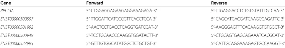
To quantify the expression of four lincRNAs (linc0949,
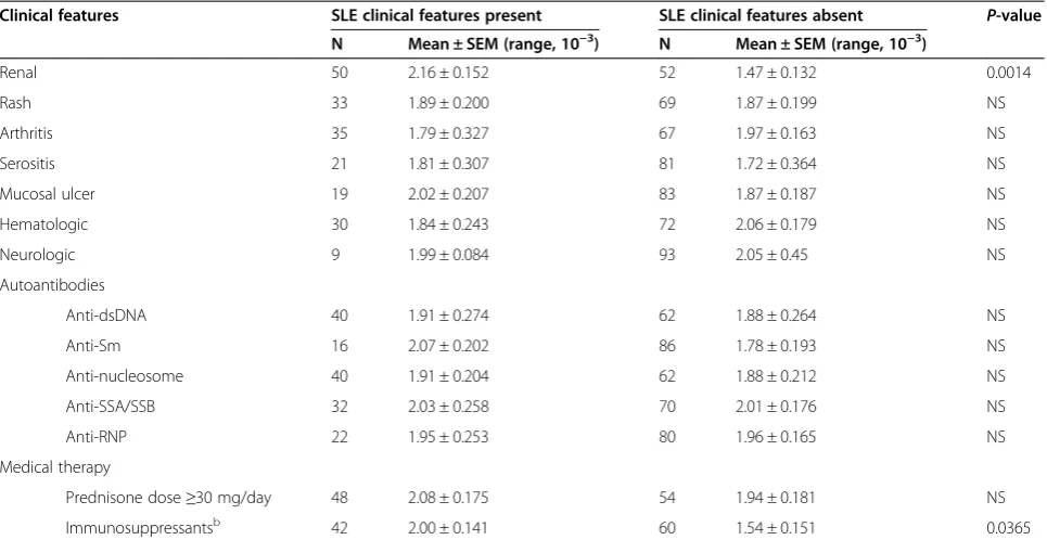
[image:3.595.57.540.111.360.2]linc0597, linc1992and linc3995), cDNA was amplified by RT-PCR with SYBR Green (SYBR Premix Ex Taq RT-PCR kit; Takara Bio). The primer sequences used for SYBR Green–based RT-PCR are given in Table 2. The ribosomal protein L13A (RPL13A) gene was used as an internal con-trol to normalize the amounts of cDNA. The SYBR Green assays were performed in duplicate using an ABI ViiA 7 Real-Time PCR System (Applied Biosystems, Foster City, CA, USA). The relative expression levels were calculated using the 2−ΔCtcomparative threshold cycle method. Table 1 Large intergenic noncoding RNAlinc0949by presence or absence of clinical features of systemic lupus erythematosusa
Clinical features SLE clinical features present SLE clinical features absent P-value
N Mean ± SEM (range, 10−3) N Mean ± SEM (range, 10−3)
Renal 50 2.16 ± 0.152 52 1.47 ± 0.132 0.0014
Rash 33 1.89 ± 0.200 69 1.87 ± 0.199 NS
Arthritis 35 1.79 ± 0.327 67 1.97 ± 0.163 NS
Serositis 21 1.81 ± 0.307 81 1.72 ± 0.364 NS
Mucosal ulcer 19 2.02 ± 0.207 83 1.87 ± 0.187 NS
Hematologic 30 1.84 ± 0.243 72 2.06 ± 0.179 NS
Neurologic 9 1.99 ± 0.084 93 2.05 ± 0.45 NS
Autoantibodies
Anti-dsDNA 40 1.91 ± 0.274 62 1.88 ± 0.264 NS
Anti-Sm 16 2.07 ± 0.202 86 1.78 ± 0.193 NS
Anti-nucleosome 40 1.91 ± 0.204 62 1.88 ± 0.212 NS
Anti-SSA/SSB 32 2.03 ± 0.258 70 2.01 ± 0.176 NS
Anti-RNP 22 1.95 ± 0.253 80 1.96 ± 0.165 NS
Medical therapy
Prednisone dose≥30 mg/day 48 2.08 ± 0.175 54 1.94 ± 0.181 NS
Immunosuppressantsb 42 2.00 ± 0.141 60 1.54 ± 0.151 0.0365
a
dsDNA, Double-stranded DNA; NS, Not significant; RNP, Ribonucleoprotein; SEM, Standard error of the mean; SLE, Systemic lupus erythematosus; Sm, Smith; SSA, Sjögren’s syndrome–related antigen A; SSB, Sjögren’s syndrome–related antigen B.b
Statistical analysis
Data were analyzed with GraphPad Prism version 5.0 software (GraphPad Software, La Jolla, CA, USA). The nonparametric Mann–WhitneyUtest was used to com-pare gene expression between two groups. The correl-ation between groups was evaluated using Spearman’s rank correlation coefficient test. The strength of the cor-relation was graded using Cohen’s criteria as follows: 0.3 to 0.5 = weak, 0.5 to 0.7 = moderate and >0.7 = strong [34].P-values (two-tailed) <0.05 were considered statisti-cally significant.
Results
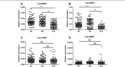
Decreasedlinc0949andlinc0597levels in patients with systemic lupus erythematosus
The expression levels of four lincRNAs (linc0949, linc0597,
linc1992andlinc3995) in PBMCs taken from 102 patients with SLE, 54 patients with RA and 76 healthy donors were measured using RT-qPCR. Patients with SLE and healthy donors did not differ significantly with respect to mean age or sex distribution (Table 3). The average disease duration of patients enrolled in our study was 4.98 years for those with SLE and 5.12 years for those with RA. In general, the patients with SLE had mild to moderate flares of disease activity and severity, with a mean SLEDAI-2K score of 7 and a mean SDI of 0.78 (Table 3).
As shown in Figure 1A, patients with SLE had signifi-cantly lower linc0949 levels than healthy donors and patients with RA (both P< 0.0001). Also, the expression of linc0597 was decreased dramatically in patients with
SLE compared with healthy donors and patients with RA (P= 0.0001 and P< 0.0001, respectively) (Figure 1B). Figure 1C and Figure 1D, however, show no significant differences inlinc1992andlinc3995levels between pa-tients with SLE and healthy donors or papa-tients with RA. These results revealed that lower expression oflinc0949 and linc0597was specific to SLE, so we selected these lincRNAs for further research.
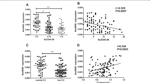
Association oflinc0949level with disease activity in patients with systemic lupus erythematosus
To investigate whether the expression of linc0949 and
[image:4.595.56.539.102.184.2]linc0597is related to SLE disease activity, we compared the relative expression levels of the lincRNAs in patients with SLE with different levels of disease activity, assessed on the basis of SLEDAI-2K score and the level of com-plement C3. In accordance with the SLEDAI-2K flare system, patients with SLE were divided into those with stable disease (SLEDAI-2K scores from 0 to 4), those with a mild flare (SLEDAI-2K scores from 5 to 10) and those with a moderate to severe disease flare (SLEDAI-2K scores >10).linc0949was significantly lower in patients with SLE who had a mild flare or a moderate to severe flare of disease than in patients without a flare (P= 0.0032 and P= 0.0004, respectively) (Figure 2A). In addition, a correlation betweenlinc0949and SLEDAI-2K score was observed in that decreased levels of linc0949coincided with increased SLEDAI-2K score in patients with SLE (r=−0.329,P= 0.0007) (Figure 2B).
Table 2 Primers used to amplify transcripts of large intergenic noncoding RNAsa
Gene Forward Reverse
RPL13A 5′-CTGGAGGAGAAGAGGAAAGAGA-3′ 5′-TTGAGGACCTCTGTGTATTTGTCAA-3′
ENST00000500597 5′-TTGGATTCATCCCGTTCACCTCCA-3′ 5′-CAGCATGACGATCAAGCGAGATTC-3′
ENST00000501992 5′-AACTCCTGACCTCAGGTGATCCAT-3′ 5′-AAGGGAGTTTCAGAAGGTGTGGCT-3′
ENST00000500949 5′-TCCTGCAACCCAAGGTGGATACTT-3′ 5′-CTGCAGTGAGCAGAAATCACGCAT-3′
ENST00000523995 5′-GTTTGTGGCATATGGCTCTGCTGT-3′ 5′-CATTGCAGGAAAGAGTGCCAAGGT-3′
a
[image:4.595.57.545.589.716.2]The four large intergenic noncoding RNAs primers are derived from the literature [26].
Table 3 Demographic dataa
Patients with SLE (n = 102) Patients with RA (n = 54) Healthy donors (n = 76)
Age (yr) 34.3 ± 1.3 (16 to 65) 38.7 ± 2.3 (20 to 65) 34.0 ± 1.2 (20 to 63)
Sex (n)
Female 93 46 70
Male 9 8 6
Disease duration (yr) 4.98 ± 0.76 (0.04 to 28) 5.12 ± 0.84 (0.5 to 20) –
ANA (%) 95.0 – –
SLEDAI-2K 6.82 ± 0.52 (1 to 15) – –
SDI 0.78 ± 0.14 (0 to 3) – –
a
C3 level is also an indicator of disease activity. Hypo-complementemia is often observed in patients with SLE with active disease. In the present research,linc0949 ex-pression was significantly decreased in patients with SLE who had a reduced level of complement C3 (<80 mg/dl) compared with those with normal levels of C3 (P< 0.0001) (Figure 2C). Further analysis revealed a positive correl-ation betweenlinc0949level and C3 level (r= 0.348,P= 0.0003) (Figure 2D). However, the level oflinc0597did not correlate with SLEDAI-2K and complement C3 level (data not shown). These results indicate that the abnormal ex-pression oflincRNA0949may be a key indicator of disease activity in patients with SLE.
Reduced expression oflinc0949in patients with systemic lupus erythematosus with organ damage
SLE is a chronic multisystem autoimmune disease that can affect virtually every organ system and may lead to significant morbidities. Assuming that lincRNAs are in-volved in tissue damage and inflammation, we investigated whetherlinc0949andlinc0597were associated with differ-ent levels of chronic and irreversible organ damage in the patients with SLE. The results revealed significantly lower
levels oflinc0949in patients with SLE with SDI scores of 1 to 2 and in those with scores >2 versus those without organ damage (P= 0.0059 and P= 0.0009, respectively) (Figure 3A). linc0597expression was not significantly re-duced in patients with organ damage (SDI ≥1) versus those who remained damage-free (data not shown).
LN is one of the most common clinical manifestations and causes of organ damage in patients with SLE, so we wanted to know whether lincRNA levels are related to LN. In our cohort, nearly 51% of patients (52 of 102 patients with SLE) had either previous or current LN (Table 1). Patients with LN had lower linc0949 expres-sion levels than those without renal manifestations (P= 0.0014) (Figure 3B). Expression levels of linc0949 were decreased in the group of patients with active LN com-pared with those without LN (P= 0.0009) (Figure 3C), whereas linc0949 levels were not significantly different in patients with inactive LN compared with patients without LN at the time of blood drawing (P= 0.0739) (Figure 3C). These data suggest thatlinc0949expression is related with cumulative organ damage in SLE and that
[image:5.595.59.536.89.346.2]linc0949may be useful in predicting long-term outcome and prognosis in patients with SLE.
Relationship of large intergenic noncoding RNA levels with clinical manifestations and medical therapies To assess the association between lincRNA levels and clinical manifestations, autoantibody profiles and med-ical treatments, linc0949and linc0597 levels were com-pared between patients with certain clinical features and
[image:6.595.58.538.90.355.2]those without certain clinical features. We identified no association between linc0949 or linc0597 expression and clinical manifestations such as rash, arthritis, serositis, mu-cosal ulcer, hematologic involvement or neurologic mani-festations (linc0949: Table 1; linc0597: data not shown). We also found that neitherlinc0949norlinc0597appeared Figure 2Association of large intergenic noncoding RNAlinc0949expression with disease activity in patients with systemic lupus erythematosus.
(A)Patients with systemic lupus erythematosus (SLE) with a moderate to severe flare of disease (Systemic Lupus Erythematosus Disease Activity Index 2000 (SLEDAI-2K) score >10) or a mild flare of disease (SLEDAI-2K score from 5 to 10) had significantly lower large intergenic noncoding RNAlinc0949expression than did those without a disease flare (SLEDAI-2K score <4) at the time of blood donation.(B)linc0949expression was negatively correlated with SLEDAI-2K score.(C)linc0949expression was significantly decreased in patients with SLE with a reduced level of complement C3 (<80 mg/dl) compared with those with normal C3levels.(D)A significantly positive correlation was observed betweenlinc0949 expression and C3 level in patients with SLE. **P< 0.01, ***P< 0.001.
[image:6.595.56.540.538.652.2]to be associated with autoantibody production, includ-ing anti-double-stranded DNA, anti-Smith antibodies, antinucleosome antibodies, anti–Sjögren’s syndrome– related antigen A and B antibodies and antiribonucleo-protein antibodies (linc0949: Table 1;linc0597: data not shown).
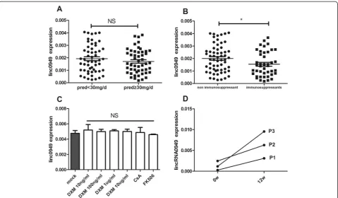
When medical therapies were considered, the expres-sion oflinc0949exhibited no significant difference in pa-tients receiving medium to high doses of prednisone (>30 mg/day) compared with patients treated with low doses of prednisone (Figure 4A). By contrast, the expres-sion oflinc0949in patients with SLE being treated with immunosuppressants (AZA, CYC, CsA, LEF, MMF, MTX and FK506) was significantly reduced compared with those not receiving immunosuppressants at the time of blood donation (P= 0.0365) (Figure 4B).
We next sought to assess whether SLE disease activity or antirheumatic drugs affect the expression oflinc0949, as it was found that some lncRNAs can be induced in response to the anti-inflammatory agent dexamethasone [35]. Thus, we treated PBMCs of two healthy donors with different concentrations of dexamethasone, CsA
and FK506. FoxP3 level was significantly reduced after treatment (Additional file 1: Figure S1), which supported the fact that dexamethasone and immunosuppressive agents worked effectivelyin vitro. As shown in Figure 4C, dexamethasone, CsA and FK506 did not affect the ex-pression of linc0949. This result relates to the effects of antirheumatic drugs on the expression of linc949, which confirms that linc0949is intrinsically underexpressed in patients with SLE.
[image:7.595.59.539.364.644.2]We next investigated whetherlinc0949was responsive to treatment and changes over time in conjunction with dis-ease activity. We chose three patients with SLE(P1,P2,P3), as described in Figure 4D, P1 and P3 had initial onset of biopsy-proved type IV LN, P2 had neuropsychological lupus, and their peripheral blood samples were collected at both the beginning of treatment and after 12 weeks of treatment. P1 and P3 used high-dose prednisone (1 mg/kg per day) plus mycophenolate mofetil (1.5-2 g/day), whereas P2 used repeated pulses of glucocorticoid (500 mg intra-venous methylprednisolone per day) for three days and then prednisone (1 mg/kg per day) plus monthly pulse of cyclophosphamide (0.8 g/month). After treatment, all of
Figure 4Association of large intergenic noncoding RNAlinc0949level with medical therapies in patients with systemic lupus erythematosus.
the three patients achieved clinical remission, with the urinary protein level dropping to less than 0.5 g/24 hour in P1 and P3; and with cerebrospinal fluid inspection and head MRI restoring to normal in P2. SLEDAI-2K score of the three patients reduced to a stable level (Additional file 1: Figure S1). In concordance with the clinical im-provement, linc0949expression in these three patients also significantly increased (Figure 4D).
Abnormal regulation of lincRNAs during innate activation of peripheral blood mononuclear cells in patients with systemic lupus erythematosus
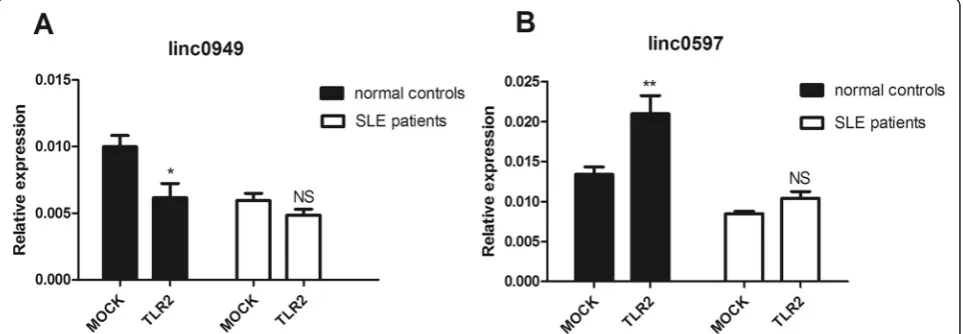
To investigate the different response to innate immunity oflinc0949 andlinc0597 in healthy donors and patients with SLE, we stimulated PBMCs from healthy donors and patients with SLE with TLR2 ligands. We chose TLR2 ligands because it was reported thatlinc0949and
linc0597 were regulated in THP-1 macrophages follow-ing Pam3CSK4 stimulation [26] and TLR2 was required for the production of prototypical lupus autoantibodies and the development of renal disease in murine lupus [36,37]. To prepare cell samples, PBMCs obtained from 5 healthy donors and 5 patients with SLE were stimu-lated with Pam3CSK4 for 4 hours. Then we performed RT-qPCR to identify changes in lincRNA expression. In PBMCs of healthy controls, linc0949 was suppressed (Figure 5A), whilelinc0597was increased (Figure 5B) after treatment with Pam3CSK4. But in patients with SLE,
linc0949 and linc0597 could not response to the stimuli compared with healthy donors (Figure 5A and B). These
results demonstrated that lincRNAs were indeed involved in the complex regulatory network of innate immunity.
Discussion
In recent years, an increasing body of evidence has shown that lncRNAs play major biological roles in em-bryogenesis, stem cell biology and cellular development and show developmental and tissue-specific expression patterns [11,38,39]. Studies have also suggested that abnormal expression of lncRNAs might be associated with numerous diseases, indicating that these RNAs may open a new avenue for diagnostic and therapeutic targets by recognition of their roles in human disease.
[image:8.595.58.540.467.634.2]In the present study, we detected four lincRNAs (linc0949, linc0597, linc1992 and linc3995) and investi-gated the association between their expression levels and specific clinical features of SLE. Two of these lincRNAs (linc0949 and linc0597) were significantly decreased in patients with SLE compared with healthy donors and disease controls.linc0949was associated with disease ac-tivity, as assessed using the SLEDAI-2K score and C3 level in patients with SLE. Moreover,linc0949expression was reduced in patients with SLE with ongoing or cu-mulative organ damage, as assessed based on SDI score or the presence of active LN. linc0949 expression does not participate in clinical manifestations other than LN, which demonstrates that it has very good detection speci-ficity for LN. Lower levels oflinc0949may thus be helpful to identify patients with SLE who have active and severe disease. To evaluate the effect of antirheumatic drugs on
the expression oflinc949, we usedin vitrostudies to test whether the addition of dexamethasone, CsA or FK506 to cultured PBMCs would affect the expression oflinc0949. As shown in Figure 4C, these antirheumatic drugs did not affect the expression oflinc0949in PBMCs, which con-firmed thatlinc0949was intrinsically underexpressed in patients with SLE.linc0949expression of three patients with severe disease flares significantly increased after treatment (Figure 4D), indicating thatlinc0949might be responsive to treatment and might change in conjunc-tion with disease activity and severity and suggesting thatlinc0949might be used to monitor disease progres-sion and guide therapy.
Over the past several decades, tremendous enthusiasm and efforts have been devoted to biomarkers for SLE because the diagnosis of SLE requires a combination of clinical manifestations and biomarkers and no single test is sufficiently sensitive and specific to be diagnostic. The traditional antibodies fail to identify the pathogenic pro-cesses, organ damage and biological responses to a thera-peutic intervention. Many groups, including the members of our laboratory, have found a set of potential biomarkers for SLE. For example, interferon (IFN)-induced genes and IFN-inducible chemokines may serve as new biomarkers for active and severe disease in patients with SLE [40,41]. Some limitations of these biomarkers are revealed grad-ually, however. Several studies have shown that overex-pressed transcripts of the type I IFN pathway are also identified in patients with myositis, RA, Sjögren’s syn-drome and scleroderma [42-44], so an IFN signature or chemokine is not sufficiently specific. In two longitudinal studies to date, researchers have reported conflicting re-sults on the correlations between type I IFN gene signa-ture score and diseases activity [45,46]. There is an urgent need for SLE biomarkers that can help enhance compre-hension of the mechanisms of diseases or effects of ther-apies by relating the changes of molecular and cellular pathways to disease status or clinical responses. In our present study, we demonstrate that lower expression of
linc0949is specific for SLE and that it is helpful in identi-fying disease activity, monitoring disease progression and guiding therapy. However,linc0949needs to be further in-vestigated in large-scale multicenter trials.
On the basis of our present observations, we believe that linc0949 could be a potentially readily accessible biomarker useful for diagnosing SLE. As a novel bio-marker, lincRNAs have the following characteristics. First, lincRNAs display a wide range of stabilities in the samples comparable to those of mRNAs of protein-coding genes [47]. Second, they show a greater tissue specificity com-pared with protein-coding mRNAs and miRNAs, which are frequently expressed in multiple tissues, and they show highly increased or decreased expression levels in disease [15]. In addition, lincRNAs are also detectible in body
fluids such as plasma and urine [48-50], diagnostic sam-ples of which are easy to collect using noninvasive methods. Moreover, detection of the lincRNAs is simple, inexpensive and has high throughput, making it a suitable approach to gaining an overview of disease activity and se-verity in patients with SLE. These features make lincRNAs very suitable as biomarkers, and many studies have been published on this matter in recent years, both in cancer and in other human diseases such as cardiovascular dis-eases [50] and neurological disorders [51].
Most lncRNAs described to date have been found to be related to transcriptional regulation or mRNA pro-cessing, characteristics that they share with microRNAs. However, unlike microRNAs, lncRNAs show a greater complexity of their functions and have a wider spectrum of biological contexts, such as epigenetic regulation, enhancer-like function and RNA splicing, editing and export [52]. In our ongoing experiments, we found that
linc0949 and linc0597 could be induced by TLR2 in PBMCs of healthy donors, but they did not respond to the stimuli in patients with SLE as compared with healthy donors (Figure 5). These results validate that lincRNAs were indeed involved in the complex regulatory network of innate immunity. We hypothesized that the regulation defect of linc0949 and linc0597 could contribute to the pathogenesis of SLE and that lincRNAs may provide potential novel strategies for therapeutic intervention, although their function and mechanism of action need further exploration.
We have suggested the abnormal expression oflinc0949 in patients with SLE, as well as the association of lincRNA level with disease activity and organ damage; however, in this study, we did not conduct a functional study of this lincRNA, and the underlying mechanism needs further in-vestigation. We did not detect the expression oflinc0949 released in the local target tissues and in specific cell sub-sets in PBMCs. Future studies are needed to investigate the lincRNA expression level in specific organ and cell types as well.
Conclusions
We found that the expression of two lincRNAs was dra-matically reduced in patients with SLE and that the de-creasing level of linc0949 was correlated with disease activity, degree of organ damage and medical therapies in patients with SLE. linc0949 may serve as a potential biomarker for diagnosis, disease activity and therapeutic interventions in patients with SLE.
Additional file
Abbreviations
ACR:American College of Rheumatology; ANA: Antinuclear antibody; AZA: Azathioprine; C3: Complement component 3; CsA: Cyclosporine A; CYC: Cyclophosphamide; dsDNA: Double-stranded DNA; DXM: Dexamethasone; EDTA: Ethylenediaminetetraacetic acid; FBS: Fetal bovine serum;
FK506: Tacrolimus;GAS5: Growth arrest–specific 5; hpf: High-power field; IFN: Interferon; IL: Interleukin; LEF: Leflunomide;linc0597:ENST00000500597; linc0949:ENST00000500949;linc1992:ENST00000501992;
linc3995:ENST00000523995; lincRNA: Large intergenic noncoding RNA; LN: Lupus nephritis; lncRNA: Long noncoding RNA; miRNA: microRNA; MMF: Mycophenolate mofetil; MTX: Methotrexate; NS: Not significant; nt: Nucleotide; PBMC: Peripheral blood mononuclear cell; RA: Rheumatoid arthritis; RNP: Ribonucleoprotein;RPL13A: Ribosomal protein L13A gene; RT-qPCR: Real-time quantitative polymerase chain reaction; SDI: Systemic Lupus International Collaborating Clinics/American College of Rheumatology Damage Index; SEM: Standard error of the mean; SLE: Systemic lupus erythematosus; SLEDAI-2K: Systemic Lupus Erythematosus Disease Activity Index 2000; SLICC: Systemic Lupus International Collaborating Clinics; Sm: Smith; SSA: Sjögren’s syndrome–related antigen A; SSB: Sjögren’s syndrome–related antigen B; TLR: Toll-like receptor; TNF: Tumor necrosis factor; SEM: Standard error of the mean.
Competing interests
The authors declare that they have no competing interests.
Authors’contributions
YFW, YJT and NS conceived of and designed the experiments and analyzed the data. YFW, FFZ, YJT and NS wrote the paper. YFW, FFZ, JYM, XYZ, LLW, BQ, SWX and SLC performed the experiments, collected blood samples and contributed reagents, materials and analytic tools. JYM, XYZ, LLW, BQ, SWX and SLC helped to draft the manuscript. All authors read and approved the final manuscript.
Acknowledgments
This work was supported by the National Basic Research Program of China (973 Program) (2014CB541901 and 2014CB541902), the National Natural Science Foundation of China (81025016, 81230072, 31370880 and No. 81421001), the State Key Laboratory of Oncogenes and Related Genes (91-14-05), the Key Research Program of the Chinese Academy of Sciences (KJZD-EW-L01-3) and the Program of the Shanghai Commission of Science and Technology (12ZR1435900, 12JC1406000 and 12431900703). The funders had no role in study design, data collection and analysis, the decision to publish or the preparation of the manuscript. The authors extend special thanks to Yuting Qin and Ting La of the Chinese Academy of Sciences for providing technological guidance during the course of this study.
Author details
1Shanghai Institute of Rheumatology, Department of Rheumatology, Renji Hospital, School of Medicine, Shanghai Jiao Tong University, Shan Dong Middle Road, Shanghai 200001, People’s Republic of China.2Institute of Health Sciences, Shanghai Jiao Tong University School of Medicine (SJTUSM) and Shanghai Institutes for Biological Sciences (SIBS), Chinese Academy of Sciences (CAS), Yue Yang Road, Shanghai 200031, People’s Republic of China. 3Division of Rheumatology and the Center for Autoimmune Genomics and Etiology (CAGE), Cincinnati Children’s Hospital Medical Center, 3333 Burnet Avenue, Cincinnati, OH 45229, USA.
Received: 29 November 2014 Accepted: 20 April 2015
References
1. Rahman A, Isenberg DA. Systemic lupus erythematosus. N Engl J Med. 2008;358:929–39.
2. Baccala R, Hoebe K, Kono DH, Beutler B, Theofilopoulos AN. TLR-dependent and TLR-independent pathways of type I interferon induction in systemic autoimmunity. Nat Med. 2007;13:543–51.
3. Marshak-Rothstein A. Toll-like receptors in systemic autoimmune disease. Nat Rev Immunol. 2006;6:823–35.
4. Wapinski O, Chang HY. Long noncoding RNAs and human disease. Trends Cell Biol. 2011;21:354–61. A published erratum appears in Trends Cell Biol. 2011;21:561.
5. Shen N, Liang D, Tang Y, de Vries N, Tak PP. MicroRNAs—novel regulators of systemic lupus erythematosus pathogenesis. Nat Rev Rheumatol. 2012;8:701–9.
6. Pauley KM, Cha S, Chan EK. MicroRNA in autoimmunity and autoimmune diseases. J Autoimmun. 2009;32:189–94.
7. Chafin CB, Reilly CM. MicroRNAs implicated in the immunopathogenesis of lupus nephritis. Clin Dev Immunol. 2013;2013:430239.
8. Wang H, Peng W, Ouyang X, Li W, Dai Y. Circulating microRNAs as candidate biomarkers in patients with systemic lupus erythematosus. Transl Res. 2012;160:198–206.
9. Wang G, Tam LS, Kwan BC, Li EK, Chow KM, Luk CC, et al. Expression of miR-146a and miR-155 in the urinary sediment of systemic lupus erythematosus. Clin Rheumatol. 2012;31:435–40.
10. Tang Y, Luo X, Cui H, Ni X, Yuan M, Guo Y, et al. MicroRNA-146a contributes to abnormal activation of the type I interferon pathway in human lupus by targeting the key signaling proteins. Arthritis Rheum. 2009;60:1065–75. 11. Guttman M, Amit I, Garber M, French C, Lin MF, Feldser D, et al. Chromatin
signature reveals over a thousand highly conserved large non-coding RNAs in mammals. Nature. 2009;458:223–7.
12. Zaratiegui M, Irvine DV, Martienssen RA. Noncoding RNAs and gene silencing. Cell. 2007;128:763–76.
13. Ponting CP, Oliver PL, Reik W. Evolution and functions of long noncoding RNAs. Cell. 2009;136:629–41.
14. Gibb EA, Brown CJ, Lam WL. The functional role of long non-coding RNA in human carcinomas. Mol Cancer. 2011;10:38.
15. Gupta RA, Shah N, Wang KC, Kim J, Horlings HM, Wong DJ, et al. Long non-coding RNAHOTAIRreprograms chromatin state to promote cancer metastasis. Nature. 2010;464:1071–6.
16. Haywood MEK, Rose SJ, Horswell S, Lees MJ, Fu G, Walport MJ, et al. Overlapping BXSB congenic intervals, in combination with microarray gene expression, reveal novel lupus candidate genes. Genes Immun. 2006;7:250–63. 17. Wang P, Xue Y, Han Y, Lin L, Wu C, Xu S, et al. The STAT3-binding long
noncoding RNA lnc-DC controls human dendritic cell differentiation. Science. 2014;344:310–3.
18. Carpenter S, Aiello D, Atianand MK, Ricci EP, Gandhi P, Hall LL, et al. A long noncoding RNA mediates both activation and repression of immune response genes. Science. 2013;341:789–92.
19. Hu G, Tang Q, Sharma S, Yu F, Escobar TM, Muljo SA, et al. Expression and regulation of intergenic long noncoding RNAs during T cell development and differentiation. Nat Immunol. 2013;14:1190–8.
20. Song J, Kim D, Han J, Kim Y, Lee M, Jin EJ. PBMC and exosome-derived Hotair is a critical regulator and potent marker for rheumatoid arthritis. Clin Exp Med. 2015;15:121–6.
21. Stuhlmüller B, Kunisch E, Franz J, Martinez-Gamboa L, Hernandez MM, Pruss A, et al. Detection of oncofetal H19 RNA in rheumatoid arthritis synovial tissue. Am J Pathol. 2003;163:901–11. A published erratum appears in Am J Pathol. 2003;163:2645.
22. Ban Y, Hirano T. Association studies of theSAS-ZFAT,IL-23R,IFIH1andFOXP3 genes in autoimmune thyroid disease. Expert Rev Endocrinol Metab. 2009;4:325–31.
23. Johanneson B, Lima G, von Salomé J, Alarcón-Segovia D, Alarcón-Riquelme ME. the Collaborative Group on the Genetics of SLE, the BIOMED II Collaboration on the Genetics of SLE and Sjögren’s Syndrome. A major susceptibility locus for systemic lupus erythematosus maps to chromosome 1q31. Am J Hum Genet. 2002;71:1060–71.
24. Tsao BP. The genetics of human systemic lupus erythematosus. Trends Immunol. 2003;24:595–602.
25. Tsao BP. Update on human systemic lupus erythematosus genetics. Curr Opin Rheumatol. 2004;16:513–21.
26. Li Z, Chao TC, Chang KY, Lin N, Patil VS, Shimizu C, et al. The long noncoding RNATHRILregulates TNFαexpression through its interaction with hnRNPL. Proc Natl Acad Sci U S A. 2014;111:1002–7.
27. Linker-Israeli M, Deans R, Wallace D, Prehn J, Ozeri-Chen T, Klinenberg J. Elevated levels of endogenous IL-6 in systemic lupus erythematosus: a putative role in pathogenesis. J Immunol. 1991;147:117–23.
28. Nagafuchi H, Suzuki N, Mizushima Y, Sakane T. Constitutive expression of IL-6 receptors and their role in the excessive B cell function in patients with systemic lupus erythematosus. J Immunol. 1993;151:6525–34.
30. Tan EM, Cohen AS, Fries JF, Masi AT, McShane DJ, Rothfield NF, et al. The 1982 revised criteria for the classification of systemic lupus erythematosus. Arthritis Rheum. 1982;25:1271–7.
31. Aletaha D, Neogi T, Silman AJ, Funovits J, Felson DT, Bingham 3rd CO, et al. 2010 rheumatoid arthritis classification criteria: an American College of Rheumatology/European League Against Rheumatism collaborative initiative. Arthritis Rheum. 2010;62:2569–81.
32. Gladman DD, Ibañez D, Urowitz MB. Systemic Lupus Erythematosus Disease Activity Index 2000. J Rheumatol. 2002;29:288–91.
33. Gladman D, Ginzler E, Goldsmith C, Fortin P, Liang M, Urowitz M, et al. The development and initial validation of the Systemic Lupus International Collaborating Clinics/American College of Rheumatology damage index for systemic lupus erythematosus. Arthritis Rheum. 1996;39:363–9.
34. Cohen J. Statistical power analysis for the behavioral sciences. 2nd ed. Hillsdale, NJ: Lawrence Erlbaum Associates; 1988.
35. Rapicavoli NA, Qu K, Zhang J, Mikhail M, Laberge RM, Chang HY. A mammalian pseudogene lncRNA at the interface of inflammation and anti-inflammatory therapeutics. Elife. 2013;2:e00762.
36. Lartigue A, Colliou N, Calbo S, François A, Jacquot S, Arnoult C, et al. Critical role of TLR2 and TLR4 in autoantibody production and glomerulonephritis inlprmutation-induced mouse lupus. J Immunol. 2009;183:6207–16. 37. Urbonaviciute V, Starke C, Pirschel W, Pohle S, Frey S, Daniel C, et al. Toll-like
receptor 2 is required for autoantibody production and development of renal disease in pristane-induced lupus. Arthritis Rheum. 2013;65:1612–23. 38. Rinn JL, Kertesz M, Wang JK, Squazzo SL, Xu X, Brugmann SA, et al.
Functional demarcation of active and silent chromatin domains in human HOXloci by noncoding RNAs. Cell. 2007;129:1311–23.
39. Pauli A, Rinn JL, Schier AF. Non-coding RNAs as regulators of embryogenesis. Nat Rev Genet. 2011;12:136–49.
40. Feng X, Wu H, Grossman JM, Hanvivadhanakul P, FitzGerald JD, Park GS, et al. Association of increased interferon-inducible gene expression with disease activity and lupus nephritis in patients with systemic lupus erythematosus. Arthritis Rheum. 2006;54:2951–62.
41. Fu Q, Chen X, Cui H, Guo Y, Chen J, Shen N, et al. Association of elevated transcript levels of interferon-inducible chemokines with disease activity and organ damage in systemic lupus erythematosus patients. Arthritis Res Ther. 2008;10:R112.
42. Walsh RJ, Kong SW, Yao Y, Jallal B, Kiener PA, Pinkus JL, et al. Type I interferon-inducible gene expression in blood is present and reflects disease activity in dermatomyositis and polymyositis. Arthritis Rheum. 2007;56:3784–92. 43. van der Pouw Kraan TC, Wijbrandts CA, van Baarsen LG, Voskuyl AE,
Rustenburg F, Baggen JM, et al. Rheumatoid arthritis subtypes identified by genomic profiling of peripheral blood cells: assignment of a type I interferon signature in a subpopulation of patients. Ann Rheum Dis. 2007;66:1008–14.
44. Higgs BW, Liu Z, White B, Zhu W, White WI, Morehouse C, et al. Patients with systemic lupus erythematosus, myositis, rheumatoid arthritis and scleroderma share activation of a common type I interferon pathway. Ann Rheum Dis. 2011;70:2029–36.
45. Petri M, Singh S, Tesfasyone H, Dedrick R, Fry K, Lal P, et al. Longitudinal expression of type I interferon responsive genes in systemic lupus erythematosus. Lupus. 2009;18:980–9.
46. Landolt-Marticorena C, Bonventi G, Lubovich A, Ferguson C, Unnithan T, Su J, et al. Lack of association between the interferon-αsignature and longitudinal changes in disease activity in systemic lupus erythematosus. Ann Rheum Dis. 2009;68:1440–6.
47. Lee JT. Epigenetic regulation by long noncoding RNAs. Science. 2012;338:1435–9.
48. Spizzo R, Almeida MI, Colombatti A, Calin GA. Long non-coding RNAs and cancer: a new frontier of translational research? Oncogene. 2012;31:4577–87. 49. Saad F. UPM3: review of a new molecular diagnostic urine test for prostate
cancer. Can J Urol. 2005;12:40–3. discussion 99–100.
50. Kumarswamy R, Bauters C, Volkmann I, Maury F, Fetisch J, Holzmann A, et al. Circulating long noncoding RNA, LIPCAR, predicts survival in patients with heart failure. Circ Res. 2014;114:1569–75.
51. Guennewig B, Cooper AA. The central role of noncoding RNA in the brain. Int Rev Neurobiol. 2014;116:153–94.
52. Van Roosbroeck K, Pollet J, Calin GA. miRNAs and long noncoding RNAs as biomarkers in human diseases. Expert Rev Mol Diagn. 2013;13:183–204.
Submit your next manuscript to BioMed Central and take full advantage of:
• Convenient online submission
• Thorough peer review
• No space constraints or color figure charges
• Immediate publication on acceptance
• Inclusion in PubMed, CAS, Scopus and Google Scholar
• Research which is freely available for redistribution