REVIEW
Harnessing developmental processes for vascular engineering
and regeneration
Kyung Min Park1and Sharon Gerecht1,2,*
ABSTRACT
The formation of vasculature is essential for tissue maintenance and regeneration. During development, the vasculature forms via the dual processes of vasculogenesis and angiogenesis, and is regulated at multiple levels: from transcriptional hierarchies and protein interactions to inputs from the extracellular environment. Understanding how vascular formation is coordinatedin vivocan offer valuable insights into engineering approaches for therapeutic vascularization and angiogenesis, whether by creating new vasculature in vitro or by stimulating neovascularizationin vivo. In this Review, we will discuss how the process of vascular development can be used to guide approaches to engineering vasculature. Specifically, we will focus on some of the recently reported approaches to stimulate therapeutic angiogenesis by recreating the embryonic vascular microenvironment using biomaterials for vascular engineering and regeneration.
KEY WORDS: Angiogenesis, Biomaterials, Stem cells, Tissue engineering, Vasculogenesis
Introduction
The vasculature plays a crucial role in delivering oxygen and nutrients to the body tissues and in removing the waste matter generated by those tissues. Vascular diseases that affect the circulatory system pose an enormous public health problem. In 2010, the global economic burden of vascular disease was estimated to cost US$863 billion and it is estimated to rise to US$1044 billion by 2030 (Bloom et al., 2011). Peripheral artery, cardiovascular, carotid artery, renal artery and venous diseases can all result in serious illness, disability and often death. Despite advances in pharmaceutical interventions and surgical therapies (Kappetein et al., 2013; Mohr et al., 2013; Thielmann et al., 2013), the treatment of vascular diseases using therapeutic vascularization strategies remains very challenging.
With advances in materials science, new approaches to treat vascular disorders have emerged that involve the induction of new vasculaturesin vivo.This can be achieved by means of material-tissue interface (see Glossary, Box 1) or by the targeted delivery of therapeutic agents, such as growth factors (GFs), cytokines or cells, via engineered materials. In addition, materials have been employed to create tissue-engineered vasculature grafts (see Glossary, Box 1) or to generate whole-engineered vasculature beds (see Glossary, Box 1) for physiological studies and regenerative applications. One approach to building scaffolds for vascular applications is to mimic the microenvironment that exists during normal vascular developmentin vivo. In doing this, numerous physical, chemical and biological parameters must be considered, such as the presence
of specific GFs/cytokines and adhesion molecules, the composition of the extracellular matrix (ECM), whether matrix remodeling occurs and the specific oxygen tension. A better understanding of how these parameters affect vascular formationin vivowill therefore enable us to develop more effective methods to engineer therapies to treat vascular disorders.
The current state-of-the-art technology in engineering and cellular therapies allow an unprecedented level of control over the patient specificity of vascular therapeutics (Sun et al., 2011; Chen et al., 2012; Cuchiara et al., 2012; Kusuma et al., 2013). Tailor-made biomaterials and patient-specific stem cells are only part of the promise held by regenerative medicine, but whereas advanced engineering approaches can provide effective therapies and implantable vasculatures, obstacles
Box 1. Glossary
Acellular matrix.Matrices, either natural or synthetic, that can stimulate tissue repair without the use of cells.
Chemical crosslinking reactions.A mechanism of hydrophilic network formation achieved via chemical reactions, such as photocrosslinking, Michael-type addition, Diels-Alder reaction, click chemistry, imine formation and enzyme-mediated reaction.
Engineered vasculature bed.Vascular network structure formed from cells encapsulated within a three-dimensional (3D) matrix. Engineered vascular beds can be used to study fundamentals in vascular biology or for regenerative medicine applications if using human cells. Engineered vasculature is a type of tissue-engineered vasculature.
Hydrogel matrix.A 3D hydrophilic network of polymers that is swollen with water. They are classified into physical and chemical hydrogels based on the crosslinking chemistry that constructs 3D network formation. Material-tissue interface. Following implantation of biomaterials, a materials-tissue interface is immediately created through nonspecific protein absorption of blood and tissue fluid onto the surface of the biomaterial.
Microfabrication.A technology used to create microscaled devices, which has been used in many research fields.
Microparticle.A microsphere made of synthetic or natural biomaterials for the encapsulation and delivery of therapeutic agents. Poly(lactic-co-glycolic) acid (PLGA) co-polymer microparticles are approved as therapeutic devices by the Food and Drug Administration (FDA), owing to their biodegradability and biocompatibility.
Physical crosslinking reaction.A mechanism of hydrophilic network formation achieved via physical interaction, such as ionic and hydrophobic interaction, stereocomplexation and host-guest reactions. Physicochemical properties.Refers to both the physical and chemical properties of a given substance.
Sequential delivery. Successive release of multiple factors in a temporally controlled manner from the biomaterial.
Sustained release.The slow and steady release of factors facilitated by specific physical and/or chemical properties of the biomaterial. Tissue-engineered vascular graft.An artificial vascular graft that can be used as prosthetic blood vessels for vascular engineering. Types of grafts include extracellular matrix (ECM)-based vascular grafts, cell sheets with/without scaffold, decellularized tissue and biodegradable scaffold-based vascular grafts.
1
Department of Chemical and Biomolecular Engineering, Johns Hopkins Physical Sciences-Oncology Center, and The Institute for NanoBioTechnology, Johns Hopkins University, Baltimore, MD 21218, USA.2Department of Materials Science and Engineering, Johns Hopkins University, Baltimore, MD 21208, USA.
*Author for correspondence (gerecht@jhu.edu)
DEVEL
O
for translational use still remain, such as long-term efficacy and functionality. In this Review, we discuss how the fundamental principles of vascular development are currently being applied to improve approaches for vascular engineering and regeneration. Specifically, we focus on some of the recently reported techniques for stimulating new blood vessel formationin vivo and for creating engineered vasculaturein vitrousing a range of different cell types and techniques. As our understanding of the mechanisms that drive vascular development increases, our approaches to vascular engineering become more advanced, creating new and exciting opportunities for tissue regeneration and vascular therapies.
Vascular development and engineering parameters
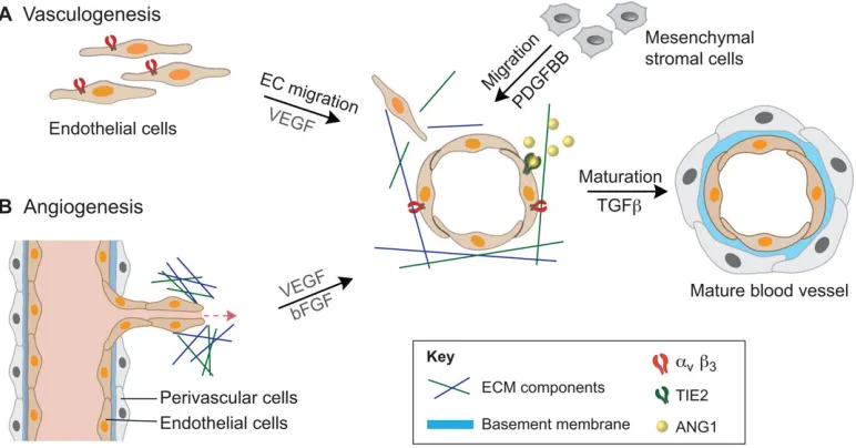
The cells that make up the adult vasculature originate from the mesoderm, one of three germ layers derived from the embryonic epiblast by the process of gastrulation. After formation of the mesoderm, blood islands formed by angioblasts and hematopoietic progenitor cells give rise to endothelial cells (ECs), which coalesce to form lumen structures within the ECM, resulting in a small network of blood vessels called the primary capillary plexus (Risau, 1997). The primary capillary plexus forms the basis for further vessel assembly, as well as for expansion of the vasculature network. In this way, the developing vasculature can be described as forming via two distinct mechanisms: vasculogenesis, whereby vessels form de novo; and angiogenesis, whereby new vessels sprout off from pre-existing vessels (see Fig. 1) (Eilken and Adams, 2010; Geudens and Gerhardt, 2011). The process of vasculogenesis occurs through three steps: first, the induction of hemangioblasts and angioblasts and their commitment to vasculogenesis; second, the assembly of the developing vessels; and third, the induction of the transition from vasculogenesis to angiogenesis. The angiogenic process, which serves to expand the vascular network, also occurs via three steps. Initially, the pre-existing, quiescent vessel is activated and a specific EC, called the tip cell, is specified to lead the
sprouting process. Second, cells neighboring the tip cells are specified as stalk cells, which undergo proliferation, elongation and lumenization in order to shape the nascent vessel. The process of EC activation and tip/stalk cell selection is controlled to a large extent by crosstalk between vascular endothelial growth factors (VEGF) and Notch signaling, and the entire process of activation, migration and vessel elongation involves considerable cell-cell rearrangements, basement membrane decomposition and remodeling (for a detailed review, see Geudens and Gerhardt, 2011). The final stages of angiogenesis involve the recruitment of cells from the surrounding stroma, either perivascular cells that are destined for microvasculature or smooth muscle cells (SMCs) that are destined for larger vessels. This recruitment is stimulated by the secretion of platelet-derived growth factor (PDGF) from the nearby ECs. These cells then differentiate to form a single layer of cells, called mural cells, around the EC vessel. Fusion of the newly formed vessel with neighboring vessels then allows perfusion of blood and stabilization of the developing vascular network.
[image:2.612.114.500.445.647.2]Both vasculogenesis and angiogenesis are driven by intrinsic genetic regulatory networks (reviewed by Carmeliet and Jain, 2011) and the specific physicochemical properties (see Glossary, Box 1) of their immediate microenvironment. These include cell-cell interactions (Stratman and Davis, 2012), cell interactions with the surrounding ECM through specific cell-adhesion molecules (Estrach et al., 2011) and cell signaling via secreted or ECM-sequestered GFs or cytokines (Stratman et al., 2011). Other physicochemical parameters include proteolytic matrix remodeling and matrix stiffness, which affect vascular morphogenesis and network formation (Hanjaya-Putra et al., 2011; Busnadiego et al., 2013; Turturro et al., 2013). Oxygen availability has also been shown to play a crucial role in early vascular development (Imanirad et al., 2014). This range of physicochemical properties that regulate vascular developmentin vivo represents the many variables that must be considered for vascular engineering and regeneration approaches.
Fig. 1. Schematic representation of vasculogenesis and angiogenesis.(A) During vasculogenesis, the mesoderm-derived endothelial cells (ECs) migrate and form lumen structures, a process mediated via the vascular endothelial growth factor (VEGF)/VEGF receptor (VEGFR) signaling pathway. Platelet-derived growth factor (PDGF) secretion by ECs recruits nearby mesenchymal stem cells (MSCs), which in turn release ANG1 (angiopoietin 1). Release of ANG1 stimulates mural coverage and basement membrane deposition, which in turn promotes vessel tightness via the ANG1/TIE2 (endothelial-specific receptor tyrosine kinase; TEK) signaling system. Transforming growth factorβ(TGFβ) is activated through the interaction of ECs and MSCs, which induces mural cell differentiation and inhibits EC proliferation. Finally, the vessel is covered with perivascular cells. (B) Angiogenesis occurs when a quiescent vessel of the existing vasculature undergoes sprouting, elongation and lumen formation to create new vessels. Pro-angiogenic factors, such as basic fibroblast growth factor (bFGF) and VEGF, stimulate initial EC sprouting before perivascular cells stabilize the nascent vessel to establish a perfusablede novoblood vessel.αv,β3, a type of integrin; ECM, extracellular matrix; PDGFBB, platelet-derived growth factor subunit B homodimer;
DEVEL
O
Cells sources for generating vasculature
Engineering vasculature in vitrorequires multiple cell sources to generate both the lining of the vessels and the supporting cellular network. Whereas the cell types that form the vasculaturein vivoare limited to ECs and mural cells, approaches to generate de novo
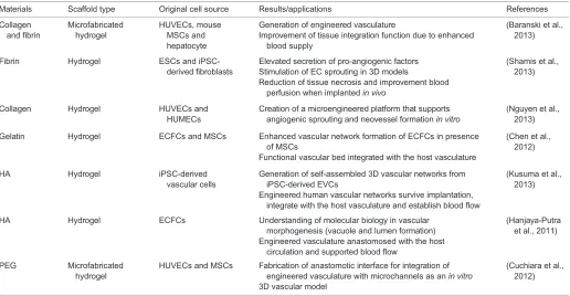
human vasculature have used a much wider variety of cell types (see Table 1). These include primary ECs (Baranski et al., 2013) and endothelial progenitor cells (EPCs) (Hanjaya-Putra et al., 2011, 2012) as cell sources for the vessel inner lining, as well as mesenchymal stem cells (MSCs) (Stratman and Davis, 2012) and pluripotent stem cells (PSCs) (Kusuma et al., 2013; Samuel et al., 2013), which have a wider lineage potential. Primary ECs can be derived from various tissues, including umbilical cord blood vessels, the dermal vasculature, the lungs and the pulmonary artery. These primary ECs have been widely used in vascular engineering for many years because they already possess the functional attributes of ECs and thus require little manipulation prior to transplantation. In the past decade, EPCs, such as endothelial colony-forming cells (ECFCs) (Critser and Yoder, 2010) and human outgrowth ECs (Lin et al., 2000; Glynn and Hinds, 2013), have also been investigated for vascular tissue engineering. These cells circulate in the bloodstream and can be differentiated into ECs, contributing to vasculogenesis and angiogenesis (Asahara et al., 1997; Richardson and Yoder, 2011). To this end, several researchers have recently isolated and expanded EPCs from peripheral blood monocytes in much higher numbers than those present in the systemic circulation (Colombo et al., 2013; Yuan et al., 2013). Despite the attractiveness of both primary ECs and EPCs for vascular tissue engineering, a crucial limitation of these cells is the number that can be harvested at any one time and the limited expansion capabilitiesin vitro.
More recently, PSCs, such as embryonic stem cells (ESCs) and induced pluripotent stem cells (iPSCs), have attracted substantial
attention as a cell source for vascular engineering and for regeneration (reviewed by Leeper et al., 2010). These cells have the ability to renew indefinitely and to differentiate into any cell type, including ECs, SMCs and perivascular cells (Vazao et al., 2011; Dar et al., 2012; Wanjare et al., 2013). In many cases, the approach for directing the differentiation of PSCs into vascular cells in vitro recapitulates the in vivo developmental process (Kraehenbuehl et al., 2011b). These cells are used for generating engineered vasculatures to treat vascular disease in stem cell-based vascular regeneration, which is an important milestone towards clinical applications (Samuel et al., 2013). For example, using ECs derived from human PSCs, Levenberg and colleagues were able to construct a three-dimensional (3D) vascularized human cardiac tissue with enhanced stabilization and functionality compared with non-vascularized constructs (Caspi et al., 2007). In another example, Shusta and colleagues recently demonstrated for the first time the derivation of brain ECs from PSCs, thus enabling research into the development and function of the human blood-brain barrier (Lippmann et al., 2012). Human iPSCs (hiPSCs), which can be generated by reprogramming somatic cells to pluripotency (Takahashi et al., 2007), can also undergo directed differentiation into vasculature cells (Taura et al., 2009; Feng et al., 2010). hiPSCs open up possibilities for personalized medicine, as each patient could in theory have their own hiPSC cells created and used for vascular therapy (Takahashi and Yamanaka, 2013).
[image:3.612.49.565.442.710.2]Creating a stable and functional vasculature requires in addition to ECs a second cell type, the perivascular cell, to support the primary EC capillary plexus, which allows the formation of a mature vascular structure. MSCs have been shown to differentiate into SMCs and perivascular cells in the presence of transforming growth factorβ(TGFβ) (Hirschi et al., 1998; Kinner et al., 2002) as in the in vivo vascular developmental process. Co-culture of mesenchymal stem cells or progenitor cells with ECs or EPCs
Table 1. Hydrogel materials and cell sources for engineered vasculature
Materials Scaffold type Original cell source Results/applications References
Collagen and fibrin
Microfabricated hydrogel
HUVECs, mouse MSCs and hepatocyte
Generation of engineered vasculature
Improvement of tissue integration function due to enhanced blood supply
(Baranski et al., 2013)
Fibrin Hydrogel ESCs and iPSC-derived fibroblasts
Elevated secretion of pro-angiogenic factors Stimulation of EC sprouting in 3D models
Reduction of tissue necrosis and improvement blood perfusion when implantedin vivo
(Shamis et al., 2013)
Collagen Hydrogel HUVECs and HUMECs
Creation of a microengineered platform that supports angiogenic sprouting and neovessel formationin vitro
(Nguyen et al., 2013)
Gelatin Hydrogel ECFCs and MSCs Enhanced vascular network formation of ECFCs in presence of MSCs
Functional vascular bed integrated with the host vasculature
(Chen et al., 2012)
HA Hydrogel iPSC-derived
vascular cells
Generation of self-assembled 3D vascular networks from iPSC-derived EVCs
Engineered human vascular networks survive implantation, integrate with the host vasculature and establish blood flow
(Kusuma et al., 2013)
HA Hydrogel ECFCs Understanding of molecular biology in vascular morphogenesis (vacuole and lumen formation) Engineered vasculature anastomosed with the host
circulation and supported blood flow
(Hanjaya-Putra et al., 2011)
PEG Microfabricated hydrogel
HUVECs and MSCs Fabrication of anastomotic interface for integration of engineered vasculature with microchannels as anin vitro 3D vascular model
(Cuchiara et al., 2012)
All cell sources are human unless specified otherwise.
ECFC, endothelial colony-forming cell; ESC, embryonic stem cell; HA, hyaluronic acid; HUMECs, human microvascular endothelial cells; HUVEC, human umbilical vein endothelial cell; iPSC, induced pluripotent stem cells; MSC, mesenchymal stem cell; PEG, polyethylene glycol.
DEVEL
O
supports the perivascular role of mesenchymal-derived cells, which is necessary for proper vessel network formation (Koike et al., 2004; Chen et al., 2012). In a seminal approach to build vascular networks in vitro, Koike and colleagues showed that human umbilical vein endothelial cells (HUVECs) co-cultured with mesenchymal progenitor cells could give rise to prevascular networks, in which the mesenchymal progenitor cells adopted a perivascular morphology and were necessary for vessel stabilization (Koike et al., 2004). Since this early approach, many studies have further confirmed the perivascular-like role of mesenchyme-derived cells in supporting blood vessel formation bothin vitroandin vivo, and have demonstrated that the presence of these cells is necessary for proper vessel formation (Melero-Martin et al., 2008; Traktuev et al., 2009).
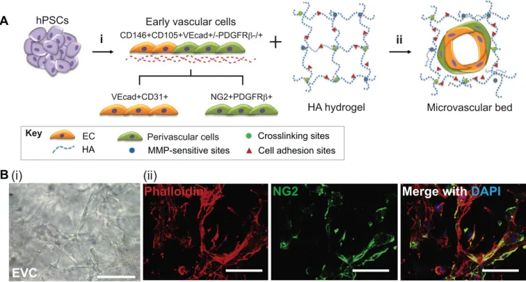
The task of creating two distinct cell types, the ECs and the perivascular or SMCs, from separate progenitor populations is a laborious and largely inefficient procedure. Ideally, vasculature would be created from only a single progenitor cell that gives rise to both cell types needed. Recently, we characterized a unique bipotential cell population derived from human PSCs (hPSCs) called early vascular cells (EVCs) that can differentiate into both ECs and perivascular cells, which then self-organize into microvascular networks within an engineered matrix (Kusuma et al., 2013). The ability to generate both ECs and perivascular cells from a single vascular progenitor population may help to reduce the time, effort and cost of vascular differentiation protocols, as well as to generate more stable engineered vasculature.
Growth factors are crucial during vascular formation
Various GFs have been implicated in the regulation of vascular development, including basic fibroblast growth factor (bFGF), VEGFs, PDGF and TGFβ (Carmeliet and Jain, 2011). Of these, VEGFs and their receptors (Flk and Flt) are considered to be master determinants of vasculogenesis and angiogenesis, as together they regulate the survival, differentiation, proliferation, morphogenesis and migration of ECs (Gentile et al., 2013; Hang et al., 2013; Nakayama et al., 2013). Given their crucial role in vascular development, genes encoding VEGFs have been widely used as therapeutic pro-angiogenic factors in clinical trials or to regulate vascular differentiation and morphogenesis to create vascular network structuresin vitro(Hanjaya-Putra et al., 2010; Yee et al., 2011). PDGFs are also essential for vascular development, as they recruit mural cell precursors that promote vessel tightness and stabilization (Zhou et al., 2014). Like PDGFs, TGFβ, angiopoietin 1 (ANG1 or ANGPT1) and FGFs are also important for vessel stabilization (Ramsauer and D’Amore, 2007; Murakami et al., 2008). TGFβ inhibits EC proliferation and stimulates mural cell differentiation into SMCs or perivascular cells, as well as affecting matrix deposition (Heimark et al., 1986; D’Amore and Smith, 1993). ANG1, which activates ligands for TIE2 (endothelial-specific receptor tyrosine kinase; TEK), is expressed by mural cells and stimulates mural coverage and basement membrane deposition, thereby promoting vessel tightness, whereas angiopoietin 2 (ANG2 or ANGPT2), which is an antagonist to ANG1 and TIE2 signaling, promotes mural cell detachment and EC sprouting, resulting in angiogenesis (Fagiani et al., 2011; Yu et al., 2013). Accumulating evidence demonstrates that stromal cell-derived factor 1 (SDF1) and insulin-like GF (IGF) stimulate angiogenesis through recruitment of EPCs from bone marrow and enhanced EPC mobilization (Maeng et al., 2009; Jujo et al., 2010). Given their pivotal role in orchestrating
in vivovascularization, many of these GFs have been used either as therapeutics to treat vascular disease or as additives to promote
vascular network formation from cells in engineered matrices
in vitro. One of the major barriers to this approach is the very short half-life of GFsin vivo. Thus, a recent focus to improve GF-based therapies has been the use of engineered materials to better maintain GF activityin vivo, and to control timing of GF activity in order to mimic the bona fide stepwise developmental processes.
The vascular ECM
The ECMs present during vascular differentiation and development are mainly composed of fibronectin, laminin, collagen and hyaluronic acid (HA) (Nelson and Bissell, 2006; Estrach et al., 2011; Kashima et al., 2013). The ECM not only provides a structural framework to support cellular function, but also regulates cellular behavior, including adhesion, proliferation, migration and differentiation of vascular cells through both cell-to-cell and cell-to-matrix interactions. Integrin-mediated cell adhesion, (Hodivala-Dilke, 2008; Desgrosellier and Cheresh, 2010), matrix metalloproteinase (MMPs)-mediated matrix remodeling (Iruela-Arispe and Davis, 2009; Stratman et al., 2009; Hanjaya-Putra et al., 2011) and matrix stiffness (Sieminski et al., 2007; Moon et al., 2010; Hanjaya-Putra et al., 2012) have been implicated as key parameters supporting vascular development, including vascular morphogenesis, differentiation and network formation. We and others have demonstrated that incorporating an integrin-binding peptide [Arg-Gly-Asp (RGD)] within the synthetic matrix promotes vascular lumen and capillary formation via integrin-mediated cell adhesion and morphogenesis (Moon et al., 2010; Hanjaya-Putra et al., 2011; Kusuma et al., 2013). In addition, Davis and collaborators reported that a specific MMP, membrane-type 1 MMP (MT1-MMP), plays a crucial role in the formation of lumens and the network formation of ECs within the 3D HA and collagen matrix (Stratman et al., 2009; Hanjaya-Putra et al., 2011). In this study, MT1-MMP allows for the activation and localization of other MMPs (e.g. MMP1 and MMP2) to the migratory end of the cell, resulting in degradation of the matrix. This proteolytic degradation allows cells to migrate and undergo morphogenesis in a hydrogel matrix (see Glossary, Box 1) through a specially designed vascular guidance tunnel. Matrix stiffness can also influence vascular morphogenesis and microcapillary network formation (Sieminski et al., 2007; Kniazeva and Putnam, 2009; Hanjaya-Putra et al., 2010; Moon et al., 2010).In vitro-derived microvasculatures were shown to become more extensive as mechanical stiffness decreased, due to larger pore sizes that facilitate cell migration and morphogenesis, and decrease the hydrogel complexity. It has become clear from these studies that the ECM plays an important role in modulating vascular formation
in vitro. Parameters such as matrix stiffness and composition are therefore key components to consider when designing an engineered scaffold to support vascular formation, and are likely to emerge as major targets for vascular regenerative therapies. However, designing the optimal 3D microenvironment for supporting vascular development remains fairly challenging, as there are a multiple factors to be considered, such as the intrinsic properties of materials and cells themselves.
Oxygen tension is crucial during vascular formation
In the developing embryo, oxygen levels range from 2% to 9%, with low oxygen tension being defined as below 5% partial pressure of oxygen, a state also known as hypoxia (Simon and Keith, 2008). Low oxygen tension has been implicated in the promotion of vascular differentiation and growth during embryonic development, in tumor progression, and in tissue regeneration (Majmundar et al., 2010; De Bock et al., 2011; Keith et al., 2012). The cellular response to hypoxia is regulated by hypoxia-inducible factors (HIFs) that stimulate a series
DEVEL
O
of vascular developmental processes, including angiogenesis and vasculogenesis. Accumulating evidence suggests that hypoxia not only promotes expression of pro-angiogenic factors and their receptors, but also regulates ECM remodeling (Deschene et al., 2012; Gilkes et al., 2013). Semenza and colleagues demonstrated that hypoxia promotes ECM remodeling by inducing the expression of the collagen hydroxylase family (Gilkes et al., 2013). Feng and colleagues (Feng et al., 2010) suggested that hypoxia in MSCs enhanced their angiogenic potential by upregulation of VEGF and bFGF. They found that MSCs preconditioned in a hypoxic environment promoted the expression and secretion of VEGF and bFGF, which resulted in extensive vascular network formation (Liu et al., 2013). Hypoxia has also been shown to enhance angioblast specification and differentiation into ECs (Ramirez-Bergeron et al., 2004). More recently, Liehn and colleagues demonstrated that hypoxia can induce an upregulation of cytokines and chemokines that promote EPC-induced angiogenesis, suggesting that the effects of hypoxia may operate at multiple stages of vascular formation (Kanzler et al., 2013). Manipulating oxygen tension is therefore an attractive approach for enhanced vascular engineering strategies. However, it has so far been difficult to study the effects of oxygen tensionin vitro, due to our limited ability to monitor and control dissolved oxygen levels within the microenvironment. Recently, our laboratory developed an oxygen-controlling hydrogel that can be used as a 3D hypoxic microenvironment. We demonstrated that these hypoxia-inducible hydrogels steer vascular morphogenesisin vitrovia HIF activation of MMPs and encourage neovascularization during subcutaneous wound healing (Park and Gerecht, 2014).
Engineering materials for vascular regeneration
Current approaches to the treatment of vascular disease are primarily aimed at either creating new vasculaturein vitroor at stimulating host vasculature to undergo neovascularizationin vivothrough the delivery of therapeutic pro-angiogenic factors and/or the use of specialized biomaterials. In order to achieve this, it is necessary to engineer a scaffold upon which to deliver the therapeutic factors, much like the bona fide ECM found in vivo. Various kinds of engineered materials can mimic the ECM in order to support vascular differentiation, morphogenesis and tube formation, with the aim of recapitulating thein vivomicroenvironment of vascular formation. The engineered ECM-like scaffold provides a structural framework and is capable of presenting various physiologically relevant signals, including cell-adhesion sites and proteolytically degradable sites (Cuchiara et al., 2012; Hanjaya-Putra et al., 2012; Kusuma et al., 2013; Turturro et al., 2013). In this section, we discuss the application of fundamental theories of vascular development either to create engineered vasculatures in vitro, or to promote neovascularization in situ. Specifically, we focus on emerging approaches using either of the two aforementioned strategies: using engineered cellular constructs to support the
in vitroformation of vasculatures for transplantation into injured or diseased tissues, or using an acellular matrix (see Glossary, Box 1) to stimulate therapeutic vascularizationin vivo.
Engineered cellular constructs: creating new vasculaturein vitro
With recent advances in materials science and cell biology, tissue engineering approaches to create engineered human vascular constructs have been widely used to study vascular development and disease modeling, as well as to explore potential therapies in regenerative medicine. Researchers have tried to generate engineered vasculature
in vitroby recreating thein vivobiological microenvironments. To do this, it is important to consider not only the different cell types and
structural components, but also the biochemical milieu, as well as physical parameters, such a fluid flow and tensile strength (Huebsch and Mooney, 2009). A number of biomaterials have been investigated as artificial microenvironments to support vascular differentiation, morphogenesis and tube formation. Specifically, hydrogels have been widely used as cellular microenvironments for 3D assembly of vascular networks, due to their structural similarity to the natural ECM and to their ability to be tailor-made for specific requirements (McKinnon et al., 2013; Tokuda et al., 2014). A hydrogel is a hydrophilic polymer network swollen with water. These 3D networks can be fabricated via physical and/or chemical crosslinking reactions (see Glossary, Box 1) of various natural materials, such as collagen, fibrinogen, dextran and HA, as well as synthetic polymers, e.g. polyethylene glycol (PEG) and its derivatives (Park et al., 2011; Koehler et al., 2013; Sandker et al., 2013; Stahl et al., 2014; Tokuda et al., 2014). In addition, hydrogels can be easily tailored with bioactive molecules (Mosiewicz et al., 2013), and their physicochemical properties are easily controlled (Tibbitt et al., 2013).
Recapitulating vascular formation: beyond the ECM
To create a cellular microenvironment for vascular development, the hydrogel scaffold should provide cell adhesion domains to control vascular cell behavior. The cell-adhesion moieties are incorporated into matrices during hydrogel formation and allow cell attachment and vascular morphogenesis through cell-matrix interaction. The RGD peptides have been widely used as a cell adhesion domain, as integrin-mediated cell adhesion plays a key role in vascular development, for example in EC attachment, migration, differentiation and vascular morphogenesis (Hanjaya-Putra et al., 2011; Le Saux et al., 2011). In addition, MMP-sensitive degradable sites, which allow cleaving of the biomacromolecular components of the ECMs, should be included, as ECM remodeling is an important aspect of vascular morphogenesis and tube formation, as well as chemotactic migration and proliferation of ECs (Stratman et al., 2009). These signaling domains are key parameters in the design of emerging hydrogel materials in vascular engineering.
HA, a high-molecular-weight polysaccharide, is well-known as an important ECM component; it regulates vascular development by stimulating EC proliferation and cytokine/GF secretion (Takahashi et al., 2005; Mo et al., 2011). HA can be used in hydrogel scaffolds in conjunction with RGD and MMP-sensitive peptides to support vascular morphogenesis and network formation (Hanjaya-Putra et al., 2011, 2012; Yee et al., 2011; Kusuma et al., 2013). ECFCs, which are progenitors of ECs, have been shown to undergo a wide range of coordinated morphogenic processes, including vacuole and lumen formation, as well as branching and sprouting, when placed within a synthetic HA hydrogel tailored with integrinα5β1- andαvβ3-mediated cell adhesion domains, and MMP-sensitive sites (Hanjaya-Putra et al., 2011). The resulting engineered vasculature was able to functionally couple with the host blood circulation, supporting blood flow through the hydrogels after transplantation. These results are significant, as tissue vascularization and integration with host blood circulation remains a key challenge for the translation of engineered tissues into clinically relevant therapies. The effect of matrix stiffness and remodeling of HA hydrogels on vascular morphogenesis and the migration of ECFCs has also recently been investigated (Hanjaya-Putra et al., 2012). For this study, the authors created patterned hydrogel constructs of different degrees of matrix stiffness using photo-polymerization. Encapsulated ECFCs within the HA hydrogel were irradiated with UV using a patterned photomask, which resulted in varying degrees of stiffness within one hydrogel. The encapsulated ECFCs within the softer regions underwent vascular morphogenesis
DEVEL
O
and formed a vascular network, whereas the cells encapsulated in stiffer regions exhibited limited cell spreading. Furthermore, the softer region of the hydrogel appeared to promote greater ECFC sprouting into the matrix compared with the stiffer regions. These results demonstrate that matrix stiffness plays an important role in the migration and vascular morphogenesis of ECFCs, and that HA hydrogels can either support or inhibitin vitrovasculogenesis and angiogenesis of ECFCs by controlling matrix degradation cues. Other cell types have also shown success in generating engineered vascular networks in HA hydrogels. Recently, we demonstrated how human EVCs, a bipotential vascular cell population with both EC and pericyte lineage potential, can be used to engineer human vascular networks when encapsulated within HA hydrogels (Kusuma et al., 2013). The encapsulated EVCs were capable of maturing into ECs and perivascular cells and furthermore were able to self-organize into multicellular microvascular networks within HA hydrogels. The engineered human vascular networks survived and integrated with the host vasculature to establish blood flow (Fig. 2). These integrated approaches, including novel cell sources and the use of advanced hydrogel materials to create implantable human microvasculature, have great potential for the study of vascular biology and will also be useful in studying a range of vascular diseases and therapeutic approaches to tissue regeneration.
Defining vasculature architecturein vitro
The vascular network is highly organized and has a distinctive architecturein vivothat is important for its function. Recently, Chen and colleagues generated engineered vessels using a microtissue molding approach to create highly aligned‘cords’of ECs, which then facilitated the proper formation of new capillaries along the patterned cords (Baranski et al., 2013). The authors used collagen and fibrin hydrogels containing vascular cells, including HUVECs and mouse mesenchymal cells (C3H10T1/2), to create the engineered vasculature. Upon transplantation, the vessels integrated with host blood vessels as early as 3 days and progressively matured for 28 days, demonstrating that implanting cords with a prescribed geometry provides a template for defined neovascular formation
in vivo. Such well-defined vascular architecture may help to guide tissue formation in other tissue engineering settings. Indeed, the authors used primary hepatocyte constructs containing either random or organized EC networks and implanted these into mice to assess whether organized vascular architecture can modulate tissue viability. They found that organized engineered vessels enhanced hepatic cell viability and function when compared with randomly distributed vessels, thus demonstrating the significance of multicellular architecture in tissue integration and function. The possibility of creating pre-vascularized tissue constructs is an exciting application of vascular engineering and regeneration. Biomimetic models to simulate angiogenic sprouting morphogenesis in vivo have also been developed using a tissue-molding technique (Morgan et al., 2013; Nguyen et al., 2013). The development of highly organized
in vitro vascular constructs also presents a powerful screening platform for studying the effects of different angiogenic factors.
Other emerging approaches to control vascular architecture have combined collagen hydrogels with microfabrication (see Glossary, Box 1) techniques to create in vitro microenvironments. The microfabrication technique has been widely used to create artificial tissue constructs, including microvascularized networks embedded within a 3D hydrogel, which can be applied as a platform to mimic
in vivo vascular microenvironments. Fischbach-Teschel and colleagues recently developed a novel approach to create fully enclosed and perfusable blood vessels in collagen I hydrogels using
microfabrication, which allows the control of nutrients and oxygen transfer, and of blood flow rate and pressure (Zheng et al., 2012; Morgan et al., 2013). This technique can be used to study vascular function across multiple parameters, such as mass transfer, which is the transfer of nutrients and oxygen across the vessel wall, physical parameters, such as blood flow and pressure, and drug screening (Rodriguez-Rodriguez et al., 2012; Griep et al., 2013; Westein et al., 2013). Thus, microfabrication is a valuable technique that enables the study of basic vascular and blood biology, as well as a biologically meaningful platform for modeling vascular-related diseases and for generating vascular constructs for regenerative medicine.
A dual approach to create highly functionalized and architecturally sound vasculature was recently developed by West and colleagues, who used a PEG hydrogel tailored with RGD- and MMP-sensitive peptides to support vascular network formation. They then applied the hydrogel to a microfluidic system in order to mimic native vascular organization and function (Moon et al., 2010). PEG-based hydrogels tailored with RGD- and MMP-sensitive peptides have been widely used for supporting vascular network formation (Moon et al., 2009, 2010; Cuchiara et al., 2012; Turturro et al., 2013). The authors first encapsulated HUVECs and mesenchymal progenitors (10T1/2 cells) that exhibited a perivascular cell morphology within PEG hydrogels containing both RGD peptides and MMP-sensitive sites (Cuchiara et al., 2012). They found that the hydrogel supported rapid vascular morphogenesis of the HUVEC and 10T1/2 cells, and that the synthetic matrix was soon replaced with newly formed basement membrane proteins, such as laminin and collagen type IV. The authors then applied the engineered vasculatures to perfusable microfabricated channels, thus culturing the HUVECs and 10T1/2 cells encapsulated within the synthetic hydrogels inside the microchannels (Cuchiara et al., 2012). The use of microchannels represents an innovative approach to perfusing and integrating the vasculature with nutrient-containing fluids in vitro. Using this approach, they found that nutrient transport within the system shifts from simple diffusion to vessel-supported convective transport and extra-vessel diffusion, meaning that the transport of nutrients was more efficient and more closely replicated that of bona fide vascular beds. This integrated approach has considerable value because it mimics the complex in vivo environment using emerging microfabrication technologies, and therefore holds great potential for tissue-engineering applicationsin vivo.
Engineering acellular materials to promote vasculature regeneration
Thede novogeneration of vasculature using exogenous cell sources may be required for some medical applications, but in other cases it might be possible to coax existing endogenous cells to undergo neovascularizationin vivo. In the past decade, biomaterials have been used as a carrier to deliver single or multiple GFs for therapeutic neovascularization (Lee et al., 2011). These strategies were met with limited success due to the very rapid release and short half-life of the GFs, as well as an inability to control sequential release. More recently, researchers have focused on developing advanced materials that allow much finer control over the spatial and temporal release of different GFs. Such developments include the sustained release (see Glossary, Box 1) and sequential delivery (see Glossary, Box 1) of multiple GFs, approaches that allow a much closer approximation of the precise signaling events that occur during vascular formation
in vivo. In addition, engineered materials can play a role not only as delivery vehicles for therapeutic agents, but also to directly stimulate neovascularization through physicochemical interactions between the
biomaterials and the host tissue.
DEVEL
O
Combinatorial and sequential growth factor delivery
Recent studies have demonstrated that delivering multiple pro-angiogenic GFs either in tandem or sequentially can help to stimulate rapid neovascularization during wound healing. Recently, our group used dextran hydrogels to deliver a combination of VEGF, ANG1, SDF1 and IGF during subcutaneous wound healing (Sun et al., 2011). The rats transplanted with these four factors displayed significantly more and larger functional vessels in the hydrogel compared with other groups. We also examined individual GFs and various combinations of these GFs and found that VEGF delivery is required for functional neovascularization, which is enhanced by supplementary pro-angiogenic GFs.
A similar approach was reported by Mooney and colleagues (Brudno et al., 2013). They investigated the effects of a temporally regulated presentation of pro-angiogenic factors VEGF and ANG2 plus additional pro-maturation factors PDGF and ANG1 on angiogenesis both in vitro and in vivo. Initially, they found that, whereas VEGF and ANG2 stimulated EC sprouting and perivascular cell detachment within a fibrin gel, simultaneous delivery of PDGF and ANG1 inhibited this process. Similarly, whereas VEGF and ANG2 enhanced microvascular density in a subcutaneous model of blood vessel formation, simultaneous injection of PDGF and ANG1 significantly inhibited microvessel formation. However, the sequential delivery of PDGF and ANG1 after delivery of VEGF and ANG2 promoted both vessel maturation and remodeling without inhibiting sprout angiogenesis. These results demonstrated that sequentially controlled delivery of multiple GFs could be an important strategy for promoting vessel maturation and remodeling.
Other pro-angiogenic factors
In addition to pro-angiogenic and pro-maturation GFs, new protein and peptide drugs have recently been investigated for their potential use in treating vascular disease. Lee and colleagues successfully used a sequential delivery system of an anti-apoptotic agent plus VEGF in order to treat ischemia in the mouse hind limb (Shin et al., 2013). The
anti-apoptotic agent consisted of a heat shock protein (HSP) fused with a transcriptional activator (TAT; TAT-HSP), whereas the VEGF was encapsulated within a porous poly(lactic-co-glycolic) acid (PLGA) microparticle (see Glossary, Box 1). Both elements were incorporated into an alginate hydrogel, in which the combination of the responsive TAT element and the porous microparticle allowed the controlled release of HSP and VEGF, respectively. The authors observed an initial high burst of TAT-HSP and a complete release within 7 days, whereas VEGF release was sustained for 4 weeks. This finding demonstrated that the release of both elements from hybrid delivery systems could be controlled in a sequential manner. Mice treated with this hybrid system delivering both elements exhibited an enhanced rate of limb salvage and reduced rate of limb loss at up to 4 weeks, suggesting that sequential delivery is pivotal for therapeutic angiogenesis at the ischemic sites.
[image:7.612.117.495.61.264.2]Another potential therapeutic agent is thymosin β4 (Tβ4), a 43 amino acid peptide that has been shown to induce angiogenesis and sustain vascular stability via the recruitment of ECs and SMCs (Smart et al., 2007). Several groups have studied the local delivery of Tβ4 using PEG- and collagen-based hydrogel materials to treat cardiac infarction (Chiu and Radisic, 2011; Kraehenbuehl et al., 2011a; Chiu et al., 2012a,b). Radisic and colleagues developed collagen hydrogel materials for controlled release of Tβ4 over 28 days (Chiu and Radisic, 2011). They observed increased EC migration and tube formation in a hydrogel containing Tβ4 and enhanced vascularization when injectedin vivo(Chiu et al., 2012a). Moreover, they applied the hydrogel/Tβ4 system to myocardial infarction in rats, which resulted in improved angiogenesis, reduced tissue loss and retained left ventricular wall thickness after infarction compared with Tβ4-free and soluble controls (Chiu et al., 2012b). Interestingly, when arterial or venous vessels were culturedex vivoon Tβ4-containing collagen hydrogels, capillary sprouting was observed and was further accelerated by treatment of the cultures with soluble VEGF and hepatocyte growth factor (HGF), the former being a key factor in regulating vasculogenesis and angiogenesisin vivo.
Fig. 2. Engineered vascular network from pluripotent stem cells within a hyaluronic acid hydrogel matrix.(A) Human pluripotent stem cells (PSCs) differentiate into bipotential early vascular cells (EVCs; i) that can mature into functional endothelial cells (ECs) and perivascular cells (ii) when placed within a synthetic hyaluronic acid (HA) hydrogel. The HA hydrogel supports the self-organization and differentiation of the bipotential EVCs into a vascular network. (B) PSC-derived EVCs self-assemble into networks in HA hydrogels (i). Multilayered structures can be detected (ii), as demonstrated by 3D projection images of NG2 (green) and phalloidin (red), and by nuclei (blue) showing NG2+ perivascular cells integrated into hollow structures. Adapted, with permission, from Kusuma et al. (2013). Scale bars: 50μm. CD31, cluster of differentiation 31; CD105, cluster of differentiation 105; CD146, cluster of differentiation 146; DAPI, 40 ,6-diamidino-2-phenylindole; MMP, matrix metalloproteinase; NG2, neural/glial antigen 2; PDGFRβ, platelet-derived growth factor receptorβ; VEcad, VE-cadherin.
DEVEL
O
Sustained and controlled release of pro-angiogenic factors
Whereas microencapsulation enables the sustained release of GFs over time, this strategy is largely dependent on physicochemical parameters, such as pore size and particle size, to control the release of the GFs. By comparison, GF binding within the ECM is a dynamic process that is sensitive to the presence and specificity of binding sites. Thus, ECM-regulated GF release may represent a more advanced way to fine-tune temporal and even spatial delivery of pro-angiogenic factors. This was demonstrated by Hubbell and colleagues, who developed a novel strategy to regulate the interaction between synthetic materials and GFs to control sequential delivery of pro-angiogenic GFs during regeneration (Martino et al., 2011, 2013; Traub et al., 2013). The pro-angiogenic effects of VEGFA and platelet-derived growth factor subunit B homodimer (PDGFBB) were greatly enhanced by creating a PEG-based hydrogel tailored with both integrin-binding domains and promiscuous GF-binding domains derived from fibronectin (Martino et al., 2011). The synergistic signaling between integrin and the GF receptors improved regeneration in vivo, whereas delivery of the GFs VEGFA and PDGFBB alone had no significant effect. More recently, it has been demonstrated that fibrinogen can also bind GFs through its heparin-binding domain, and that incorporation of the heparin-heparin-binding site into the synthetic PEG-based hydrogels results in the sustained and controlled release of GFs from the fibrin matrix (Martino et al., 2013). In this study, the authors identified 15 unique binding interactions between fibrin and various GFs, including bFGF, VEGF, PDGF and TGF. This is in addition to the heparin-binding domain of fibrin, which can maintain GF activities and enable a controlled release (Martino et al., 2013). To confirm this variable binding activity in a hydrogel system, the authors incorporated the heparin-biding sites into PEG hydrogels and applied this to a diabetic mouse model of impaired wound healing. They demonstrated that the synthetic matrix could release GFs in a sustained manner that promoted wound healing. An alternative way to control the release of GFs is via physical containment. Recently, Park and colleagues developed a double-layered core-shell microcapsule, whereby PDGF was encapsulated in the core of the particle and VEGF was encapsulated in the shell. This arrangement meant that VEGF was released faster than PDGF, thereby allowing control over the timing and sequence of GF release. Using this system, the authors were able to show that a controlled release of VEGF followed by PDGF resulted in enhanced angiogenic activity of ECs (Choi et al., 2013).
Concluding remarks
This Review has discussed how the fundamental principles of vascular development can be applied to develop tissue-engineering strategies for promoting therapeutic vascularization and angiogenesis. The complex series of processes that occur during vascular development provide important information for the engineering of materials for vascular growth and regeneration, as well as provision of the biologically relevant GFs and other microenvironmental cues. Researchers have designed and investigated numerous engineered materials that deliver essential pro-angiogenic factors to enhance therapeutic angiogenesis, both to stimulate and promote rapid neovascularization, and to provide a cellular microenvironment that will generate engineered vasculatures.
By integrating vascular biology and materials science, engineered vascular constructs can provide various therapeutic tools and vascular tissue models in order to improve vascular regeneration and understand disease. However, further improvements are required to enhance the ability of these engineering approaches to mimic the physiological environment. For example, a major challenge lies in recapitulating the
complex geometries and mechanical properties of the native environments. This is particularly important, as such an architecture allows very fine spatial and temporal control over the instructive cues that regulate neo-vascularization. To overcome these limitations, many studies are beginning to use new techniques to generate 3D geometrically or mechanically controlled microenvironments (West, 2011; Culver et al., 2012; Gandavarapu et al., 2014). In addition, integration of emerging techniques, such as microfluidics devices, with advances in biomaterials may help to further elucidate the complex process of vascular development and regeneration. Thus, current engineering approaches and emerging techniques will provide the necessary platform to support extensive research, ranging from basic vascular biology to clinical applications for vascular engineering and regeneration.
Acknowledgements
We thank Quinton Smith for his assistance with the editing of the manuscript.
Competing interests
The authors declare no competing financial interests.
Funding
Work presented here was partially supported by the National Institutes of Health and a National Science Foundation grants. Deposited in PMC for release after 12 months.
References
Asahara, T., Murohara, T., Sullivan, A., Silver, M., van der Zee, R., Li, T., Witzenbichler, B., Schatteman, G. and Isner, J. M.(1997). Isolation of putative progenitor endothelial cells for angiogenesis.Science275, 964-967.
Baranski, J. D., Chaturvedi, R. R., Stevens, K. R., Eyckmans, J., Carvalho, B., Solorzano, R. D., Yang, M. T., Miller, J. S., Bhatia, S. N. and Chen, C. S.(2013). Geometric control of vascular networks to enhance engineered tissue integration and function.Proc. Natl. Acad. Sci. U.S.A.110, 7586-7591.
Bloom, D. E., Cafiero, E. T., Jané-Llopis, E., Abrahams-Gessel, S., Bloom, L. R., Fathima, S., Feigl, A. B., Gaziano, T., Mowafi, M., Pandya, A. et al.(2011).The Global Economic Burden of Noncommunicable Diseases. Geneva: World Economic Forum.
Brudno, Y., Ennett-Shepard, A. B., Chen, R. R., Aizenberg, M. and Mooney, D. J.
(2013). Enhancing microvascular formation and vessel maturation through temporal control over multiple pro-angiogenic and pro-maturation factors.
Biomaterials34, 9201-9209.
Busnadiego, O., Gonzalez-Santamaria, J., Lagares, D., Guinea-Viniegra, J.,
Pichol-Thievend, C., Muller, L. and Rodriguez-Pascual, F.(2013). LOXL4 is
induced by transforming growth factor beta1 through Smad and JunB/Fra2 and contributes to vascular matrix remodeling.Mol. Cell. Biol.33, 2388-2401.
Carmeliet, P. and Jain, R. K. (2011). Molecular mechanisms and clinical
applications of angiogenesis.Nature473, 298-307.
Caspi, O., Lesman, A., Basevitch, Y., Gepstein, A., Arbel, G., Habib, I. H. M.,
Gepstein, L. and Levenberg, S.(2007). Tissue engineering of vascularized
cardiac muscle from human embryonic stem cells.Circ. Res.100, 263-272.
Chen, Y.-C., Lin, R.-Z., Qi, H., Yang, Y., Bae, H., Melero-Martin, J. M. and
Khademhosseini, A.(2012). Functional human vascular network generated in
photocrosslinkable gelatin methacrylate hydrogels. Adv. Funct. Mater. 22, 2027-2039.
Chiu, L. L. Y. and Radisic, M.(2011). Controlled release of thymosin beta4 using
collagen-chitosan composite hydrogels promotes epicardial cell migration and angiogenesis.J. Control. Release155, 376-385.
Chiu, L. L. Y., Montgomery, M., Liang, Y., Liu, H. and Radisic, M.(2012a).
Perfusable branching microvessel bed for vascularization of engineered tissues.
Proc. Natl. Acad. Sci. U.S.A.109, E3414-E3423.
Chiu, L. L. Y., Reis, L. A. and Radisic, M.(2012b). Controlled delivery of thymosin beta4 for tissue engineering and cardiac regenerative medicine.Ann. N. Y. Acad. Sci.1269, 16-25.
Choi, D. H., Subbiah, R., Kim, I. H., Han, D. K. and Park, K.(2013). Dual Growth
Factor Delivery Using Biocompatible Core-Shell Microcapsules for Angiogenesis.
Small9, 3468-3476.
Colombo, E., Calcaterra, F., Cappelletti, M., Mavilio, D. and Della Bella, S.
(2013). Comparison of fibronectin and collagen in supporting the isolation and expansion of endothelial progenitor cells from human adult peripheral blood.
PLoS ONE8, e66734.
Critser, P. J. and Yoder, M. C.(2010). Endothelial colony-forming cell role in
neoangiogenesis and tissue repair.Curr. Opin. Organ Transplant.15, 68-72.
DEVEL
O
Cuchiara, M. P., Gould, D. J., McHale, M. K., Dickinson, M. E. and West, J. L.
(2012). Integration of self-assembled microvascular networks with microfabricated PEG-based hydrogels.Adv. Funct. Mater.22, 4511-4518.
Culver, J. C., Hoffmann, J. C., Poché, R. A., Slater, J. H., West, J. L. and
Dickinson, M. E.(2012). Three-dimensional biomimetic patterning in hydrogels to guide cellular organization.Adv. Mater.24, 2344-2348.
Dar, A., Domev, H., Ben-Yosef, O., Tzukerman, M., Zeevi-Levin, N., Novak, A.,
Germanguz, I., Amit, M. and Itskovitz-Eldor, J. (2012). Multipotent
vasculogenic pericytes from human pluripotent stem cells promote recovery of murine ischemic limb.Circulation125, 87-99.
De Bock, K., Mazzone, M. and Carmeliet, P.(2011). Antiangiogenic therapy,
hypoxia, and metastasis: risky liaisons, or not?Nat. Rev. Clin. Oncol.8, 393-404.
Deschene, K., Céleste, C., Boerboom, D. and Theoret, C. L.(2012). Hypoxia
regulates the expression of extracellular matrix associated proteins in equine dermal fibroblasts via HIF1.J. Dermatol. Sci.65, 12-18.
Desgrosellier, J. S. and Cheresh, D. A.(2010). Integrins in cancer: biological
implications and therapeutic opportunities.Nat. Rev. Cancer10, 9-22.
D’Amore, P. A. and Smith, S. R.(1993). Growth factor effects on cells of the
vascular wall: a survey.Growth Factors8, 61-75.
Eilken, H. M. and Adams, R. H.(2010). Dynamics of endothelial cell behavior in
sprouting angiogenesis.Curr. Opin. Cell Biol.22, 617-625.
Estrach, S., Cailleteau, L., Franco, C. A., Gerhardt, H., Stefani, C., Lemichez, E.,
Gagnoux-Palacios, L., Meneguzzi, G. and Mettouchi, A. (2011).
Laminin-binding integrins induce Dll4 expression and Notch signaling in endothelial cells.
Circ. Res.109, 172-182.
Fagiani, E., Lorentz, P., Kopfstein, L. and Christofori, G.(2011). Angiopoietin-1 and -2 exert antagonistic functions in tumor angiogenesis, yet both induce lymphangiogenesis.Cancer Res.71, 5717-5727.
Feng, Q., Lu, S.-J., Klimanskaya, I., Gomes, I., Kim, D., Chung, Y., Honig, G. R.,
Kim, K.-S. and Lanza, R. (2010). Hemangioblastic derivatives from human
induced pluripotent stem cells exhibit limited expansion and early senescence.
Stem Cells28, 704-712.
Gandavarapu, N. R., Azagarsamy, M. A. and Anseth, K. S.(2014). Photo-click
living strategy for controlled, reversible exchange of biochemical ligands.Adv. Mater.26, 2521-2526.
Gentile, C., Muise-Helmericks, R. C. and Drake, C. J.(2013). VEGF-mediated
phosphorylation of eNOS regulates angioblast and embryonic endothelial cell proliferation.Dev. Biol.373, 163-175.
Geudens, I. and Gerhardt, H.(2011). Coordinating cell behaviour during blood
vessel formation.Development138, 4569-4583.
Gilkes, D. M., Bajpai, S., Chaturvedi, P., Wirtz, D. and Semenza, G. L.(2013).
Hypoxia-inducible factor 1 (HIF-1) promotes extracellular matrix remodeling under hypoxic conditions by inducing P4HA1, P4HA2, and PLOD2 expression in fibroblasts.J. Biol. Chem.288, 10819-10829.
Glynn, J. J. and Hinds, M. T.(2013). Endothelial outgrowth cells: function and
performance in vascular grafts.Tissue Eng. Part B Rev.
Griep, L. M., Wolbers, F., de Wagenaar, B., ter Braak, P. M., Weksler, B. B., Romero, I. A., Couraud, P. O., Vermes, I., van der Meer, A. D. and van den
Berg, A. (2013). BBB on chip: microfluidic platform to mechanically and
biochemically modulate blood-brain barrier function.Biomed. Microdevices15, 145-150.
Hang, T.-C., Tedford, N. C., Reddy, R. J., Rimchala, T., Wells, A., White, F. M.,
Kamm, R. D. and Lauffenburger, D. A.(2013). Vascular endothelial growth
factor (VEGF) and platelet (PF-4) factor 4 inputs modulate human microvascular endothelial signaling in a three-dimensional matrix migration context.Mol. Cell. Proteomics12, 3704-3718.
Hanjaya-Putra, D., Yee, J., Ceci, D., Truitt, R., Yee, D. and Gerecht, S.(2010).
Vascular endothelial growth factor and substrate mechanics regulate in vitro tubulogenesis of endothelial progenitor cells.J. Cell. Mol. Med.14, 2436-2447.
Hanjaya-Putra, D., Bose, V., Shen, Y.-I., Yee, J., Khetan, S., Fox-Talbot, K., Steenbergen, C., Burdick, J. A. and Gerecht, S.(2011). Controlled activation of morphogenesis to generate a functional human microvasculature in a synthetic matrix.Blood118, 804-815.
Hanjaya-Putra, D., Wong, K. T., Hirotsu, K., Khetan, S., Burdick, J. A. and
Gerecht, S. (2012). Spatial control of cell-mediated degradation to regulate
vasculogenesis and angiogenesis in hyaluronan hydrogels. Biomaterials 33, 6123-6131.
Heimark, R. L., Twardzik, D. R. and Schwartz, S. M. (1986). Inhibition of
endothelial regeneration by type-beta transforming growth factor from platelets.
Science233, 1078-1080.
Hirschi, K. K., Rohovsky, S. A. and D’Amore, P. A.(1998). PDGF, TGF-beta, and
heterotypic cell-cell interactions mediate endothelial cell-induced recruitment of 10T1/2 cells and their differentiation to a smooth muscle fate.J. Cell Biol.141, 805-814.
Hodivala-Dilke, K.(2008).αvβ3 integrin and angiogenesis: a moody integrin in a changing environment.Curr. Opin. Cell Biol.20, 514-519.
Huebsch, N. and Mooney, D. J.(2009). Inspiration and application in the evolution of biomaterials.Nature462, 426-432.
Imanirad, P., Solaimani Kartalaei, P., Crisan, M., Vink, C., Yamada-Inagawa, T., de Pater, E., Kurek, D., Kaimakis, P., van der Linden, R., Speck, N. et al.
(2014). HIF1α is a regulator of hematopoietic progenitor and stem cell development in hypoxic sites of the mouse embryo.Stem Cell Res.12, 24-35.
Iruela-Arispe, M. L. and Davis, G. E.(2009). Cellular and molecular mechanisms of vascular lumen formation.Dev. Cell16, 222-231.
Jujo, K., Hamada, H., Iwakura, A., Thorne, T., Sekiguchi, H., Clarke, T., Ito, A.,
Misener, S., Tanaka, T., Klyachko, E. et al.(2010). CXCR4 blockade augments
bone marrow progenitor cell recruitment to the neovasculature and reduces mortality after myocardial infarction.Proc. Natl. Acad. Sci. U.S.A.107, 11008-11013.
Kanzler, I., Tuchscheerer, N., Steffens, G., Simsekyilmaz, S., Konschalla, S.,
Kroh, A., Simons, D., Asare, Y., Schober, A., Bucala, R. et al.(2013).
Differential roles of angiogenic chemokines in endothelial progenitor cell-induced angiogenesis.Basic Res. Cardiol.108, 310.
Kappetein, A. P., Head, S. J., Morice, M.-C., Banning, A. P., Serruys, P. W., Mohr,
F.-W., Dawkins, K. D. and Mack, M. J.;Syntax Investigators(2013). Treatment
of complex coronary artery disease in patients with diabetes: 5-year results comparing outcomes of bypass surgery and percutaneous coronary intervention in the SYNTAX trial.Eur. J. Cardiothorac. Surg.43, 1006-1013.
Kashima, Y., Takahashi, M., Shiba, Y., Itano, N., Izawa, A., Koyama, J.,
Nakayama, J., Taniguchi, S., Kimata, K. and Ikeda, U.(2013). Crucial role of
hyaluronan in neointimal formation after vascular injury.PLoS ONE8, e58760.
Keith, B., Johnson, R. S. and Simon, M. C.(2012). HIF1alpha and HIF2alpha: sibling rivalry in hypoxic tumour growth and progression.Nat. Rev. Cancer12, 9-22.
Kinner, B., Zaleskas, J. M. and Spector, M.(2002). Regulation of smooth muscle
actin expression and contraction in adult human mesenchymal stem cells.Exp. Cell Res.278, 72-83.
Kniazeva, E. and Putnam, A. J.(2009). Endothelial cell traction and ECM density
influence both capillary morphogenesis and maintenance in 3-D.Am. J. Physiol. Cell Physiol.297, C179-C187.
Koehler, K. C., Anseth, K. S. and Bowman, C. N.(2013). Diels-Alder mediated
controlled release from a poly(ethylene glycol) based hydrogel.Biomacromolecules 14, 538-547.
Koike, N., Fukumura, D., Gralla, O., Au, P., Schechner, J. S. and Jain, R. K.
(2004). Tissue engineering: creation of long-lasting blood vessels.Nature428, 138-139.
Kraehenbuehl, T. P., Ferreira, L. S., Hayward, A. M., Nahrendorf, M., van der Vlies, A. J., Vasile, E., Weissleder, R., Langer, R. and Hubbell, J. A.(2011a). Human embryonic stem cell-derived microvascular grafts for cardiac tissue preservation after myocardial infarction.Biomaterials32, 1102-1109.
Kraehenbuehl, T. P., Langer, R. and Ferreira, L. S.(2011b). Three-dimensional
biomaterials for the study of human pluripotent stem cells.Nat. Methods8, 731-736.
Kusuma, S., Shen, Y.-I., Hanjaya-Putra, D., Mali, P., Cheng, L. and Gerecht, S.
(2013). Self-organized vascular networks from human pluripotent stem cells in a synthetic matrix.Proc. Natl. Acad. Sci. U.S.A.110, 12601-12606.
Le Saux, G., Magenau, A., Böcking, T., Gaus, K. and Gooding, J. J.(2011). The
relative importance of topography and RGD ligand density for endothelial cell adhesion.PLoS ONE6, e21869.
Lee, K., Silva, E. A. and Mooney, D. J.(2011). Growth factor delivery-based tissue engineering: general approaches and a review of recent developments.J. R. Soc. Interface8, 153-170.
Leeper, N. J., Hunter, A. L. and Cooke, J. P.(2010). Stem cell therapy for vascular regeneration: adult, embryonic, and induced pluripotent stem cells.Circulation 122, 517-526.
Lin, Y., Weisdorf, D. J., Solovey, A. and Hebbel, R. P.(2000). Origins of circulating endothelial cells and endothelial outgrowth from blood.J. Clin. Invest.105, 71-77.
Lippmann, E. S., Azarin, S. M., Kay, J. E., Nessler, R. A., Wilson, H. K., Al-Ahmad, A., Palecek, S. P. and Shusta, E. V.(2012). Derivation of blood-brain barrier endothelial cells from human pluripotent stem cells.Nat. Biotechnol.30, 783-791.
Liu, L., Gao, J., Yuan, Y., Chang, Q., Liao, Y. and Lu, F. (2013). Hypoxia
preconditioned human adipose derived mesenchymal stem cells enhance angiogenic potential via secretion of increased VEGF and bFGF.Cell Biol. Int. 37, 551-560.
Maeng, Y.-S., Choi, H.-J., Kwon, J.-Y., Park, Y.-W., Choi, K.-S., Min, J.-K.,
Kim, Y.-H., Suh, P.-G., Kang, K.-S., Won, M.-H. et al.(2009). Endothelial
progenitor cell homing: prominent role of the IGF2-IGF2R-PLCbeta2 axis.
Blood113, 233-243.
Majmundar, A. J., Wong, W. J. and Simon, M. C.(2010). Hypoxia-inducible factors
and the response to hypoxic stress.Mol. Cell40, 294-309.
Martino, M. M., Tortelli, F., Mochizuki, M., Traub, S., Ben-David, D., Kuhn, G. A.,
Muller, R., Livne, E., Eming, S. A. and Hubbell, J. A.(2011). Engineering the
growth factor microenvironment with fibronectin domains to promote wound and bone tissue healing.Sci. Transl. Med.3, 100ra89.
Martino, M. M., Briquez, P. S., Ranga, A., Lutolf, M. P. and Hubbell, J. A.(2013). Heparin-binding domain of fibrin(ogen) binds growth factors and promotes tissue repair when incorporated within a synthetic matrix.Proc. Natl. Acad. Sci. U.S.A. 110, 4563-4568.
McKinnon, D. D., Kloxin, A. M. and Anseth, K. S.(2013). Synthetic hydrogel
platform for three-dimensional culture of embryonic stem cell-derived motor neurons.Biomater. Sci.1, 460.