RESEARCH ARTICLE
A regulatory receptor network directs the range and output of the
Wingless signal
Sabine Schilling*,‡, Sarah Steiner‡, Dario Zimmerli‡and Konrad Basler§
ABSTRACT
The potent activity of Wnt/Wingless (Wg) signals necessitates sophisticated mechanisms that spatially and temporally regulate their distribution and range of action. The two main receptor components for Wg – Arrow (Arr) and Frizzled 2 (Fz2) – are transcriptionally downregulated by Wg signaling, thus forming gradients that oppose that of Wg. Here, we analyze the relevance of this transcriptional regulation for the formation of the Wg gradient in theDrosophilawing disc by combiningin vivoreceptor overexpression with anin silicomodel of Wg receptor interactions. Our experiments show that ubiquitous upregulation of Arr and Fz2 has no significant effects on Wg output, whereas clonal overexpression of these receptors leads to signaling discontinuities that have detrimental phenotypic consequences. These findings are supported by ourin silicomodel for Wg diffusion and signal transduction, which suggests that abrupt changes in receptor levels causes discontinuities in Wg signaling. Furthermore, we identify a 200 bp regulatory element in thearrlocus that can account for the Arr gradient, and we show that this is indirectly negatively controlled by Wg activity. Finally, we analyze the role of Frizzled 3 (Fz3) in this system and find that its expression, which is induced by Wg, contributes to the establishment of the Arr and Fz2 gradients through counteracting canonical signaling. Taken together, our results provide a model in which the regulatory network of Wg and the three receptor components account for the range and shape of this prototypical morphogen system.
KEY WORDS: Wingless receptors, Wnt pathway, Model for Wg diffusion
INTRODUCTION
Communication between the cells in a tissue depends on extracellular signaling molecules. The vast majority of these are produced by defined subsets of cells. This restricted production provides spatial information that is needed for the signals to orchestrate patterning and growth. The signaling output is dependent on the range of action and level of activity of the secreted signaling protein–determinants that depend not only on the biochemical nature of the signal but also on the receptor systems that are employed by the receiving cells. The glycoproteins of the Wnt family serve as an interesting model to understand the interplay between these determinants.
Mechanisms that precisely and tightly control Wnt signal reception and transduction, both spatially and temporally, are absolutely necessary – Wnt signaling is crucial in many developmental processes, and improper activation of the canonical signaling cascade
can have severe pathological consequences, such as cancer (Clevers, 2006). Altering receptor levels is often the first step in attenuating the cellular response to a signaling molecule. Receptor downregulation can be achieved on the transcriptional level through repression of the receptor-encoding gene, or on the protein level through internalization and subsequent lysosomal degradation. Both processes lead to a diminution of active receptors from the cell membrane and, thereby, to an attenuation of the signaling output.
Wingless (Wg), the founding member of the highly conserved Wnt family, plays an important role in the primordium of theDrosophila melanogasterwing (Zecca et al., 1996; Neumann and Cohen, 1997). In this tissue, Wg is expressed in a thin stripe at the dorsal-ventral boundary from where it spreads to either side, producing a symmetrical concentration gradient. The canonical Wnt/Wg transduction pathway is initiated by the binding of Wnt/Wg ligands to a receptor of the Frizzled family and a co-receptor, the low-density lipoprotein receptor-related LRP5/6 [Arrow (Arr) in D. melanogaster]. This binding transduces the extracellular signal into an intracellular cascade that ultimately results in the cytoplasmic stabilization and nuclear localization of β-catenin/Armadillo (Arm) and its subsequent association with TCF/LEF [Pangolin (Pan) in D. melanogaster] DNA binding proteins. The activation of Wnt target genes is promoted by the interaction of the bipartite Arm-Pan complex with various auxiliary co-factors (Mosimann et al., 2009).
InDrosophila, the genes encoding the Wg receptor Fz2 and the co-receptor Arr are transcriptionally downregulated by Wg signaling (Cadigan et al., 1998; Wehrli et al., 2000). However, the details of the underlying molecular mechanism, and an understanding of its physiological significance, are still lacking. Wnt-induced transcriptional repression could be either direct or indirect; only a detailed analysis of the cis-regulatory elements (CREs) and the corresponding transcription factors can distinguish between these modes of repression (Affolter et al., 2008). In addition to signaling-induced transcriptional downregulation, there is evidence for a collateral post-transcriptional layer of regulation of Wg receptors. Both Fz2 and Arr contain endocytosis signals and have been shown to localize to endocytic compartments together with their ligand Wg (Rives et al., 2006), which suggests that both the receptor and co-receptor are internalized upon Wg binding. The contribution of this to Wg internalization, however, is less clear, as it can also occur in their absence (Baeg et al., 2004).
Here, we experimentally address the contribution of the transcriptional regulation of the Wg receptors Arr and Fz2 to wing development and Wg gradient formation. We show that Wg-mediated patterning is, remarkably, unaffected by a ubiquitous upregulation of the levels of the receptor. However, local interruptions of the transcriptional gradient ofarrandfz2lead to detrimental outcomes caused by an imbalance between receptor and ligand levels at the borders of signaling discontinuity. Furthermore, we uncover a second, but less important, layer of control–in high-signaling regions the decoy receptor Fz3 is expressed, dampening the signal. By dissecting
Received 7 February 2014; Accepted 24 April 2014
Institute of Molecular Life Sciences, University of Zurich, 8057 Zurich, Switzerland. *Present address: Institute of Molecular Systems Biology, ETH Zurich, 8093 Zurich, Switzerland.
‡
These authors contributed equally to this work
§
Author for correspondence (basler@imls.uzh.ch)
DEVEL
O
the regulatory control regions that are responsible for arrand fz3 expression, we find fz3to be a direct positive, andarran indirect negative, target of the Wg pathway.
RESULTS
Transcriptional downregulation of Arr and Fz2 by Wg signaling
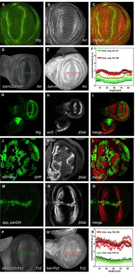
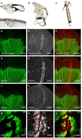
In theDrosophila melanogasterwing imaginal disc, the expression pattern of Arr and the inferred Wg protein gradient are inverse (Fig. 1A-C)–low Arr levels close to the Wg source are contrasted
[image:2.612.49.326.179.738.2]by high Arr levels at sites of little or no Wg. We explored the potential relevance of the Arr expression pattern by experimentally perturbing it, by using a heterologous ubiquitously active promoter. Surprisingly, animals that expressed Arr under the control of the tubulinα1promoter (tubArr) were viable and did not show any wing phenotypes. To monitor Arr expression intubArranimals, we used an antibody against Arr for immunohistochemical analysis. Arr expression was elevated intubArrwing pouches, but still graded, with reduced levels towards the Wg expression domain (Fig. 1D-F). We quantified the fluorescent intensities of Arr in tubArr and
Fig. 1. Wg represses its receptors Arrow and Frizzled 2–graded expression of the receptors is maintained upontubulinα1 promoter-mediated misexpression.In all panels showing imaginal discs, the posterior compartment is oriented towards the top, the anterior compartment is towards the bottom of the image. The green (D,P) and red (E,Q) rectangles displayed on these example discs show the ROI, sized 100 µm×16 µm, that was used to quantify the level of receptor in F and R. (A-C) Wingless (Wg) and Arrow (Arr) exhibit an inverse relationship in their expression levels. Immunofluorescence staining for Wg (green in A and C) and Arr (gray in B and red in C) revealed the inverse expression levels of the two proteins (C). (D,E) Graded Arr expression was maintained in Arr-overexpressing discs. Wild-type wing discs (D), and wing discs that misexpressed Arr, controlled by thetubulinα1promoter (tubArr) (E), were stained for Arr. (G-L) Wg repressesarrtranscription. The transcriptionalarrreporter
arr-lacZ(arrZ) (gray, H) was repressed in Wg-expressing areas of the wing imaginal disc. It was also repressed in randomly induced somatic clones (gray, K), as revealed by immunostaining against β-galactosidase (β-Gal), that overexpressed membrane-attached NRT-Wg [marked by green fluorescent protein (GFP) in J]. (M-O) Dominant-negative pan ( panDN) derepresses Arr. Expression of dominant-negativeUAS-panDNwithdppGAL4(M), marked by GFP, led to derepression of thearrZreporter (gray, N). (P,Q) Graded Fz2 expression is maintained in Fz2-overexpressing discs.tubFz2 -expressing wing discs (Q) that were immunostained for Fz2 showed a higher intensity profile of Fz2 than that of wild-type controls (P). Note that the receptor upregulation intubFz2was much stronger than in the correspondingtubArrdiscs. (F,R) Receptor level intensity plots. In both panels, intensities of four wild-type (wt, green-shaded crosses) and four receptor-overexpressing discs (OE, red-shaded crosses) are projected on the longer axis of the ROI, perpendicular to the dorsal-ventral compartment boundary. The corresponding moving averages (defined by a half window size of three micrometer) are denoted by solid green (wt) and red (overexpression) lines. Green- and red-shaded areas denote standard deviations of the mean of the data from wild-type and mutant imaginal discs, respectively. Intensity plots were normalized to the maximum of the moving average of the wild-type discs. a.u., arbitrary units.
DEVEL
O
control animals. The absolute Arr levels in tubArranimals were higher, whereas the shapes of the gradients were similar to those in the wild type (Fig. 1F; supplementary material Table S1).
In order to further analyze the arr transcriptional gradient, we used the recessive lethallacZP-element insertionl(2)k08131, which we refer to asarrZ(Wehrli et al., 2000) and is uncharacterized at the molecular level. We mapped the insertion site and found that it is positioned in the 50UTR region ofarrin the short first untranslated exon (data not shown). Owing to its insertion site and its expression pattern, arrZ can be used as a molecular read-out to study the transcription of thearrgene.arrZis expressed in a graded manner and is inactive in areas of high Wg signaling (Fig. 1G-I). To investigate this negative regulation of arr by Wg, we clonally overexpressed NRT-Wg, which is a membrane-tethered non-diffusible fusion protein of Wg (Zecca et al., 1996). All over the wing imaginal disc, clones that expressed NRT-Wg showed repression of arrZ, as did adjacent wild-type neighboring cells (Fig. 1J-L). We next asked whether the nuclear mediator of Wg signaling Pan is involved inarrZrepression. Overexpression of a dominant-negative form of Pan (UAS-panDN-HA), which lacks the Armadillo-binding domain and was tagged with hemagglutinin (HA), resulted in a strong and strictly cell-autonomous derepression ofarrZexpression (Fig. 1M-O). Hence,arrZis a negative target of Wg signaling in the wing pouch and is repressed as a consequence of signaling events downstream of Pan.
Negative transcriptional regulation by Wg signaling has also been reported for the fz2 gene, which encodes the main Wg receptor (Cadigan et al., 1998). To study the physiological relevance of this regulation, we misexpressed Fz2 under the control of thetubulinα1 promoter (tubFz2) in the wing imaginal disc. Similar to the case of tubArr,tubFz2animals had wings that appeared wild type. Also, as observed for Arr, Fz2 protein levels were elevated in tubFz2 animals, but remained graded with a slightly steeper slope, as observed for that in corresponding wild-type discs (Fig. 1P-R; supplementary material Table S1). Although weaker, a gradient could still be observed in larvae that harboredtubFz2but lacked endogenous Fz2 (supplementary material Fig. S1A-D and Table S1). These observations suggest that post-transcriptional regulation plays a rather minor role in gradient formation.
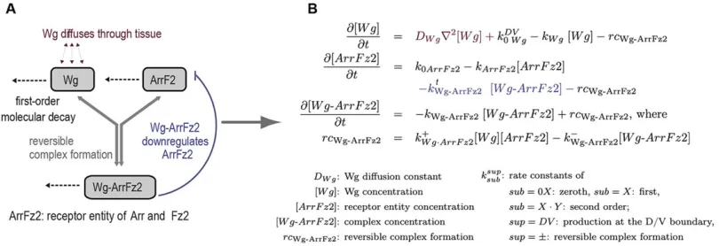
Modeling Wg receptor interactions
To formalize, and systematically study, the mechanisms of Wg-receptor interaction, we developed an in silico model that reproduces the negative regulation of Arr and Fz2 by Wg in cells of the wing pouch (Fig. 2). We first reduced the complexity of the
system by summarizing the similar biological roles of the receptor Arr and its co-receptor Fz2 into the receptor entity‘ArrFz2’. Next, we set up a system of coupled ordinary and partial differential equations that described the production and decay of the Wg ligand and its receptors, the reverse complex formation between ligand and the receptor entity, the diffusion of Wg, as well as the transcriptional downregulation of the receptor entity by Wg (Fig. 2).
A simplified analytical model suggests that most Wg is not bound to its receptors
Neglecting the Wg-induced transcriptional receptor repression (kt
WgArrFz2¼0 in Fig. 2) allowed us to study the effect of receptor
upregulation analytically in one dimension (Schwank et al., 2011; details in the supplementary methods). Supplementary material Fig. S2 shows the dependence of the amplitude of total Wg (black) in regions that overexpressed the receptor on the relative amount of ligand that was not bound to the receptor,a¼½Wg=½WgT. In such a simplified model, the higher the value ofa, the less the amplitude of the total Wg distribution is affected by changes in receptor levels.
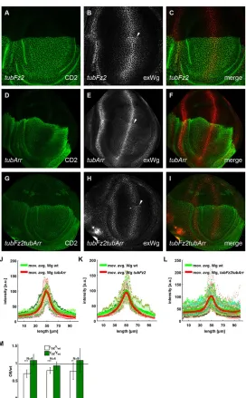
To study the consequences of apposing wild-type and receptor-overexpressing cells, as well as to have a direct comparison of the Wg distribution in receptor-overexpressing cells to wild-type cells in the same disc, we generatedtubulinα1>CD2,y+>arrand tubulinα1>CD2,y+>fz2transgenes that harbored flp-out cassettes between the promoter and coding region. Exposure to Flp recombinase during development leads to clones that overexpress the corresponding receptor. In discs, where the receptor overexpression was compartmental (Fig. 3; supplementary material Fig. S3 and Table S1), we found no significant difference between the Wg amplitudes in wild-type compartments and those that overexpressed the receptor. If the staining revealed the total amount of Wg, then the analytical model (supplementary material Fig. S2 and supplementary methods) suggests that the majority of Wg is not receptor bound.
A simplified analytical model predicts narrowing of Wg distribution upon receptor overexpression
[image:3.612.104.509.564.703.2]Assuming further that Wg decay occurs only in complex with its receptors (leading to kWg¼0), the analytical model predicts a narrowing of the Wg gradient in regions of receptor overexpression– ann-fold upregulation of the receptor would lead to a decrease in the Wg decay length (the distance from the Wg source, where the Wg concentration is reduced by a factor of 1/e from its maximal value) by a factor 1=pffiffiffiffiffiffiffiðnÞ in the receptor-overexpression (OE) region (supplementary methods). Remarkably, theory and experiment coincided within the measurement error for compartmental Arr
Fig. 2. Model representing Wg-induced negative regulation of Arr and Fz2.(A) Dotted arrows denote the first order decay of either single molecules or complexes of molecules; the reversible formation of complexes is denoted by groups of arrows. This mechanistic model can be translated into a set of differential
equations (B).
DEVEL
O
overexpression experiments; we found lOE;Arr=lwt¼0:7+0:1 (Fig. 3M; supplementary material Table S1), whereas the simple model predicted lOEth ;Arr=lwtth¼1=pffiffiffin¼1=pffiffiffi2¼0:7 for the upregulation of Arr by a factor ofn=2.
Receptors hinder diffusion of Wg
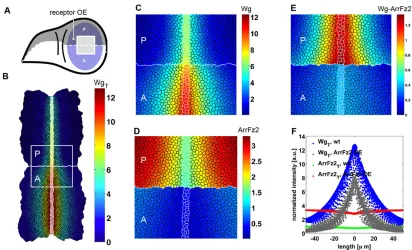
Extending our Wg receptor interaction model to two dimensions, we numerically solved the corresponding system of differential equations (displayed in Fig. 2) on a mesh, which resembled the cell shapes at the apical side of the wing disc. The vertex model describes cells and their contact surfaces as polygons that are composed from connected vertices with positions that are defined by the local minimum of an energy function (Farhadifar et al., 2007). Using these polygons as local control volumes, we discretized the diffusion of Wg by using the Finite Volume Method. Our modeling approach allowed us to study the consequences of changing receptor levels with cellular resolution in patches of cells, disc compartments or the entire wing pouch.
Simulating receptor overexpression in entire compartments (Fig. 4) led to a narrower Wg distribution than that in the corresponding
wild-type compartments (Fig. 4B,C,F; supplementary material Table S1). These findings are also supported by our measurements of the extracellular Wg gradients (Fig. 3J-L; supplementary material Fig. S3). In our model, cells that overexpressed ArrFz2 hindered the spreading of Wg, the only diffusing protein of the model; thus, these cells serve as a‘sink’for Wg if placed next to wild-type cells (Fig. 3)– cells that overexpressed ArrFz2, positioned immediately adjacent to wild-type cells, exhibited pathway activity (simulated by the amount of Wg-ArrFz2 complex formation) further away from the Wg source than the overexpressing cells that were not adjacent to wild-type cells (Fig. 4E). In our model, these cells receive unbound Wg from neighboring wild-type cells (see Discussion).
Abrupt changes in receptor levels lead to detrimental phenotypic effects
[image:4.612.50.322.291.730.2]The clonal abrupt upregulation of the receptor levels by using tubulinα1>CD2,y+>arr or tubulinα1>CD2,y+>fz2 transgenes caused detrimental phenotypic defects in the wings and legs of adult flies; in wings, we detected clones that caused typical Wg gain-of-function phenotypes, such as blistering and ectopic sensory-margin
Fig. 3. Compartmental misexpression of Arr and Fz2 leads to accumulation of Wg at the edge that is shared between wild-type and receptor-overexpressing cells and to a slightly decreased distribution of Wg.The posterior (towards the top of the image) and anterior compartments of the wing imaginal discs are marked by the absence and presence of CD2, respectively. Arrowheads indicate the point of Wg accumulation. (A-C) Expression oftubFz2in the P compartment led to an accumulation of extracellular Wg, exactly at the boundary between the A and P compartments. (D-I) Expression of
tubArrin the P compartment, ortubArrandtubFz2together, led to a similar extracellular Wg accumulation. (J-L) Wg level intensity plots in discs that compartmentally overexpressed Arr (J,n=4), Fz2 (K,n=4) and simultaneous overexpression of Arr and Fz2 (L,n=9), wheren
denotes the number of discs. Green-shaded crosses denote Wg intensities in the A compartment, red-shaded dots denote Wg intensities in the receptor-overexpressing P compartment. The moving average and standard error are defined as described in Fig. 1. a.u., arbitrary units. (M) Quantification of the change in Wg decay length (λ) and amplitude (y) upon receptor overexpression. Within each disc, we calculated the ratio between the decay length (white) and amplitude (green) in the receptor-overexpression (OE) and wild-type (wt) compartments. The bar charts display the mean and standard deviation for each experimental set up. We tested the null hypothesis, that decay length and amplitude were unchanged upon receptor overexpression, ***P<0.001, **P<0.01.
DEVEL
O
bristles (Fig. 5A). In legs, we found limb bifurcations that created supernumerary appendages (Fig. 5B), or an absence of tarsal segments and claws (Fig. 5C).
As an additional line of experimental validation, and to reach a higher level of reproducibility, we obtained clones that overexpressed Arr and Fz2 simultaneously through the activation of the tubulinα1>CD2, y+>fz2andtubulinα1>CD2,y+>arrcassettes upon ahedgehog-driven Flp recombinase, which was specifically expressed in the posterior compartment. They showed similar defects to the flies described above. Accordingly, when probed with an antibody against the prototypical Wg target geneSenseless(Sens), cells that co-overexpressedtub>fz2 andtub>arrand were adjacent to wild-type cells displayed ectopic expression of Sens (Fig. 5D-F). We observed a similar phenotype withtub>fz2flp-out clones (Fig. 5G-I), but not withtub>arrclones (Fig. 5J-L). If we induced clones overexpressing Arr with the stronger GAL4-UAS expression system (Fig. 5M-O), again, we observed upregulation of Sens at the dorsal-ventral compartment border. Thus, we conclude that perturbations of Wg receptor levels cause ectopic pathway activity in cells with high amounts of receptor, if juxtaposed with cells that have lower levels of receptor levels.
Receptor-overexpressing cells neighboring wild-type cells act as a Wg sink
To test our model–that the receptor-overexpressing cells apposed to wild-type cells, indeed, serve as a Wg sink and benefit from this situation by increased signaling–we monitored extracellular Wg in discs that misexpressed the receptors in all posterior (P) compartment
cells. Consistent with our model, such discs exhibited an accumulation of extracellular Wg, exactly at the border between wild-type anterior (A) and misexpressing P cells (arrows in Fig. 3A-I). These findings also explain the very restricted upregulation of Sens, noted above, exactly at the border between wild-type cells and cells that overexpressed the receptor.
The dual role of Fz3–stabilization of Wg, and attenuation of Fz2 and Arr
[image:5.612.99.511.55.305.2]Although Arr and Fz2 constitute the main Wg receptors, Fz3 has also been implicated in modulating the Wg response. Fz3 is expressed in the wing pouch (Sivasankaran et al., 2000) and has been proposed to serve as a negative-feedback regulator of the Wg pathway (Sato et al., 1999). Unlike Fz2 and Arr, however, the expression of Fz3 seems to be positively regulated by Wg signaling (Sivasankaran et al., 2000; Sato et al., 1999). To clarify the role of Fz3 in Wg gradient formation, we overexpressed Fz3 in the P compartment and monitored Sens levels as a read-out for Wg pathway activity. Indeed, we observed decreased Sens levels in the P territory, suggesting that overexpressed Fz3 attenuates Wg signaling (Fig. 6A-C). Consistent with this finding, Sens levels are enhanced infz3mutant mitotic clones (Fig. 6D-F). We wondered whether the alterations in Sens levels infz3mutant tissue, or tissue that overexpressedfz3, were mediated by changes in the levels of Wg, Fz2 or Arr. We first monitored extracellular Wg protein in compartments that overexpressed Fz3 and observed a notably enhanced and broadened extracellular distribution of the Wg protein (Fig. 6G-J). Interestingly, Fz2 and Arr levels were reduced in such Fig. 4. Modeling reveals ectopic pathway activation in receptor-overexpressing cells juxtaposed with wild-type ells.(A) Schematic view of a wing disc. The posterior (P) compartment of the wing pouch is marked in dark blue, and the anterior (A) compartment is marked in light blue.(B) An example simulation result shows the total Wg concentration of a disc, where the ArrFz2 production rate was increased by a factorn=3in all cells of the upper (P) compartment. Concentrations are displayed using Matlab’s jet algorithm, where red corresponds to high, and blue corresponds to low concentrations. All concentrations were normalized to the maximum value of the receptor entity ArrFz2 in wild-type discs and are given in arbitrary units. The white square corresponds to a zoomed-in region of 80 µm×80 µm centered around the zoomed-intersection of the dorsal-ventral and anterior-posterior compartment boundaries. (C-E) Wg (C), ArrFz2 (D) and Wg-ArrFz2 (E) distributions of the zoomed-in region of B. Wg-expressing cells are marked by white cell edges, the anterior-posterior compartment boundary is marked by a white (horizontal) line. Note the narrowing of the total Wg distribution in the receptor-overexpressing compartment. (C) The model predicted reduced levels of diffusible Wg in anterior cells that were adjacent to the compartment boundary. (E) The distribution of Wg-ArrFz2 broadened in posterior cells that were adjacent to the compartment boundary. (F) The total ArrFz2 concentrations (defined as the sum of free and Wg-bound receptor) in a wild-type wing disc (green) and in an ArrFz2-overexpressing wing disc (red) was projected onto the anteroposterior axis. Each cell in the tissue corresponds to one dot in the graphic. Total Wg levels in a wild-type simulation are shown in blue and in an ArrFz2-overexpression simulation in gray. a.u., arbitrary units.
DEVEL
O
tissue (supplementary material Fig. S4A-F); however, the activity of a transcriptional arr reporter (ArrC19, described below and in supplementary material Fig. S7) did show a small increase in the area over which it was expressed (supplementary material Fig. S4G-I), suggesting that Fz3 post-translationally regulates the levels of Fz2 and Arr. By contrast,fz3mutant wing discs exhibited increased levels of Fz2 and, to a lesser extent, Arr (supplementary material Fig. S4M-N), but no discernible effect on the distribution of Wg was observed (Fig. 6K). Remarkably, we only observed the downregulation of Arr and Fz2 upon overexpression of Fz3, if the overexpressing cells included the Wg source (supplementary material Fig. S4A-F). In Fz3-overexpressing clones that were distant from the Wg source, Arr levels were higher, as we expected for a negative target, following attenuation of signaling (supplementary material Fig. S4J-L). This also suggests that downregulation of the receptor is not the primary cause for the observed attenuation of Wg signaling. We did not detect any upregulation of Fz2 levels in this situation. We conclude from these findings that the binding of Wg to Fz3 mediates the degradation of Arr and Fz2, presumably lowering receptor levels at the
Wg-expressing stripe even further (supplementary material Fig. S4M-N), as well as broadening the area of Wg distribution through stabilizing the protein (Fig. 6G-J). These findings are supported by the observation that compartmental Fz3 overexpression, combined with RNA interference against Wg, reduces the downregulation of Arr in this compartment (data not shown).
[image:6.612.50.323.57.515.2]To further define and rationalize the above observations, we incorporated Fz3 into ourin silicomodel (supplementary material Fig. S5). We modeled Fz3 production to depend on the concentration of the Wg-ArrFz2 complex (our approximation of Wg pathway activity). Furthermore we assumed–as in the case of the receptor entity ArrFz2–a reversible complex formation between Wg and Fz3. Our experimental data (supplementary material Fig. S4J-L) suggest that the Fz3-induced repression of Arr and Fz2 is mediated by Wg (albeit the exact mechanism still has to be investigated). Therefore, we assume in our model that Fz3 represses Arr and Fz2 expression only when Fz3 is interacting with Wg. The resulting extended set of differential equations is provided in the supplementary methods. Scanning the corresponding parameter Fig. 5. Clonal overexpression of Arr and Fz2 leads to severe border effects.(A) Adult wing phenotypes that arose from random clones that expressedtubFz2ortubArrshowed Wg gain-of-function defects, such as blistering and ectopic sensory bristles (arrowheads). (B,C) RandomtubFz2- andtubArr-expressing mitotic clones resulted in bifurcations and truncations in legs. (D-F) Third-instar imaginal discs that containedhhGal4, UAS-Flp, tubFz2, tubArrclones (D-F) showed ectopic expression of the Wg target Sens (gray in E, red in F), predominantly in cells that were adjacent to wild-type neighbors at the border of the clone, close to the Wg source [tubFz2- andtubArr-expressing clones were marked by the absence of CD2 (green in D and F)]. (G-I) Similar to cells that expressed both receptor components, cells that only expressedtubFz2exhibited ectopic expression of Sens at the border of the clone (gray in H, red in I). CD2 expression is shown in green in G and I. (J-O)hhGal4, UAS-Flp, tubArrclones did not show strong Sens upregulation at the border with wild-type cells (J-L); however, in random heatshock-inducedUAS-GFP, UAS-Arr-expressing clones (M-O), ectopic Sens expression at the border of the clone facing the Wg source was observed (arrowhead). CD2 staining is shown in green in J and L; Sens staining is marked by gray in K and N, and by red in L and O; GFP in M represents the Arr expression domain; the red outline in N marks the clone outline shown in M.
DEVEL
O
space (supplementary methods), we identified a parameter set that qualitatively reproduced the experimentally observed Wg and receptor distributions in the wild-type and receptor-misexpression conditions (the exact parameters are shown in supplementary material Table S1). Remarkably, the parameter exploration suggested that the complex that was formed by Wg and Fz3 might be more stable than the corresponding Wg-ArrFz2 complex (see supplementary material Table S2 in the supplementary methods), confirming the role of Fz3 in stabilizing Wg.
In summary, we show that Fz3 acts as an attenuator of Wg signaling in the wing pouch. We propose that Fz3 reduces the amount of Arr and Fz2 protein close to the Wg source and mediates a broader distribution of Wg.
Wg signaling indirectly repressesarrand directly activatesfz3
[image:7.612.50.322.56.573.2]The existence of the transcriptional gradients of Arr and Fz2 prompted us to analyze by which mechanism(s) Wg signaling represses the transcription of their genes. Many feedback regulatory mechanisms of signaling pathways use direct transcriptional regulation. However, signaling-mediated transcriptional repression (as opposed to activation) has rarely been described (Affolter et al., 2008). Thus, we sought to isolate and characterize the regulatory DNA elements ofarr andfz2that are responsible for the Wg input. The similar expression patterns and regulatory roles ofarrandfz2led us to speculate that both genes might be transcriptionally controlled by the same molecular mechanism. Hence, we chose the arr locus as a case study and
Fig. 6. Fz3 acts as an attenuator of Wg signaling.
(A-C) Overexpression of Fz3 in the P compartment (A), marked with GFP, results in a prominent reduction of Sens levels (gray in B, red in C). (D-F) Fz3 loss-of-function clones (fz3G10), marked by the absence of GFP (clones induced with anFRT19, ubiGFPchromosome) (D), led to an enhancement of Sens expression at the dorsal-ventral compartment boundary (gray in E, red in F). (G-I) Ectopic expression of Fz3 in the P compartment (G) resulted in an enhancement and broadening of the extracellular Wg gradient, revealed by immunostaining of the extracellular (ex) Wg levels (gray in H, red in I). GFP marks the P compartment. (J) Intensity profile of the Wg gradient in Fz3-overexpressing (OE) P compartments (red) and wild-type (wt) compartments (green) in a ROI of 100 µm×20 µm.n=8. Note the higher and broader Wg gradient in Fz3-overexpressing compartments. (K) Intensity profile of the Wg gradient infz3mutant wing discs (red,
fz3−) and wild-type wing discs (green) in a ROI of 100 µm×20 µm.n=8.
fz3mutant wing discs showed reduced levels of Wg and a narrower Wg distribution. (L) Results of the simulation model showed the total Wg levels (defined as the sum of free and receptor-bound Wg) in a wild-type (green),fz3mutant (dark red) and Fz3-overexpression simulation (light red). As in the experimental intensity profiles (J,K), Wg levels were normalized to the maximum of the wild-type expression. The simulation qualitatively reproduced the behavior of the experimental situations (J,K; supplementary material Table S1). a.u., arbitrary units. (M) A simulated profile of Senseless (red) distribution in wild-type (lower compartment) andfz3−cells (upper compartment). The ROI was the same as that described in Fig. 4. Wg-expressing cells are marked by white edges, and the compartment boundary is marked by a white line. We modeled Wg-expressing cells as being deficient for Senseless expression. It was assumed that all other cells with Wg-ArrFz2 concentrations higher than 95% of the maximal Wg-ArFz2 concentration expressed Senseless. A, anterior; P, posterior.
DEVEL
O
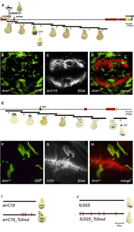
systematically dissected its regulatory region. Seven overlapping fragments (A toG) covering 25 kb were cloned into a placZ-attB reporter vector, 50to anhsp70minimal promoter. Each construct was integrated into the sameattPlanding site on the second chromosome (Bischof et al., 2007) and the expression of lacZ in imaginal discs was analyzed. FragmentarrCrecapitulated the endogenousarrexpression pattern (Fig. 7A) and also properly reacted to experimentally increased and decreased Wg pathway activity, as assessed in clones that expressedNRT-WgandpanDN, respectively (data not shown). We next narrowed down the regulatory activity ofarrCby using an extensive series of reporter constructs (supplementary material Fig. S7), resulting in a 200 bp fragment termedarrC19, which could not be shortened further without compromising activity. Clones that expressed a constitutively active version of Arm (Armact) repressed arrC19 reporter expression, confirming that it retained the element(s) responsible for the negative Wg input (Fig. 7B-D). To check if this regulatory Wg input was mediated directly by Pan, we mutated the four highly conserved Pan binding sites in the arrC19 enhancer. The expression pattern of the resultingarrC19_Tcf_muttransgene was not
derepressed, suggesting that Pan is not directly involved in regulating arrC19(Fig. 7I). Thus, we conclude that Wg-mediated repression of arrC19occurs independently of the direct binding of Pan.
Furthermore, we analyzed the nature of the Wg input onfz3by using a similar approach – dissecting the fz3 locus into seven overlapping fragments,AtoG. The expression pattern of fragment Fz3G was the only one that was reminiscent of the pattern offz3. Further dissection yielded fz3G5, a fragment of approximately 290 bp that retained fz3-like expression (Fig. 7E). Clones that expressedArmact(Fig. 7F-H) caused upregulation offz3G5reporter gene activity. This regulation seems to be directly exerted by the canonical Wg pathway because it was completely abrogated upon mutation of the Pan-binding site (Fig. 7I).
[image:8.612.50.320.276.743.2]We conclude that Wg signaling indirectly regulates arr (and presumablyfz2), whereasfz3is a direct positive-target gene of the pathway. Thus, the sign of regulation by Wg ofarrandfz2differs from that of fz3 (to impact negatively and positively on their expression, respectively), as well as the mode of regulation (indirect versus direct).
Fig. 7.arris an indirect negative Wg target, andfz3is a direct positive Wg target.A map of the genomic region ofarr(A). Translated exons are shown in red, untranslated regions in yellow. FragmentsA-G
were tested for their ability to activate a heterologous promoter driving
lacZexpression (blue stain). Only fragmentCrecapitulated thearr
expression pattern in the wing imaginal disc. Insertion of the P-element
arrZis shown on top. Further dissection of fragmentCrevealed the minimal enhancer fragmentarrC19(see supplementary material Fig. S4 for further details). (B-D) Randomly induced clones that overexpressed the constitutively activeUAS-Armact, marked with GFP in (B), led to a
cell-autonomous repression ofarrC19expression, revealed by immunostaining ofβ-galactosidase (gray in C, red in D) (C,D). (E) A map of the genomicfz3locus. FragmentsA-Gwere tested for their ability to activate a heterologous promoter drivinglacZexpression. Only fragmentGrecapitulated thefz3expression pattern in the wing pouch. (F-H) Overexpression ofUAS-Armact(green) (F) results in a cell-autonomous derepression offz3G, as shown by by immunostaining ofβ-galactosidase (gray in G, red in H) (G,H). (I) Mutation of TCF sites in conserved regions ofarrC19did not affect the expression of the reporter. By contrast, mutation of TCF sites in thefz3G5reporter completely abolished reporter expression (J).
DEVEL
O
DISCUSSION
During the development of a metazoan organism, signaling events are precisely regulated. One frequently employed mode of regulation is feedback loops. Here, we analyzed a network of feedback loops in theDrosophilawing pouch that regulate receptor abundance, and thus the range of distribution and signaling output of Wg.
Receptors sequester their ligands and, thereby, impact upon the range of the signal (supplementary material Fig. S6). A transcriptional regulatory link between receptor expression and signaling activity, causing up- or downregulation of receptor levels in cells in response to the signal, can thus restrict or extend the signaling range. For example, the Hedghog (Hh) signal induces the expression of its receptor Patched (Ptc), a regulatory link which severely narrows the Hh activity gradient (Chen and Struhl, 1996). In the case of Wg, we observed the opposite–Wg signaling appeared to extend the range of Wg distribution by transcriptionally downregulating expression ofarr and fz2; downregulation of the receptors hinders the formation of Wg-Arr-Fz2 complexes. This allows superfluous Wg to diffuse further away from the source without being immobilized by its receptors. In agreement with this notion, we observed a slightly narrower distribution of extracellular Wg in discs that expressed Fz2 or Arr under the tubulinα1 promoter (Fig. 3; supplementary material Fig. S2). Quantifying these observations in discs that compartmentally overexpressed the receptor (Fig. 3; supplementary material Fig. S2 and Table S1), we observed a subtle reduction of the decay length (corresponding to a slightly steeper Wg distribution) in compartments that overexpressed the receptor compared with that in wild-type compartments.
In apparent contradiction, a previous study (Cadigan et al., 1998) has shown that high levels of Fz2 can stabilize Wg and promote Wg spreading; accordingly, we observed an accumulation of Wg when repeating this experiment by overexpressing Fz2 using the GAL4-UAS system. These contradictory findings can be reconciled by taking into account the different strength of Fz2 upregulation in the two experimental setups – Fz2 expression that is driven by the tubulinα1promoter leads to a relatively mild upregulation of the receptor by, approximately, a factor of 2 (Fig. 1; supplementary material Table S1), whereas overexpression by using the GAL4-UAS system causes a much stronger overexpression. Presumably, Arr becomes the limiting factor inUAS-Fz2-overexpressing cells, a situation that might prevent the surplus Wg-Fz2 complexes from being internalized, thus causing an extracellular accumulation of Wg (Marois et al., 2006). If Fz2 is only moderately overexpressed, sufficient Arr protein might be available to allow this extra Fz2 to form Wg-Arr-Fz2 complexes, which are subsequently internalized, leading to a slight narrowing of the gradient because there is less free and diffusible Wg. Consistent with this notion, simultaneous strong overexpression of both of the receptors Fz2 and Arr, by means of Gal4, leads to a reduction of extracellular Wg levels (Piddini et al., 2005).
Receptors act as a Wg sink
Although Wg signaling transcriptionally represses both Arr and Fz2, ubiquitous overexpression of Arr, or Fz2, had no phenotypic consequences. Unexpectedly, however, severe phenotypes arose upon mosaic expression of the tub>arr or tub>fz2 transgenes. Theoretical modeling (Fig. 4) and reporter gene analysis indicated that cells that had elevated receptor levels ectopically activated the pathway when situated close to wild-type cells. Apparently, the ‘high-receptor-level state’allowstub-fz2ortub-arrcells to engage in ligand-receptor interactions that depend on the ‘ low-receptor-level state’of their neighbors. One plausible explanation might be
that tub-fz2, or tub-arr, cells bind to Wg that diffuses in from neighboring wild-type cells.
The different outcome of clonal versus uniform alteration of the Wg pathway is reminiscent of observations that have been reported by Piddini and Vincent (Piddini and Vincent, 2009), where loss of Wg signaling in the entire P compartment had no impact on the expression of low-threshold target genes but resulted in their repression, and in patterning defects, when Wg signaling was only clonally abolished. Piddini and Vincent also used different patterns of Wg receptor expression for their experiments, and they explained their findings by postulating that there is a Wg-induced, still to be identified, inhibitory signal that negatively regulates target gene expression in surrounding cells.
The negative feedback regulator Fz3
In an additional layer of negative-feedback regulation in the wing pouch, Wg signaling activates the expression of the Frizzled family member Fz3 (this work; Sato et al., 1999; Sivasankaran et al., 2000). Fz3 seems to act as a negative regulator of Wg signaling by repressing Wg signaling readouts and downregulating Wg receptor levels. Various models could be envisaged of how Fz3 acts as an inhibitor of Wg signaling. As it has been demonstrated that Fz3 is able to bind Wg (Wu and Nusse, 2002), Fz3 could work as a decoy receptor that acts as a molecular trap by binding to Wg without eliciting a signal. Decoy receptors are often part of negative-feedback mechanisms. In the Drosophila epidermal growth factor (EGF) system, the pathway inhibitor Argos is a target of EGF signaling and functions as a decoy receptor (Klein et al., 2004). In vertebrates, decoy Frizzled receptors have been identified that modulate Wnt signaling–secreted Frizzled-related molecules (sFRPs) have strong homology to the Frizzled extracellular domains. sFRPs inhibit signaling by directly binding to the Wnt ligands (Mii and Taira, 2011). No sFRP gene has been identified in theDrosophilagenome.
In another scenario, Fz3 could work as a negative regulator of Wg receptors. Its function could be analogous to that of ZNRF3 and RNF43 in crypt base columnar intestinal stem cells. These related E3 ubiquitin ligases have been shown to regulate the stability and levels of cell-surface Fz and LRP5/6, through internalization and lysosomal degradation of the receptor components in the presence of Wnt signaling (Hao et al., 2012; Koo et al., 2012). Several of our experimental findings indicate that Fz3 might work as an inhibitor of Wg feedback at the receptor level–firstly, we observed decreased Arr and Fz2 levels in compartments that overexpressed Fz3 (supplementary material Fig. S2B,E), and secondly, Arr and Fz2 levels were increased in fz3 mutant wing discs (supplementary material Fig. S4M,N). Most probably, Fz3 acts by more than one mechanism–cells that overexpressed Fz3 in the Wg stripe lead to Arr downregulation, whereas cells that overexpressed Fz3 outside of the Wg stripe lead to Arr upregulation (supplementary material Fig. S4J-L). Furthermore, extracellular Wg was stabilized upon Fz3 overexpression (Fig. 6G-I). In a wild-type situation, this stabilization of Wg might contribute to a broader Wg gradient and promote signaling in the outskirts of the wing pouch. Taken together, these findings suggest that only Wg-bound Fz3 causes inhibition of the pathway.
The role of transcriptional Wg receptor regulation
The post-translational regulation of Wg receptor levels was not the focus of this study, but we undertook substantial efforts to further characterize the transcriptional regulation of the receptor genes. In particular, we sought to identify the regulatory elements of these genes that mediated the feedback loops. The isolation of a 200 bp
DEVEL
O
fragment of thearrlocus and a 300 bp fragment of thefz3gene– each of which was responsive to Wg signaling and drove reporter gene expression in a pattern that was reminiscent of the endogenous expression pattern – allowed us to investigate whether the Wg pathway controls these genes directly or indirectly; fz3 appeared to be a direct target of canonical Wg signaling, whereasarrdid not. Pan-binding sites were dispensable in the minimalarrenhancer, indicating that either Arm regulates the transcriptional activity of arr through another DNA-binding protein, or that Arm and/or Pan transcriptionally induce one (or more) negative regulators that, in turn, regulate arr expression. Hence, although the Wg pathway has been reported to possess the capacity to directly negatively regulate transcription (Blauwkamp et al., 2008; Affolter et al., 2008), it apparently does not use this mechanism to attenuatearrexpression.
Including transcriptional Wg receptor downregulation in our model led to a broader distribution of Wg (supplementary material Fig. S6A)–receptor downregulation by ligand-induced endocytosis consumes the ligand, this was not the case for transcriptional repression (supplementary material Fig. S6B). The broadening of the Wg distribution area under a mechanism of transcriptional receptor repression might facilitate a robust signaling readout for high-threshold Wg target genes.
A recent study indicates that the long-range Wg gradient might be less important for imaginal disc patterning than assumed previously (Alexandre et al., 2014). Hence, it is also conceivable that the receptor gradients are not essential, a notion supported by our finding that uniform misexpression of Arr or Fz2 in the wing imaginal disc had no phenotypic consequences. Nevertheless, it remains to be determined whether the Arr and Fz2 gradients are dispensable; our tubArr transgene is not able to rescue arr loss-of-function mutants.
A cell-based ligand-receptor model
So far, most quantitative models of the Wnt-Wg pathway have focused on intracellular events (reviewed in Kofahl and Wolf, 2010), and only a few models have taken into account the spatial aspects of this signaling system (Sick et al., 2006; Ramis-Conde et al., 2008; van Leeuwen et al., 2009; Zhu, 2011). Our model is the first to systematically study the roles of Wg-receptor complexes – Wg-ArrFz2 and Wg-Fz3–in the spatial profile of Wg signaling, as well as being the first to be challenged experimentally by manipulations of the receptor levels. Our cell-based modeling approach of ligand receptor interactions allowed us to vary all parameters in a cell-autonomous manner, which has not been done in previous studies (Eldar and Barkai, 2005; Dalessi et al., 2012; Schwank et al., 2011). This technique is, thus, an ideal tool to predict the impact of clonal conditions with cellular precision, which have historically formed the basis of experimental approaches inDrosophilabut have also become increasingly available in vertebrates.
MATERIALS AND METHODS
Quantifying the shape of Wg and receptor distributions
We quantified the Wg intensity projections of each disc,i, by fitting an
exponential decayyi s¼y0i sexpðx=li sÞ, wherey0i sis the corresponding
Wg amplitude andli sthe decay length, from the center of the region of
interest to both sides (s[½left;right) up to 30 µm, omitting the
Wg-producing source region (approximately 4 µm in width).
How well the data fitted to each curve was estimated by calculating the coefficient of determination,R2is, from the intensity projections and the fit of the curve. For the two fits, to the left and right within each region of interest
(ROI), of disc numberi, we calculated the meanR2i and the mean of the
absolute values of the two decay lengths,li¼ðjli leftjþjli rightjÞ=2.
We quantified the receptor intensity projections of each disciby fitting
exponential functions, linear (data not shown) and second order
polynomials yiðxÞ¼aix2þbixþci, where the latter showed the best
overall coefficients of determination. The slope of the receptor distributions was then defined as the derivation of the second order
polynomial with respect tox. Supplementary material Table S1 displays the
mean fit coefficients of n measurements.
Fly stocks
Transgenic flies were generated by using the PhiC31 transgenesis system and integrated on the landing sites ZH-51D or ZH-86Fb (Bischof et al., 2007).
The following stocks were used for genetic and clonal analysis: Sp/CyO; hhGAL4, UAS-Flp/TM6B(Pérez-Garijo et al., 2009); yw hsp-flp; UAS-HA-NRT-Wg/SM5^; MKRS/TM6B(Zecca et al., 1996); yw hsp-flp; UAS-ΔPan-HA/TM6B(van de Wetering et al., 1997); yw hsp-flp; UAS-Armact(Zecca et al., 1996);
yw hsp-flp; Sp/CyO; dpp-GAL4/TM6B(Entchev et al., 2000);
yw hsp-flp; enGAL4, UAS-GFP, tubGal80ts/CyO; MKRS/TM6B (individual components are from Bloomington);
yw hsp-flp; act>CD2, y+>GAL4, UAS-GFP(Pignoni et al., 1997); UAS-Fz3, UAS-Arr, UAS-Fz2 and UAS-PanHA(Bischof et al., 2013); yw hsp-flp; C765-GAL4(Nellen et al., 1996);
Tub-flp(Struhl et al., 1993);
P(lacZ) arrk08131(Bloomington stock 665);
FRT19(Bloomington stock 1709),Fz3G10(Sato et al., 1999); fz2(c1)(Chen and Struhl, 1999);
Df(3L)fz2(Bloomington stock 6754).
Vectors
For cloning, standard molecular biology cloning methods were used.
To generate hybridtubArrandtubFz2constructs, the coding regions of
Arr and Fz2, respectively, were cloned 30in-frame of thetubulinα1promoter
and aFRT-flanked >CD2,y+>flp-out cassette in a plasmid that contained
thetubulinα130UTR and anattBsite for phage-mediated transgenesis.
To generatelacZreporter constructs, fragments of the genomicarrand
thefz3locus were amplified fromywgenomic DNA by using PCR with
primers that harbored suitable restriction enzymes for subsequent cloning
into placZattB. A detailed list of the primers and constructs that were used is
available upon request.
The sequences of arrC19 and fz3G5 containing mutated TCF sites
(mutated from ttt to ccc, the site is in bold) were synthesized by Entelechon
GmBH as follows: arrC19, 50
-CCAACAGCCAGCAGTCTCCCCGTT-
CGACGAATTGCTAATTTATTTCACTCTTGTACCTCGGAACACGCT-TCCTATCTAGACGTTGGAAAgggA
CCAATTTAAAGTTTATTTATGG-CgggTATTAgggTATA CCCTCTCTATATTTGGCATTTTTCCTGTTGC- TCTTTTTTTTTTTGCCTTTTGCAGCTGCCTCTTCGACTGGCGTTTG-TCAATgggTAACCTGTTGTGCATACTAATTTG-30; fz3G5, 50
;-GGTAC-CCTTTCCCTGCTCTCTGGATGgggGGGC
TGCGTCAGAACACGGG-TAATTGGTATCAACCGAGGCACAATTAAgggGAT
TGAAGCTGCC-ACACCGCATGCCGCTCCAACGAACAACGAATCCATCCTACcccT
T-GCCGGCCGTCGTGGCAGGCAACAAGCCGccc
CACTGCGGCAACA-
TCGGCACAATAGCAACAGCAACTGTGCAGCAAGCGTGCAATTA-ACTCTTGTGCGCCAAGATCcccG
CGATTAATGCGGCCCCAAGTT-CCCAGACGAAGGGACTCTAGA-30.
Clone induction
tubArr- andtubFz2-expressing clones were generated by heat-shocking the larvae for 1 h at 37°C 72 h (±12 h) after egg-laying and dissected 2-3 days after clone induction. P compartments that expressed UAS constructs were generated
by shifting the crosses to 29°C for 2-3 days before dissection. NRT-Wg
-expressing clones were induced 4 days after egg-laying for 15 min at 37°C.
Immunohistochemistry
Immunostainings were performed using standard protocols. Images were taken by using a Zeiss LSM710 confocal microscope using 40× and 63× oil
objectives and analyzed with the ImageJ software. Maximumzprojections
DEVEL
O
of ROI were performed for the individual micrographs: Discs were oriented and processed in Adobe Photoshop, intensities were projected on the longer axis of the ROI, perpendicular to the dorsal-ventral compartment boundary. For each condition, we measured at least four independent discs.
The following antibodies were used against the indicated proteins:
β-galactosidase (mouse, 1:2000, Promega); Arrow (rabbit, 1:15,000, a
generous gift from Steve DiNardo, University of Pennsylvania, USA; Senseless (guinea pig, 1:800, a generous gift from Hugo Bellen, Baylor College of Medicine, Houston, TX, USA; Wg [mouse, 1:3 (for extracellular stainings, see Baeg et al., 2001), Developmental Studies Hybridoma Bank]; Fz2 (mouse, 1:30, Developmental Studies Hybridoma Bank); CD2 (rat, Alexa Fluor 488-conjugated, 1:25, Serotec); HA (rabbit, 1:200, Santa Cruz Biotechnology).
To detectβ-galactosidase activity of thearrreporters, third-instar larvae
were subjected to standard X-Gal reactions. For color detection, we used standard conditions.
The secondary antibodies used were Alexa Fluor 594-conjugated goat against mouse IgG, Alexa Fluor 488-conjugated goat against mouse IgG, Alexa Fluor 594-conjugated against rabbit IgG, Alexa Fluor 488-conjugated against rabbit IgG, Alexa Fluor 568-conjugated against guinea pig IgG. All were obtained from Molecular Probes and were used 1:400.
Acknowledgements
We thank S. DiNardo and H. Bellen for antibodies; G. Morata, G. Struhl and T. Kojima for fly stocks; F. Meyer and P. Herr for help with injections; and G. Hausmann, T. Valenta, M. Hödl and C. Cantù for critically reading the manuscript.
Competing interests
The authors declare no competing financial interests.
Author contributions
All authors developed the presented concepts and prepared the manuscript. S. Steiner and D.Z. performed the experiments; S. Schilling, S. Steiner and D.Z. performed the data analysis. S. Schilling developed thein silicomodel of Wg receptor interactions and wrote the supplementary methods.
Funding
The European Research Council, the Swiss National Science Foundation and the Canton of Zurich supported this work.
Supplementary material
Supplementary material available online at
http://dev.biologists.org/lookup/suppl/doi:10.1242/dev.108662/-/DC1
References
Affolter, M., Pyrowolakis, G., Weiss, A. and Basler, K.(2008). Signal-induced
repression: the exception or the rule in developmental signaling?Dev. Cell15, 11-22.
Alexandre, C., Baena-Lopez, A. and Vincent, J.-P.(2014). Patterning and growth
control by membrane-tethered Wingless.Nature505, 180-185.
Baeg, G. H., Lin, X., Khare, N., Baumgartner, S. and Perrimon, N.(2001).
Heparan sulfate proteoglycans are critical for the organization of the extracellular distribution of Wingless.Development128, 87-94.
Baeg, G.-H., Selva, E. M., Goodman, R. M., Dasgupta, R. and Perrimon, N.(2004).
The Wingless morphogen gradient is established by the cooperative action of Frizzled and Heparan Sulfate Proteoglycan receptors.Dev. Biol.276, 89-100.
Bischof, J., Maeda, R. K., Hediger, M., Karch, F. and Basler, K.(2007). An
optimized transgenesis system for Drosophila using germ-line-specific phiC31 integrases.Proc. Natl. Acad. Sci. U.S.A.104, 3312-3317.
Bischof, J., Björklund, M., Furger, E., Schertel, C., Taipale, J. and Basler, K.
(2013). A versatile platform for creating a comprehensive UAS-ORFeome library in Drosophila.Development140, 2434-2442.
Blauwkamp, T. A., Chang, M. V. and Cadigan, K. M.(2008). Novel TCF-binding
sites specify transcriptional repression by Wnt signalling.EMBO J.27, 1436-1446.
Cadigan, K. M., Fish, M. P., Rulifson, E. J. and Nusse, R.(1998). Wingless
repression of Drosophila frizzled 2 expression shapes the wingless morphogen gradient in the wing.Cell93, 767-777.
Chen, Y. and Struhl, G.(1996). Dual roles for patched in sequestering and
transducing hedgehog.Cell87, 553-563.
Chen, C. M. and Struhl, G.(1999). Wingless transduction by the Frizzled and
Frizzled2 proteins of Drosophila.Development126, 5441-5452.
Clevers, H.(2006). Wnt/β-catenin signaling in development and disease.Cell127,
469-480.
Dalessi, S., Neves, A. and Bergmann, S.(2012). Modeling morphogen gradient
formation from arbitrary realistically shaped sources.J. Theor. Biol.294, 130-138.
Eldar, A. and Barkai, N. (2005). Interpreting clone-mediated perturbations of
morphogen profiles.Dev. Biol.278, 203-207.
Entchev, E. V., Schwabedissen, A. and González-Gaitán, M.(2000). Gradient
formation of the TGF-beta homolog Dpp.Cell103, 981-991.
Farhadifar, R., Roper, J.-C., Aigouy, B., Eaton, S. and Julicher, F.(2007). The
influence of cell mechanics, cell-cell interactions, and proliferation on epithelial packing.Curr. Biol.17, 2095-2104.
Hao, H.-X., Xie, Y., Zhang, Y., Charlat, O., Oster, E., Avello, M., Lei, H., Mickanin,
C., Liu, D., Ruffner, H. et al.(2012). ZNRF3 promotes Wnt receptor turnover in
an R-spondin-sensitive manner.Nature485, 195-200.
Klein, D. E., Nappi, V. M., Reeves, G. T., Shvartsman, S. Y. and Lemmon, M. A.
(2004). Argos inhibits epidermal growth factor receptor signalling by ligand sequestration.Nature430, 1040-1044.
Kofahl, B. and Wolf, J.(2010). Mathematical modelling of Wnt/β-catenin signalling.
Biochem. Soc. Trans.38, 1281-1285.
Koo, B.-K., Spit, M., Jordens, I., Low, T. Y., Stange, D. E., van de Wetering, M.,
van Es, J. H., Mohammed, S., Heck, A. J. R., Maurice, M. M. et al.(2012).
Tumour suppressor RNF43 is a stem-cell E3 ligase that induces endocytosis of Wnt receptors.Nature488, 665-669.
Marois, E., Mahmoud, A. and Eaton, S.(2006). The endocytic pathway and
formation of the Wingless morphogen gradient.Development133, 307-317.
Mii, Y. and Taira, M.(2011). Secreted Wnt “inhibitors” are not just inhibitors:
regulation of extracellular Wnt by secreted Frizzled-related proteins.Dev. Growth Differ.53, 911-923.
Mosimann, C., Hausmann, G. and Basler, K.(2009). [beta]-Catenin hits chromatin:
regulation of Wnt target gene activation.Nat. Rev. Mol. Cell Biol.10, 276-286.
Nellen, D., Burke, R., Struhl, G. and Basler, K.(1996). Direct and long-range
action of a DPP morphogen gradient.Cell85, 357-368.
Neumann, C. J. and Cohen, S. M.(1997). Long-range action of Wingless organizes
the dorsal-ventral axis of the Drosophila wing.Development124, 871-880.
Pérez-Garijo, A., Shlevkov, E. and Morata, G.(2009). The role of Dpp and Wg in
compensatory proliferation and in the formation of hyperplastic overgrowths caused by apoptotic cells in the Drosophila wing disc.Development136, 1169-1177.
Piddini, E. and Vincent, J.-P. (2009). Interpretation of the wingless gradient
requires signaling-induced self-inhibition.Cell136, 296-307.
Piddini, E., Marshall, F., Dubois, L., Hirst, E. and Vincent, J.-P.(2005). Arrow
(LRP6) and Frizzled2 cooperate to degrade Wingless in Drosophila imaginal discs.Development132, 5479-5489.
Pignoni, F. and Zipursky, S. L.(1997). Induction of Drosophila eye development by
decapentaplegic.Development124, 271-278.
Ramis-Conde, I., Drasdo, D., Anderson, A. R. A. and Chaplain, M. A. J.(2008).
Modeling the influence of the e-cadherin-β-catenin pathway in cancer cell invasion: a multiscale approach.Biophys. J.95, 155-165.
Rives, A. F., Rochlin, K. M., Wehrli, M., Schwartz, S. L. and DiNardo, S.(2006).
Endocytic trafficking of Wingless and its receptors, Arrow and DFrizzled-2, in the Drosophila wing.Dev. Biol.293, 268-283.
Sato, A., Kojima, T., Ui-Tei, K., Miyata, Y. and Saigo, K.(1999). Dfrizzled-3, a new
Drosophila Wnt receptor, acting as an attenuator of Wingless signaling in wingless hypomorphic mutants.Development126, 4421-4430.
Schwank, G., Dalessi, S., Yang, S.-F., Yagi, R., de Lachapelle, A. M., Affolter, M.,
Bergmann, S. and Basler, K. (2011). Formation of the long range Dpp
morphogen gradient.PLoS Biol.9, e1001111.
Sick, S., Reinker, S., Timmer, J. and Schlake, T.(2006). WNT and DKK determine
hair follicle spacing through a reaction-diffusion mechanism. Science 314, 1447-1450.
Sivasankaran, R., Calleja, M., Morata, G. and Basler, K.(2000). The Wingless
target gene Dfz3 encodes a new member of the Drosophila Frizzled family.Mech. Dev.91, 427-431.
Struhl, G., Fitzgerald, K. and Greenwald, I.(1993). Intrinsic activity of the lin-12
and Notch intracellular domains in vivo.Cell74, 331-345.
van de Wetering, M., Cavallo, R., Dooijes, D., van Beest, M., van Es, J., Loureiro, J.,
Ypma, A., Hursh, D., Jones, T., Bejsovec, A. et al.(1997). Armadillo coactivates
transcription driven by the product of the Drosophila segment polarity gene dTCF. Cell88, 789-799.
van Leeuwen, I. M. M., Mirams, G. R., Walter, A., Fletcher, A., Murray, P., Osborne,
J., Varma, S., Young, S. J., Cooper, J., Doyle, B. et al.(2009). An integrative
computational model for intestinal tissue renewal.Cell Prolif.42, 617-636.
Wehrli, M., Dougan, S. T., Caldwell, K., O’Keefe, L., Schwartz, S.,
Vaizel-Ohayon, D., Schejter, E., Tomlinson, A. and DiNardo, S.(2000). arrow encodes
an LDL-receptor-related protein essential for Wingless signalling.Nature407, 527-530.
Wu, C.-h. and Nusse, R.(2002). Ligand receptor interactions in the Wnt signaling
pathway in Drosophila.J. Biol. Chem.277, 41762-41769.
Zecca, M., Basler, K. and Struhl, G.(1996). Direct and long-range action of a
Wingless morphogen gradient.Cell87, 833-844.
Zhu, H. (2011). Spatiotemporally modulated Vestigial gradient by Wingless
signaling adaptively regulates cell division for precise wing size control.
J. Theor. Biol.268, 131-140.