RESEARCH ARTICLE
β
-catenin-mediated adhesion is required for successful
preimplantation mouse embryo development
Daniel Messerschmidt1,2,*, Wilhelmine N. de Vries3,*, Chanchao Lorthongpanich1,4, Sathish Balu1,5, Davor Solter1,4and Barbara B. Knowles1,3,4,‡
ABSTRACT
β-catenin (CTNNB1) is integral to cell adhesion and to the canonical Wnt signaling pathway. The effects of maternal and zygotic CTNNB1 on embryogenesis have each been separately assessed, whereas the effect of its total absence has not. As the‘traditional’conditional
Ctnnb1knockout alleles give rise to truncated CTNNB1 fragments, we designed a new knockout allele incapable of CTNNB1 production. Mouse embryos lacking intact maternal/zygotic CTNNB1 from two knockout strains were examined in detail. Preimplantation embryos are formed, yet abnormalities in their size and shape were found throughout pre- and early postimplantation development. In the absence of the zona pellucida, embryos lacking CTNNB1 undergo fission and these separated blastomeres can become small trophoblastic vesicles, which in turn induce decidual reactions. Comparing the severity of this defective adhesion phenotype in embryos bearing the null allele with those carrying the‘traditional’ knockout allele suggests a hypomorphic effect of the truncated CTNNB1 protein fragment, an important observation with possible impact on previous and future studies.
KEY WORDS:β-catenin allele,Ctnnb1tm1Knw, Blastocyst,
Preimplantation, Peri-implantation, Mammalian embryogenesis, Maternal/zygotic, Null phenotype, Truncated CTNNB1
INTRODUCTION
β-catenin plays a basic role in establishing epithelial polarity in all metazoans. Indeed, even in social amoebae a β-catenin-related molecule is necessary to form the polarized epithelium that gives rise to the fruiting body (Dickinson et al., 2011). In addition, in multicellular organismsβ-catenin also mediates the Wnt signaling pathway, which controls many aspects of embryonic development and adult homeostasis at the transcriptional level (Bauer and Willert, 2012; Valenta et al., 2012).
Given the fundamental requirement forβ-catenin in multicellular forms of life (Dickinson et al., 2011), as well as the presence ofβ -catenin (CTNNB1) in the mouse oocyte and the early activation of
Ctnnb1in the 2-cell embryo (Bauer and Willert, 2012; de Vries et al., 2004; Valenta et al., 2012), it was a surprise to find that there is
no phenotypic effect inCtnnb1null offspring ofCtnnb1+/−B6.129 mice until∼5.5 days post coitum (dpc), when the well-known role of CTNNB1 in the Wnt signaling pathway becomes essential for gastrulation (Haegel et al., 1995; Huelsken et al., 2000; Rudloff and Kemler, 2012). These authors suggested that either plakoglobin, a closely related protein, or maternally contributed CTNNB1 is able to support preimplantation embryonic development. When we eliminated maternal CTNNB1, using an oocyte-specific (Zp3-Cre) Cre-lox strategy (de Vries et al., 2000) in these same mice backcrossed onto a B6 background, we found maternalCtnnb1 -deleted eggs fertilized by wild-type males could give rise to normal embryos and viable mice (de Vries et al., 2004). The blastomeres of these embryos, which in the absence of the zona pellucida fall apart at the 2-cell stage, become adherent as CTNNB1 becomes available from activation of the paternal genome. Yet, despite finding punctate paternal CTNNB1 at adherens junctions in the 4- to 8-cell embryo and normal compaction, fewer embryos developedin vitrofrom the 2-cell to blastocyst stage and significantly fewer pups/litter were born to these maternal-deleted (matΔ) Ctnnb1females (de Vries et al., 2004). The complete, i.e. maternal and zygotic, loss of CTNNB1 has never been addressed in detail. Furthermore, the conditional allele (Brault et al., 2001) that we used in our previous study (de Vries et al., 2004), which deleted the portion of CTNNB1 towards the N-terminus necessary for activation of the Wnt pathway, led to the production of a truncated protein (de Vries et al., 2004). It is conceivable that this truncated fragment could substitute for, or interferes with, full-length CTNNB1 encoded by the wild-type paternal allele, leading to the impaired fertility that we observed.
To investigate these questions we prepared a new, targetedCtnnb1 allele on a B6 background, which proved to be a true null allele. We compared mice bearing this newCtnnb1allele with mice carrying the traditionalCtnnb1conditional knockout allele (Brault et al., 2001) in a B6Zp3-Cre mating scheme designed to eliminate maternal, or both maternal and zygotic, CTNNB1 (Fig. S1). We find that maternal/ zygoticCtnnb1null blastocysts are produced and lineage allocation occurs on schedule, but the blastocysts that form tend to be small and, in the absence of the zona pellucida, undergo fission forming trophoblastic vesicles that decidualize but do not develop.
RESULTS AND DISCUSSION
Protein products of‘traditional’Ctnnb1null alleles
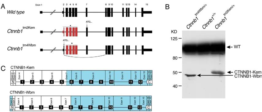
Two conditionalCtnnb1knockout alleles have been independently produced in the past and are widely used to study CTNNB1 function in a spatiotemporal manner (Brault et al., 2001; Huelsken et al., 2000). In both alleles a portion of the N-terminus of the protein is targeted.Ctnnb1tm2Kem (also referred to below asβCatN) deletes exons 2-6 (Brault et al., 2001), whereasCtnnb1tm4Wbm(Huelsken et al., 2000) targets exons 3-6 (Fig. 1A). In our previous study addressing the maternal function of CTNNB1 using the
Received 26 November 2015; Accepted 10 April 2016 1
Institute of Medical Biology, A*STAR, 8A Biomedical Grove, Immunos 06-06, 138648, Singapore.2Institute of Molecular and Cellular Biology, A*STAR, Proteos 5-02, 138673, Singapore.3The Jackson Laboratory, 600 Main Street, Bar Harbor, ME 04609, USA.4Siriraj Center of Excellence for Stem Cell Research, Mahidol University, Bangkok, 10170 Thailand.5Nanyang Polytechnic, School of Chemical and Life Sciences, 569830, Singapore.
*These authors contributed equally to this work
‡
Author for correspondence (bbk4@me.com)
B.B.K., 0000-0002-2597-5619
DEVEL
O
Ctnnb1tm2Kem allele, we were able to detect nuclear, potentially
truncated CTNNB1 protein by immunofluorescence using anti-CTNNB1 antibodies, despite efficient Cre-mediated deletion of the floxed portion of the molecule (de Vries et al., 2004). This suggested active, alternative, downstream translational start sites in transcripts produced from the‘deleted’allele.
To define the potential products of the Ctnnb1tm2Kem and
Ctnnb1tm4Wbmdeletion alleles we performed western blot analysis
on liver extracts from adult heterozygous mice. When probed with polyclonal antibody directed to the C-terminus of CTNNB1, protein products of slightly below (Ctnnb1tm4Wbm) and ∼50 kDa (Ctnnb1tm2Kem) were detected in addition to the expected ∼94 kDa band corresponding to the intact CTNNB1 molecule (Fig. 1B). In accordance with the truncated CTNNB1 product expected from the design of the floxed alleles, the band from the
Ctnnb1tm2Kemallele is of slightly higher molecular weight than that
fromCtnnb1tm4Wbm. Inspection of the construct diagrams for each targeted allele and genomic Ctnnb1 sequence indicates that the truncated product of the Ctnnb1tm2Kem allele results from an alternative translational start site in exon 7, whereas the truncated product of the Ctnnb1tm2Wbm allele is most likely the result of alternative splicing of exon 1 to 8 (Fig. 1A,C, Fig. S2). Thus, both of the traditionally used conditionalCtnnb1knockout alleles produce similar, although slightly variant, CTNNB1 fragments that fully encompass armadillo repeats 7-12 and the C-terminus. Although the levels of the truncated proteins are low compared with that of CTNNB1 in heterozygous mice, these truncated proteins have the potential to interfere with CTNNB1 function, or to mask a full knockout effect (de Vries et al., 2004).
A novel conditionalCtnnb1knockout model
The production of a truncated protein from theCtnnb1tm2Kemand
Ctnnb1tm4Wbm deletion alleles prompted us to design a novel
conditional knockout model for Ctnnb1. This novel targeted allele, Ctnnb1tm1Knw, was designed to delete exons 8-13 in the C-terminal portion of the protein (Fig. 2A, Fig. S3). Mice heterozygous for this deletion are viable and fertile and, as expected, heterozygous intercrosses did not give rise to
homozygous mutant pups whereas heterozygous and wild-type pups were born at expected ratios.
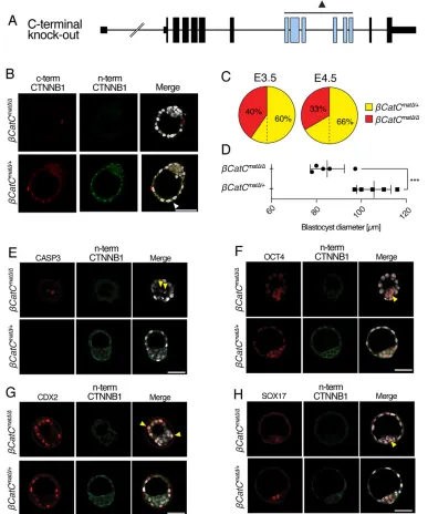
To study the effect of the novel Ctnnb1tm1Knw knockout allele (henceforth termedβCatC for clarity) on preimplantation embryonic development, we used Zp3-Cre to eliminate floxed Ctnnb1 in growing oocytes ofβCatCf/ffemale mice (de Vries et al., 2000). When these eggs (βCatCmatΔ) are fertilized by heterozygous (βCatCΔ/+) males, 50% maternal and 50% maternal/zygotic null embryos are expected (βCatCmatΔ/+and βCatCmatΔ/Δ, respectively) (Fig. S1). Blastocysts were isolated at 3.5 dpc and examined by immunofluorescence for CTNNB1 expression using specific antibodies reactive to epitopes in both the N- and C-terminal portions of the protein (Fig. 2B). As expected, membrane-associated CTNNB1 was detected in wild-type (not shown) and maternal null (βCatCmatΔ/+) embryos. In the latter, the CTNNB1 is encoded by the wild-type paternal allele. No CTNNB1 was detected in maternal/zygotic (βCatCmatΔ/Δ) blastocysts (Fig. 2B, upper panels). We conclude that Ctnnb1tm1Knw is a true null allele, in contrast to the traditionalCtnnb1tm2Kem, where a truncated protein is detectable in preimplantation embryos by immunofluorescence (de Vries et al., 2004).
Analyzing the distribution of 3.5 dpcβCatCmatΔ/+andβCatCmatΔ/Δ blastocysts (n=104) obtained from 14 females, we found under-representation ofβCatCmatΔ/Δblastocysts (42βCatCmatΔ/Δversus 62
βCatCmatΔ/+ blastocysts; a ratio of 1:1.47; Fig. 2C), representing a major deviation from the expected Mendelian ratio of 1:1. This skewing of the expected Mendelian ratio is even more pronounced a day later, at 4.5 dpc (n=27, dissected from 4 females; 9βCatCmatΔ/Δ versus 18βCatCmatΔ/+; a ratio of 1:2; Fig. 2C).
[image:2.612.91.518.54.237.2]It is of interest to note thatβCatCmatΔ/Δembryos, although still resembling blastocysts, were often smaller and misshapen compared with their heterozygous, paternal-rescued siblings (Fig. 2B,E-H). This is clearly evident in reduced blastocyst diameter at 3.5 dpc (P=0.0026,t-test; Fig. 2D). In line with this observation, a few (one to three) cells within the 3.5 dpc maternal/zygotic null blastocysts were positive for active caspase 3, an indicator of apoptosis, which in the course of this and our previous studies has rarely been observed in wild-type blastocysts (Fig. 2E) (Messerschmidt and Kemler, 2010).
Fig. 1. Traditional mouseCtnnb1knockout alleles produce truncated CTNNB1.(A) The wild-typeCtnnb1locus and the two conditional allelesCtnnb1tm2Kem andCtnnb1tm4Wbm. Exons marked in red are deleted upon Cre-mediated recombination (triangle). Arrows indicate translational start sites (ATG) used to produce the respective truncated proteins. (B) Western blot of liver lysates, from mice of the indicated genotypes, using a polyclonal antibody targeting the C-terminus of CTNNB1. Wild-type CTNNB1 (WT) and the truncated products fromCtnnb1tm2KemandCtnnb1tm4Wbmare indicated. (C) Schematic representation of CTNNB1 and the respective coding exons. Domains in blue are predicted to be translated from the deleted alleles. Fragments CTNNB1-Kem and CTNNB1-Wbm slightly vary at the N-terminus, yet are identical downstream of armadillo repeat 6. 1-12, armadillo repeats.
DEVEL
O
Given the observed defects, we examined whether normal early embryonic lineage segregation takes place in these 3.5 dpc blastocysts. The first lineage segregation gives rise to the inner cell mass (ICM) and trophectoderm (TE) (Ralston and Rossant, 2008). In the second segregation, the primitive endoderm (PE) lineage arises from the ICM (Gardner and Beddington, 1988). At 3.5 dpc, high levels of OCT4 (POU5F1) expression are characteristic for the pluripotent cells of the ICM, yet OCT4 still remains detectable in all cells of the embryo, whereas CDX2 expression is restricted to the TE (Strumpf et al., 2005). Despite the morphological defects inβCatCmatΔ/Δembryos, both markers are expressed robustly and in the expected localized manner (Fig. 2F,G). Furthermore, at 4.5 dpc OCT4 is confined, as expected, to the pluripotent epiblast and forming PE (Fig. S4A). At 3.5 dpc, early PE cells are identifiable as SOX17 positive and detectable in the ICM in the typical‘salt and pepper’distribution (Chazaud et al., 2006). As with the ICM and TE markers,
SOX17-positive cells can be detected in βCatCmatΔ/Δ embryos (Fig. 2H).
Thus, in the complete absence of maternal and zygotic CTNNB1, although lineage allocation appears to be normal, the presence of apoptotic cells within smaller blastocysts and the loss of viable
Ctnnb1 null blastocysts over time suggest that normal preimplantation development is compromised.
Peri- and postimplantation development ofβCatC maternal-zygotic null embryos
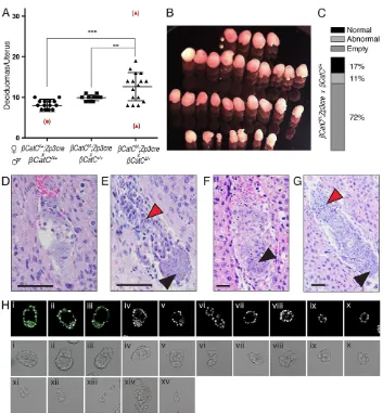
[image:3.612.45.432.60.524.2]Assuming that surviving maternal/zygotic null embryos are capable of normal implantation, we dissected deciduomas from the uteri of females at 5.5, 6.5 and 7.5 dpc, derived from the three different βCatC matings (Fig. 3A, Fig. S1). From heterozygous intercrosses (Fig. 3A, left,‘mating 1’), where 75% of the embryos are expected to carry at least one wild-type allele and 25% are zygotic knockouts, a mean of 8.2±1.07 (s.d.) deciduomas/litter was found. From
Fig. 2. Blastocyst formation in maternal-zygoticCtnnb1null mice.
(A) The novelCtnnb1tm1Knw(βCatC) knockout allele. Exons deleted by Cre-mediated recombination are in blue. (B) Immunofluorescence analysis of CTNNB1 expression in 3.5 dpc βCatCmatΔ/ΔandβCatCmatΔ/+ blastocysts after incubation with antibodies to epitopes of the N- and C-terminal portions of CTNNB1. The arrowhead highlights an adherence junction in the heterozygous embryo. (C) Quantification ofβCatCmatΔ/Δand
βCatCmatΔ/+embryos across multiple litters at 3.5 and 4.5 dpc. Only 40% or 33% of blastocysts (expected 50%) at 3.5 and 4.5 dpc areβCatCmatΔ/Δ,
respectively. (D) Blastocyst diameter analysis comparingβCatCmatΔ/Δand βCatCmatΔ/+embryos (***P=0.0026, Student’st-test). (E) Active caspase 3 staining to address apoptosis in βCatCmatΔ/ΔandβCatCmatΔ/+embryos (arrowheads). (F-H) Lineage segregation analysis inβCatCmatΔ/Δ
andβCatCmatΔ/+embryos for the inner cell mass marker OCT4 (F), trophectoderm marker CDX2 (G) and primitive endoderm marker SOX17 (H) (arrowheads). Scale bars: 50μm.
DEVEL
O
βCatCf/f;Zp3-Cre (matΔ) females mated with wild-type B6 males (Fig. 3A, middle,‘mating 2’), where all embryos carry a wild-type paternal allele but all lack maternal contribution, a mean of 9.9±0.74 implantation sites was noted. A highly variable number of deciduomas, averaging 12.63±3.48 per mating, was found in the uteri of femaleβCatCf/f;Zp3-Cre (matΔ) mated toβCatCΔ/+males (Fig. 3A, right,‘mating 3’). Here, 50% of the embryos are expected to be completeCtnnb1knockouts (i.e. maternal/zygotic) and 50% should lack the maternal contribution yet express paternalCtnnb1. In fact, the uterus of one such mated female contained 31 deciduomas of different sizes (Fig. 3B). Even after excluding this extreme case from our statistical analysis, there is a statistically highly significant increase in implantation sites comparing matings 1 and 3 (P=0.0002, unpairedt-test) and matings 2 and 3 (P=0076, unpairedt-test) (Fig. 3A).
In light of these results, we dissected uteri fromβCatCf/f;Zp3-Cre (matΔ) females mated toβCatCΔ/+males to study their decidual content. Deciduomas were fixed, embedded, serially sectioned, stained and examined microscopically to classify their phenotypes at 5.5, 6.5 and 7.5 dpc (see also Table 1). Later stages were not examined as zygotic, non-conditionalCtnnb1null embryos do not survive until this age (Haegel et al., 1995). It is remarkable that, in 237 isolated deciduomas from 18 pregnant females, only 17% (n=40) contained normal embryos; the rest were either empty (n=171) or contained embryo remnants only (n=26) (Fig. 3C). Representative sections from the 5.5 dpc implantation sites showed
abnormal embryos ranging from small aggregates of cells to small abnormal embryos with pyknotic nuclei (Fig. 3D,E). Many deciduomas contained only a few loosely connected cells. The 6.5 dpc deciduomas exhibited a range of phenotypes: some contained a few loosely connected cells, some contained small disorganized embryos with defective embryonic portions (Fig. 3F), and others contained embryos with apparently normal extra-embryonic but defective extra-embryonic portions (Fig. 3G).
What leads to the substantially increased number of implantation sites? We carefully re-examined the uterine contents of βCatCf/f;
[image:4.612.48.401.53.434.2]Zp3-Cre females (n=18) mated to βCatCΔ/+ males at 3.5 dpc (representative images of litters are shown in Fig. S5), which revealed an average of nine embryos per litter (n=18) that ranged from expanded, phenotypically normal blastocysts to the developmentally compromised blastocyst phenotypes described above (Fig. 2, Fig. S5). In addition, these litters regularly contained delayed, uncompacted or compacted morulae, next to uncharacteristic semi-compacted morulae (see description in Fig. S5). Most remarkably, in six of the 18 uteri we found not only morulae and normal hatched and unhatched blastocysts but also a large number of zona pellucida-free structures, consisting of small trophoblastic vesicles containing three or more cells and pairs of, or just single, blastomeres (Fig. 3H, Fig. S5A,B). Typing the embryos and remnants shown in Fig. 3H using anti-β-catenin antibodies revealed the three apparently normal embryos to be CTNNB1-positive blastocysts (thusβCatCmatΔ/+, i-iii), and the remaining embryos, cell aggregates and remnants to be
Fig. 3. The maternal-zygotic deletionβCatC results in an increased number of implantation sites.(A) Quantification of deciduomas isolated/uterus at 5.5, 6.5 and 7.5 dpc from females of the indicated mating schemes. (B) Thirty-one deciduomas isolated from a single uterus of aβCatCf/f;Zp3-Cre female mated with aβCatCΔ/+male at 6.5 dpc. (C) Quantification of deciduomal content from allβCatCf/f;Zp3-Cre females mated with a βCatCΔ/+male. (D) Embryo from aβCatCf/f; Zp3-Cre female mated with aβCatCΔ/+male at 5.5 dpc, with only a few cells left at the implantation site. (E) A 5.5 dpc embryo from a βCatCf/f;Zp3-Cre female mated with aβCatCΔ/+ male, with small and deformed embryo proper (black arrowhead). The extra-embryonic portion and the ectoplacental cone (red arrowhead) contain many pyknotic nuclei. (F) A 6.5 dpc embryo from aβCatCf/f;Zp3-Cre female mated with aβCatCΔ/+male. The embryo is very small and the presumed embryonic ectoderm comprises a small round clump of cells (arrowhead). (G) A 6.5 dpc embryo from a βCatCf/f;Zp3-Cre female mated with aβCatCΔ/+ male. The embryonic ectoderm is a
disorganized mass of cells (black arrowhead), whereas the ectoplacental cone (red arrowhead) and the extra-embryonic portion appear normal. Scale bars: 100μm. (H) A 3.5 dpc litter flushed from the uterus of a βCatCf/f;Zp3-Cre female mated with aβCatCΔ/+ male. Embryos and fragments were imaged after isolation and subsequently stained for CTNNB1 to determine genotypes (fragments xi to xv were lost in the procedure).
DEVEL
O
CTNNB1 negative (thusβCatCmatΔ/Δ, iv-x; remnants xi-xv were lost during the staining procedure).
Taken together, these results lead us to suggest that following hatching from, or losing, their zona pellucida, embryos lacking CTNNB1 are prone to fission. Although each embryo does not undergo complete fission, it is likely that the resulting fragments, which often display the unmistakable morphology of trophoblastic vesicles (Fig. S5B), are capable of inducing a decidual reaction but not of sustaining further embryogenesis. This could explain the increase in the total number of deciduomas (and large number of empty deciduomas) in some of the females from this cross (Table 1, Fig. 3A-C).
The extremely variable phenotype resulting from this single mutation on an inbred background indicates that the paternal CTNNB1, made following zygotic gene activation at the 2-cell stage and translation at the 4- to 8-cell stage (de Vries et al., 2004), is not always sufficient for embryo rescue. Not all maternal/zygotic null blastocysts without a zona pellucida undergo complete fragmentation. Although individual cells may be shed, the remaining embryo may still be able to form an egg cylinder stage embryo later on. In the worst case scenario (e.g. 31 implantation sites observed, Fig. 3B), the preimplantation embryos disintegrate, resulting in mostly empty deciduomas. In the case of embryos derived from maternally deletedCtnnb1null oocytes fertilized with wild-type sperm (maternal-only deletion), the phenotype depends on the timing of paternalCtnnb1activation and on its expression at the blastomere cell surface. In earlier studies we were able to detect CTNNB1 on some but not all blastomeres within a given embryo by the 6- to 8-cell stage by immunofluorescence (de Vries et al., 2004). Accordingly, from such embryos we might find a range of phenotypes from complete fragmentation to loss of individual blastomeres to normal embryos. Although we might expect each embryo to be rescued by the paternal allele this is not the case and the entire range of phenotypes from empty deciduomas to normal fertile adults was observed (Table 1). Haploinsufficiency, due to small differences in the time of paternal allele activation, or the quantity of CTNNB1 produced in each blastomere appears to have a significant impact on successful embryogenesis.
Does truncated CTNNB1 have a role in embryo rescue? Our previous results suggested that deletion of the N-terminal portion of CTNNB1 (i.e. from theβCatN allele) leads to blastocyst wastage and subfertility but not complete sterility ofβCatNf/f;Zp3-Cre females
mated to wild-type males (de Vries et al., 2004). Interestingly, the truncated CTNNB1 from theβCatN deletion theoretically retains the portion of the protein that interacts with E-cadherin at adherence junctions. To determine whether the truncated CTNNB1 in eggs and embryos of βCatNf/f;Zp3-Cre females can play a role in embryo rescue, we compared deciduomas from females bearing the complete null allele (βCatC) with those of females that express the truncated gene product (βCatN) afterZp3-Cre-induced deletion (Table 1).
First, we find an obvious difference in the number of uterine implantation sites in allCtnnb1maternally deleted mice regardless of the allele (βCatC orβCatN), ranging on average from 7.4 to 13.2 (Table 1, groups 1-8) as compared with 6.9 for normal B6 females (Table 1, group 9). No abnormal B6 embryos were detected, although two deciduomas examined did not contain embryos.
Second, since transcription of the paternal Ctnnb1 allele is activated at the 2-cell stage and the protein is translated and located at the membrane of some blastomeres at the 4- to 8-cell stage (de Vries et al., 2004), paternal rescue of the maternally deletedCtnnb1 embryos is possible. However, embryos from both groups 2 and 5 (βCatNf/f;Zp3-Cre orβCatCf/f;Zp3-Cre females mated with normal B6 males) are only partially rescued by activation of the wild-type paternal allele (group 2 versus 5, P=0.234; group 2 versus 9,
P=0.00001; and group 5 versus 9,P=0.001).
Finally, when considering the outcomes from matings where the only difference between them is in the presence of paternally activated, truncated CTNNB1 versus a total absence of CTNNB1 in the embryo (group 1 versus 4, group 3 versus 6, group 1 versus 8, group 4 versus 7 and group 7 versus 8), it becomes apparent that the presence of truncated CTNNB1 always decreases the degree of embryo fragmentation, although it is not sufficient to normalize development. One should bear in mind that the level of truncated CTNNB1 synthesized is substantially reduced compared with that of normal CTNNB1 (see relative levels in liver extracts in Fig. 1C), yet when available at cleavage stages it may provide a degree of rescue to the CTNNB1-negative embryo.
[image:5.612.49.561.69.193.2]In summary, although analysis of these data is fraught with statistical challenges due to embryo fragmentation and partial rescue scenarios, they indicate that embryos containing truncated CTNNB1 are not affected to the same extent as embryos that completely lack CTNNB1. This finding leads us to suggest that truncated CTNNB1, lacking the portion of the protein required for interaction with the Wnt pathway, can substitute, albeit incompletely, for intact CTNNB1 during preimplantation development.
Table 1. Effect on implantation of maternal/zygotic deletion ofCtnnb1alleles
Genotype No. deciduoma
(average implantation sites/mouse)
No. embryos
Mating Maternal Paternal No. females Normal Small or abnormal Empty or remnants Ratio*
1 βCatCf/f;Cre βCatCΔ/+ 18 237 (13.2) 40 26 171 1:4.9
2 βCatCf/f;Cre βCat+/+ 9 89 (9.9) 54 15 20 1.5:1
3 βCatCf/+;Cre βCatCΔ/+ 15 120 (8.0) 39 31 50 1.0:2
4 βCatNf/f;Cre βCatNΔ/+ 13 130 (10) 41 15 74 1:2.2
5 βCatNf/f;Cre βCat+/+ 10 88 (8.8) 64 10 14 2.6:1
6 βCatNf/+;Cre βCatNΔ/+ 10 74 (7.4) 38 13 23 1.0:1
7 βCatNf/βCatCf;Cre βCatCΔ/+ 7 84 (12) 12 7 65 1.0:6
8 βCatNf/βCatCf;Cre βCatNΔ/+ 12 140 (11.7) 44 14 82 1:2.2
9 βCat+/+ βCat+/+ 8 55 (6.9) 53 0 2 –
*The ratio of normal embryos to the sum of [small or abnormal and empty or remnants].
Pairwiseχ2comparative analyses: 1 versus 2,χ2=72.1,P<0.0001; 4 versus 5,χ2=40.5,P<0.0001; 1 versus 4,χ2=11.1,P=0.004; 2 versus 5,χ2=2.9,P=0.234; 7 versus 8,χ2=9.16,P=0.01; 2 versus 9,χ2=23.0,P<0.0001; 5 versus 9,χ2=13.1,P=0.001; 3 versus 6,χ2=6.84,P=0.033; 1 versus 7,χ2=0.907,P=0.635; 1 versus 8,χ2=10.9,P=0.004; 4 versus 7,χ2=9.93,P=0.007; 4 versus 8,χ2=0.180,P=0.914.
βCatC refers toCtnnb1tm1Knw;βCatN refers toCtnnb1tm2Kem.
DEVEL
O
Conclusions
We initiated these experiments to resolve a conflict in the literature regarding the requirement of β-catenin expression for normal development. Although some suggest that the major phenotypic disturbance is seen when the Wnt pathway is first activated at gastrulation (Haegel et al., 1995; Huelsken et al., 2000), others have reported that molecular inactivation of nuclear Wnt/β-catenin signaling limits blastocyst competency for implantation (Xie et al., 2008).
With regard to the view that the first embryonic phenotype in
Ctnnb1 null embryos, derived from heterozygous matings of B6.129 mice crossedinter se, is at gastrulation, we find an earlier phenotype, with an effect observable by the blastocyst stage. The differences observed here could be a consequence of the different mouse strains used, but are more likely to reflect the fact that truncated CTNNB1 at least partially prevents embryo fragmentation and masks the fission effect, which is difficult to visualize in the presence of an intact zona pellucida. In any case, these results are in accordance with our previous observations of only partial fertility of maternal-deletion females using the B6 Ctnnb1tm2Kem strains employed here (de Vries et al., 2004).
Our findings are at odds with reports that inhibitors of the Wnt/β -catenin pathway have a negative effect on implantation (Xie et al., 2008), implicating this molecular pathway in implantation.In vivo partial rescue of CTNNB1-negative embryos by truncated CTNNB1, which lacks the portion of the protein responsible for interaction with the Wnt pathway, suggests that the Wnt/β-catenin pathway is not necessary at preimplantation stages. From our results we suggest thatCtnnb1null embryos first fail because of inadequate adhesion between cells of the blastocyst, leading to embryo fragmentation. We thus propose that total loss of CTNNB1 in the embryo interferes with normal development at compaction, when its major role is to collaborate with E-cadherin to ensure cell-cell contact and interaction with the cytoskeletal network, so that the effective polarized epithelium necessary for subsequent embryonic development can form.
MATERIALS AND METHODS Mice
All mice, with the exception of those bearing theCtnnb1tm4Wbmallele, were prepared in, and/or backcrossed onto, an inbred C57BL/6J background. The mating schemes required to obtain maternal/zygoticCtnnb1null embryos is illustrated in Fig. S1. The B6Zp3-Cre line was described previously (de Vries et al., 2000). These experiments were performed under protocols approved by the IACUC at The Jackson Laboratory and at the A*STAR Institute of Medical Biology.
Preparation of the C57BL/6JCtnnb1tm1Knwmouse strain
Using a recombineering approach, we generated a construct containing loxP sites spanning exon 8 to exon 13 ofCtnnb1, resulting in the removal of amino acids 361 to 692 encompassing a part of armadillo repeat 6 to a part of the C-terminus (designatedβCatCf) to distinguish it from the N-terminally
floxed alleles (Fig. S3). The FRT-flanked neomycin resistance gene in intron 13 was inserted at a position that should not interfere with the splicing machinery, so that homozygous floxed mice could be obtained without deleting the neomycin resistance gene.
Constructs generated using C57BL/6J (B6) BACs were electroporated into the germline-competent B6/J ESC line #693 (Schuster-Gossler et al., 2001). Of 400 colonies screened by Southern blot for homologous recombination, 42 were identified, analyzed by a long-range PCR approach, and 12 clones in which recombination occurred correctly were identified. Two of ten clones containing an intact 5′loxP site were injected into C57BL/6-Tyrc-2J blastocysts, and transferred into pseudo-pregnant females. Chimeric mice were crossed with C57BL/6-Tyrc-2J mice to identify those transmitting the floxed allele through their germline. B6
offspring that inherited theβCatCffloxed allele were identified using primers on either side of the 5′loxP site, and matedinter seto generate homozygous progeny. Mice of this strain, known as C57BL/6JCtnnb1tm1Knw, are available from the Jackson Laboratory repository (JR022775).
The B6.129Ctnnb1tm2Kem(Brault et al., 2001) mouse line was previously backcrossed ten times onto the C57BL/6J background (de Vries et al., 2004). This floxed allele is referred to asβCatN, to denote the more N-terminal deletion in theCtnnb1gene that results when it is crossed with the B6Zp3-Cre strain (de Vries et al., 2000) and to distinguish it from the floxed allele in the B6.129Ctnnb1tm2Kemmouse strain. Liver protein extracts from mice bearing the heterozygous deletedCtnnb1tm4Wbmallele were obtained from Walter Birchmeier (Huelsken et al., 2000).
Western blot analysis
Western blot analysis was performed as described (de Vries et al., 2004) using a rabbit polyclonal antibody toβ-catenin (Sigma, C-2206; 1:5000).
Genotyping
Tail tip biopsies were taken at weaning age of 3-4 weeks and lyzed in 50 mM NaOH at 95°C for 20 min. After neutralization, 1μl DNA was used for PCR genotyping using the following primer combinations:
Zp3-Cre Fwd TGATGAGGTTCGCAAGAACC and Rev
CCATGAGTG-AACGAACCTGG; Ctnnb1tm2KemFwd
AAGGTAGAGTGATGAAAGT-TGTT, Rev1 CACCATGTCCTCTGTCTATCC and Rev2
TACACTATTG-AATCACAGGGACTT;Ctnnb1tm1KnwFwd
CCTGAGTCCCAGGGTGT-CTA, Rev1 GCAACAGCACCTTATGCAGCTCAA and Rev2 GGCCA-ATCACAATGCACGTTCAGA.
Embryo isolation
Female mice, at least 6 weeks of age, were placed in the cage with isolated male mice in the late afternoon and vaginal plugs, as a sign of mating, were checked the next morning; noon of the day the plug was found was considered 0.5 dpc. Blastocysts were flushed from isolated uteri at 3.5 dpc and dissected at 4.5 dpc. Embryos at 5.5, 6.5 and 7.5 dpc were dissected from decidual swellings.
Indirect immunofluorescence analysis
Embryos were washed twice in M2 medium (Millipore) and twice in PBS, then fixed in 4% paraformaldehyde in PBS overnight at 4°C or 30 min at room temperature (RT). After fixation, embryos were permeabilized with 0.5% Triton X-100 in PBS for 15 min at RT, then transferred to PBS with 0.1% Triton X-100 supplemented with 10% BSA (Sigma-Aldrich) for 1 h at RT or overnight at 4°C for blocking. The embryos were transferred to primary antibody solution (dilutions given below) containing 0.1% Triton X-100 and 10% BSA diluted in PBS. Incubation was performed for 1 h at RT or overnight at 4°C. Embryos were then washed in three changes of PBS with 0.1% Triton X-100 and 10% BSA, before secondary antibody incubation for 1 h at RT (dilutions below) in 0.1% Triton X-100 and 10% BSA diluted in PBS. Following three washes in PBS, with 10 µM DAPI (Molecular Probes) added to the last wash, the embryos were mounted in micro-drops on glass-bottom dishes and imaged using an inverted Olympus Fluoview confocal microscope.
Primary antibodies were: mouse monoclonal OCT4 (Santa Cruz
Biotechnology, SC-5279; 1:200); mouse monoclonal CDX2
(BioGenex, AM392-5M; 1:200); mouse monoclonal SOX17 (Santa Cruz Biotechnology, SC130295; 1:100); rabbit polyclonal active caspase 3 (Cell Signaling, 9661; 1:200); mouse monoclonal C-terminal β-catenin (BD Transduction Laboratories, 610154; 1:100); and rabbit polyclonal N-terminal β-catenin (Cell Signaling, 9580; 1:100). Secondary antibodies were Alexa Fluor 488 goat anti-rabbit and Alexa Fluor 594 goat anti-mouse (Molecular Probes, 421206/421203; 1:500).
Competing interests
The authors declare no competing or financial interests.
Author contributions
W.N.d.V. prepared theβ-catenin allele (βCatCf) and performed theβ-catenin western blots; mice were maintained and mated by B.B.K.; mice were genotyped by S.B.;
DEVEL
O
D.S., D.M. and C.L. isolated the pre- and postimplantation embryos; D.M. performed immunofluorescence and PCR assays; D.S., W.N.d.V., D.M. and B.B.K. designed the experiments and wrote the manuscript.
Funding
This work was supported by Institute of Medical Biology, Agency for Science, Technology and Research (A*STAR), Singapore.
Supplementary information
Supplementary information available online at
http://dev.biologists.org/lookup/suppl/doi:10.1242/dev.133439/-/DC1
References
Bauer, M. and Willert, K.(2012). Wnt signaling: theβ-cat(enin)’s meow.Genes Dev.26, 105-109.
Brault, V., Moore, R., Kutsch, S., Ishibashi, M., Rowitch, D. H., McMahon, A. P., Sommer, L., Boussadia, O. and Kemler, R.(2001). Inactivation of the beta-catenin gene by Wnt1-Cre-mediated deletion results in dramatic brain malformation and failure of craniofacial development.Development128, 1253-1264. Chazaud, C., Yamanaka, Y., Pawson, T. and Rossant, J.(2006). Early lineage
segregation between epiblast and primitive endoderm in mouse blastocysts through the Grb2-MAPK pathway.Dev. Cell10, 615-624.
de Vries, W. N., Binns, L. T., Fancher, K. S., Dean, J., Moore, R., Kemler, R. and Knowles, B. B.(2000). Expression of Cre recombinase in mouse oocytes: a means to study maternal effect genes.Genesis26, 110-112.
de Vries, W. N., Evsikov, A. V., Haac, B. E., Fancher, K. S., Holbrook, A. E., Kemler, R., Solter, D. and Knowles, B. B.(2004). Maternal beta-catenin and E-cadherin in mouse development.Development131, 4435-4445.
Dickinson, D. J., Nelson, W. J. and Weis, W. I.(2011). A polarized epithelium organized by beta- and alpha-catenin predates cadherin and metazoan origins.
Science331, 1336-1339.
Gardner, R. L. and Beddington, R. S. P.(1988). Multi-lineage“stem”cells in the mammalian embryo.J. Cell Sci.1988Suppl. 10, 11-27.
Haegel, H., Larue, L., Ohsugi, M., Fedorov, L., Herrenknecht, K. and Kemler, R. (1995). Lack of beta-catenin affects mouse development at gastrulation.
Development121, 3529-3537.
Huelsken, J., Vogel, R., Brinkmann, V., Erdmann, B., Birchmeier, C. and Birchmeier, W.(2000). Requirement for beta-catenin in anterior-posterior axis formation in mice.J. Cell Biol.148, 567-578.
Messerschmidt, D. M. and Kemler, R.(2010). Nanog is required for primitive endoderm formation through a non-cell autonomous mechanism.Dev. Biol.344, 129-137.
Ralston, A. and Rossant, J.(2008). Cdx2 acts downstream of cell polarization to cell-autonomously promote trophectoderm fate in the early mouse embryo.Dev. Biol.313, 614-629.
Rudloff, S. and Kemler, R.(2012). Differential requirements forβ-catenin during mouse development.Development139, 3711-3721.
Schuster-Gossler, K., Lee, A. W., Lerner, C. P., Parker, H. J., Dyer, V. W., Scott, V. E., Gossler, A. and Conover, J. C.(2001). Use of coisogenic host blastocysts for efficient establishment of germline chimeras with C57BL/6J ES cell lines.
BioTechniques31, 1022-1024, 1026.
Strumpf, D., Mao, C.-A., Yamanaka, Y., Ralston, A., Chawengsaksophak, K., Beck, F. and Rossant, J. (2005). Cdx2 is required for correct cell fate specification and differentiation of trophectoderm in the mouse blastocyst.
Development132, 2093-2102.
Valenta, T., Hausmann, G. and Basler, K.(2012). The many faces and functions of β-catenin.EMBO J.31, 2714-2736.
Xie, H., Tranguch, S., Jia, X., Zhang, H., Das, S. K., Dey, S. K., Kuo, C. J. and Wang, H. (2008). Inactivation of nuclear Wnt-beta-catenin signaling limits blastocyst competency for implantation.Development135, 717-727.