Development 140, 3927-3931 (2013) doi:10.1242/dev.099531 © 2013. Published by The Company of Biologists Ltd
INTRODUCTION
Neuronal diversity is a fundamental feature of the nervous system. Elucidating how this diversity arises is one of the central aims in developmental neurobiology. For the past 15 years, it has been shown that differential gene expression during development forms the basis of this diversity (Goulding et al., 2002; Jessell, 2000). Thus, knowing gene expression patterns in neuronal cells is of fundamental importance. Transgenic animals expressing fluorescent proteins in a subset of neuronal cells are powerful tools, because they allow researchers to see expression patterns of particular genes in live animals. The zebrafish possesses a strong advantage over other vertebrate model organisms because its body is transparent during development.
Here, we have generated a wide variety of transgenic zebrafish, in which reporter genes are expressed in a specific subset of neurons or neuronal progenitors. We had three specific aims in this research. The first aim was to expand a repertoire of transgenic fish that express reporter genes in neurons having a particular neurotransmitter phenotype. Because neurotransmitter properties are fundamental to neurons, such transgenic fish would be of general importance. We have previously generated transgenic fish that express GFP or RFP in glutamatergic and glycinergic neurons (McLean et al., 2007; Satou et al., 2012). These lines have been effectively used in many studies (e.g. Kinkhabwala et al., 2011). Here, we have expanded our repertoire by generating lines that are linked to a GABAergic phenotype, or lines expressing Gal4 in
glutamatergic and glycinergic neurons. The second aim was to reveal domain organization in the developing dorsal spinal cord. In the vertebrate spinal cord, the first step towards generating neuronal diversity is the subdivision of neuronal progenitor cells into distinct domains along the dorsoventral axis. In amniotes, 11 distinct domains (five in the ventral and six in the dorsal spinal cord) are formed (Zhuang and Sockanathan, 2006). In zebrafish, domain organization in the ventral spinal cord is conserved (Goulding, 2009). In the dorsal spinal cord, however, domain organization is not well understood. We sought to determine the domain organization of the dorsal spinal cord by generating transgenic fish that expressed fluorescent proteins under the promoter/enhancer of genes that are known to be expressed in a domain-like manner in amniotes. The third aim was to investigate transmitter phenotypes of neurons that are produced from each domain of the dorsal spinal cord.
We successfully expanded our repertoire of transgenic fish that label particular neurotransmitter phenotypes. We found that dorsal spinal cord domain organization is largely conserved in zebrafish. We also determined transmitter properties of neurons that are produced from some of the domains.
MATERIALS AND METHODS
Generation of transgenic fish
All the transgenic fish (Table 1) were generated using the BAC (bacterial artificial chromosome) homologous recombination technique with either the I-SceI-mediated method (Kimura et al., 2006) or the Tol2-mediated method (Suster et al., 2009). The BACs used were: zK145P24 for vglut2a
(slc17a6b – Zebrafish Information Network), zC24M22 for gad1b, zC247L22 for atoh1a, zK171N3 for neurog1, zK97C6 for gsx2, zC271B14 for gsx1, zC163N18 for dbx2and zC246F16 for dmrt3a. The glyt2(slc6a5 – Zebrafish Information Network) BAC was isolated from a BAC library with a hybridization-based screening. The GFP and lRl-GFP (loxP-DsRed-loxP-GFP) constructs for homologous recombination were described previously (Kimura et al., 2006). The Gal4 (Gal4FF) (Asakawa et al., 2008) and Cre constructs were described by Kimura et al. (Kimura et al., 2013) and Satou et al. (Satou et al., 2012), respectively. The lRl-Gal4 construct was 1National Institutes of Natural Sciences, Okazaki Institute for Integrative Bioscience,
National Institute for Physiological Sciences, Okazaki, Aichi 444-8787, Japan.
2Center for Frontier Research, National Institute of Genetics, Mishima, Shizuoka
411-8540, Japan. 3PREST, JST. 4Division of Molecular and Developmental Biology,
National Institute of Genetics, Mishima, Shizuoka 444-8540, Japan.
*Present address: Sars International Centre for Marine Molecular Biology, Thormøhlensgate 55, Bergen, Norway
‡Author for correspondence (shigashi@nips.ac.jp)
Accepted 3 July 2013 SUMMARY
The developing nervous system consists of a variety of cell types. Transgenic animals expressing reporter genes in specific classes of neuronal cells are powerful tools for the study of neuronal network formation. We generated a wide variety of transgenic zebrafish that expressed reporter genes in specific classes of neurons or neuronal progenitors. These include lines in which neurons of specific neurotransmitter phenotypes expressed fluorescent proteins or Gal4, and lines in which specific subsets of the dorsal progenitor domain in the spinal cord expressed fluorescent proteins. Using these, we examined domain organization in the developing dorsal spinal cord, and found that there are six progenitor domains in zebrafish, which is similar to the domain organization in mice. We also systematically characterized neurotransmitter properties of the neurons that are produced from each domain. Given that reporter gene expressions occurs in a wide area of the nervous system in the lines generated, these transgenic fish should serve as powerful tools for the investigation of not only the neurons in the dorsal spinal cord but also neuronal structures and functions in many other regions of the nervous system.
KEY WORDS: Spinal cord, Domain organization, Neurotransmitter phenotype, Transgenic zebrafish, Transcription factors
Transgenic tools to characterize neuronal properties of
discrete populations of zebrafish neurons
Chie Satou1, Yukiko Kimura1, Hiromi Hirata2,3, Maximiliano L. Suster4,*, Koichi Kawakami4and Shin-ichi Higashijima1,‡
D
E
V
E
LO
P
M
E
N
prepared by replacing GFP with Gal4 in the lRl-GFP. For the vglut2a :lRl-Gal4 and glyt2:lRl-Gal4 construct, mCherry was used as the RFP, whereas for the neurog1:lRl-GFP and gsx2:lRl-GFP constructs, dTomato was used.
neurog1is expressed in neuronal progenitors and also in Rohon-Beard (RB) neurons. An RB neuron enhancer was found to be located within 1 kb upstream. This 1-kb sequence was eliminated in the neurog1:lRl-GFP construct. Tg[gad1b:GFP], Tg[neurog1:GFP], Tg[gsx2:GFP] and Tg[glyt2:Gal4] lines were generated by injecting CremRNA into embryos of Tg[gad1b:lRl-GFP], Tg[neurog1:lRl-GFP], Tg[gsx2:lRl-GFP] and Tg[glyt2:lRl-Gal4] lines, respectively.
Imaging
Images were taken using Zeiss 510 or Nikon A1 confocal microscopes. For the time-lapse imaging, animals were embedded in agarose every time for imaging (after imaging, animals were removed from the agarose).
RESULTS AND DISCUSSION
Generation of transgenic fish lines associated with neurotransmitter properties
Glutamate, glycine and GABA are three major neurotransmitters in the central nervous system (CNS). For glutamatergic, glycinergic and GABAergic neurons, vglut, glyt2and gadgenes, respectively, can be used as markers. We have previously generated Tg[vglut2a:GFP], Tg[vglut2a:RFP] and Tg[glyt2:GFP] lines (McLean et al., 2007; Satou et al., 2012). To expand our repertoire, we generated Tg[glyt2:RFP] and Tg[gad1b:GFP] and Tg[gad1b:RFP] transgenic fish. We compared the fluorescent protein expression patterns in each line by crossing a GFP line to an RFP line of another neurotransmitter. The spinal cord at 3.5 days post-fertilization (dpf) was examined. Fig. 1A-C shows cross sections of Tg[vglut2a:GFP] and Tg[glyt2:RFP] (Fig. 1A), Tg[vglut2a:GFP] and Tg[gad1b:RFP] (Fig. 1B) and Tg[glyt2:GFP] and Rg[gad1b:RFP] (Fig. 1C). In each of the panels, GFP and RFP are expressed in distinctive cells. Thus, each transgenic fish successfully labeled discrete neuronal populations that have a particular neurotransmitter phenotype.
Our RFP lines were created based on the lRl-GFP (loxP-RFP-loxP-GFP) cassette (Table 1). This allowed us to induce GFP expression in the subpopulation of glutamatergic, glycinergic and GABAergic neurons by crossing the lines to Cre driver lines. Two
examples of such labeling are shown in Fig. 1D,E. In Fig. 1D, GFP is expressed in glycinergic neurons that derive from the dbx1b-positive p0 progenitor domain (V0 neurons). In Fig. 1E, GFP is expressed in GABAergic neurons that derive from the gsx1-positive pd4/pd5 progenitor domains (for clarification of the pd4/pd5 progenitor domains, see below). Neurons in which Cre-mediated recombination did not occur continued to express RFP (Fig. 1D,E).
The Gal4-UAS system is a powerful tool to express reporter/effector genes, such as genetically encoded calcium indicators and optogenetic tools. It would be useful to have lines expressing Gal4 in neurons having particular neurotransmitter phenotypes. Thus, we generated Tg[vglut2a:Gal4] and Tg[glyt2:Gal4], and these lines were capable of driving reporter gene expression in glutamatergic and glycinergic neurons, respectively, with high expression levels (supplementary material Fig. S1).
[image:2.612.322.549.63.308.2]Finally, we sought to combine the Gal4-UAS system with the Cre-loxP system. We created Tg[vglut2a:lRl-Gal4] and Tg[glyt2:lRl-Gal4]. Fig. 1F,G shows examples of the inducible expression of GFP in V0 neurons. In Fig. 1F, glutamatergic V0 neurons express GFP, whereas in Fig. 1G, glycinergic V0 neurons express GFP. Using this system, researchers will be able to drive expression of various reporter/effector genes in specific populations of neurons having a particular neurotransmitter phenotype. Fig. 1. Transgenic fish associated with neurotransmitter properties. All images were taken from fish that were 3.5 dpf. Genotypes of the transgenic fish are shown at the top of each panel. (A-C) Cross-sections of the spinal cord. Green and red cells do not overlap, indicating that each transgenic fish labels discrete cell populations. (D-G) Lateral views of the spinal cord. (D) Glycinergic neurons derived from dbx1b-positive progenitors are labeled by GFP. (E) GABAergic neurons derived from gsx1 -positive progenitors are labeled by GFP. In D and E, neurons in which Cre-mediated recombination did not occur continued to express RFP. (F,G) Glutamatergic neurons (F) and glycinergic neurons (G) derived from
dbx1b-positive progenitors are labeled by GFP. Only the green channel is shown. Scale bars: 10 μm (A-C); 20 μm (D-G).
Table 1. Transgenic fish generated in this study
Transgenic fish line
1 vglut2a:Gal4
2 vglut2a:lRl-Gal4 (vglut2a:RFP)
3 glyt2:Gal4
4 glyt2:lRl-GFP (glyt2:RFP)
5 glyt2:lRl-Gal4 (glyt2:RFP)
6 gad1b:GFP
7 gad1b:lRl-GFP (gad1b:RFP)
8* atoh1a:GFP
9 neurog1:GFP
10 neurog1:lRl-GFP (neurog1:RFP)
11 gsx2:GFP
12 gsx2:lRl-GFP (gsx2:RFP)
13 gsx1:Cre
14 gsx1:GFP
15 gsx1:lRl-GFP (gsx1:RFP)
16 dbx2:GFP
17 dmrt3a:lRl-GFP (dmrt3a:RFP)
18 dmrt3a:Gal4
The names in parentheses represent fluorescent protein expression when used as single transgenic fish (without application of Cre).
*Tg[atoh1a:GFP] was described in a previous study that investigated GFP expression in the hindbrain (Kani et al., 2010).
D
E
V
E
LO
P
M
E
N
[image:2.612.49.298.72.262.2]In summary, we have successfully expanded our repertoire of transgenic fish that are associated with neurotransmitter phenotypes by creating GFP, RFP (conditional GFP), Gal4 and conditional Gal4 lines.
Characterization of progenitor domains in the dorsal spinal cord
In amniotes, progenitors in the developing dorsal spinal cord are divided into six distinct domains (pd1-pd6) that are defined by the expression of transcription factors (Zhuang and Sockanathan, 2006). In zebrafish, domain organization in the developing dorsal spinal cord has not been well characterized. To examine domain organization, we generated transgenic fish that express fluorescent proteins under the promoter/enhancer of genes that are known to be expressed in particular progenitor domains in amniotes. Genes used include atoh1a, neurog1, gsx2, gsx1and dbx2. We examined the expression domain spatial profiles of these transcription factors (represented by GFP or RFP expression). In addition to the lines listed in Table 1, Tg[dbx1b:RFP], in which RFP is expressed in the p0 domain (Satou et al., 2012), was also used for comparison.
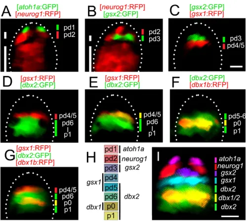
Examinations were made using embryos at 36-48 hours post-fertilization (hpf), when extensive neurogenesis occurs (Lyons et al., 2003). atoh1awas expressed in the most dorsal progenitor domain (Fig. 2A). neurog1had two expression domains: a dorsal thin neurog1 expression domain and a ventral wide neurog1 expression domain (Fig. 2A,B, short and long lines, respectively), and we only consider the former in this study. The dorsal boundary of this thin domain was adjacent to the atoh1aexpression domain (Fig. 2A). The gsx2expression domain was thin (Fig. 2B,C), and this domain was bordered dorsally by the dorsal neurog1domain (Fig. 2B) and ventrally the gsx1expression domain (Fig. 2C). dbx2 was expressed widely in the middle region of the spinal cord (Fig. 2D-G). In some sections, the dbx2expression domain was bordered dorsally by the gsx1expression domain (Fig. 2D), whereas in other sections, the dorsal-most dbx2-positive progenitors were also positive for gsx1(Fig. 2E). This indicates that gsx1-positive progenitors can be divided into two populations: one population that is negative for dbx2, and another population that is positive for dbx2. The dbx1-positive progenitors (p0 progenitors) were all positive for dbx2(Fig. 2F). The ventral boundary of the dbx2expression domain was more ventral than that of the dbx1expression domain (Fig. 2F). The dbx2-positive cells were found to be negative for the V2 neuron marker chx10(vsx2 – Zebrafish Information Network) (Kimura et al., 2008), suggesting that the ventral boundary of the dbx2 expression domain corresponds to the p1-p2 boundary (Fig. 2D-G). There was a gap between the gsx1and dbx1expression domains (Fig. 2G), indicating that a domain exists between the gsx1 expression domain and the p0 domain.
Collectively, the results are summarized in Fig. 2H,I, revealing the existence of six progenitor domains (pd1 to pd6), as in amniotes. The pd1 domain is defined by atoh1a expression, and the pd2 domain corresponds to the neurog1dorsal expression domain. The pd3 domain is defined by gsx2expression. The gsx1expression domain consists of two cell populations: dbx2-negative cells and dbx2-positive cells. The former corresponds to the pd4 domain, and the latter corresponds to the pd5 domain. The pd6 domain is defined by those cells that are positive for dbx2and negative for gsx1and dbx1.
Expression patterns of atoh1a, neurog1, gsx2, gsx1, dbx2and dbx1are consistent with those of the corresponding genes in mice (Alaynick et al., 2011), with only some minor differences. In mice, gsx2is reported to be expressed at a low level in the pd4 and pd5
domains (Kriks et al., 2005). In zebrafish, gsx2 expression, as revealed by GFP expression, is restricted to the pd3 domain (Fig. 2B,C). In addition, the pd4 and pd5 domains, which are defined by gsx1expression, might not form a sharp dorsoventral boundary, because in some sections only cells belonging to the pd4 domain (dbx2negative) are observed (Fig. 2D), whereas in other sections cells belonging to the pd5 domain (dbx2positive) are predominantly present (Fig. 2F).
Characterization of neurotransmitter phenotypes from neurons derived from each progenitor domain
We sought to determine the neurotransmitter properties of neurons that derive from each domain. Fluorescent protein expression in the transgenic lines persisted in their progeny. This allowed us to examine neurotransmitter properties of progeny neurons by crossing the transgenic lines with the lines associated with neurotransmitter properties. Examinations were made at 3.5 dpf.
[image:3.612.314.564.59.284.2]We first examined dI1 neurons (neurons that derive from the pd1 progenitors) using Tg[atoh1a:GFP] fish. Virtually, all of the GFP-labeled dI1 neurons were positive for vglut2a:RFP (Fig. 3A, arrows and arrowheads). None of GFP-labeled dI1 neurons was positive Fig. 2. Progenitor domains in the dorsal spinal cord.Images were taken from embryos that were 36-48 hpf. All images are cross-sections of the spinal cord. Genotypes of the transgenic fish are shown at the top of each panel. (A) The atoh1aexpression domain is located in the dorsal-most spinal cord. The short and long white lines show the dorsal and ventral neurog1expression domains, respectively. The dorsal neurog1
expression domain is located just ventral to the atoh1aexpression domain. (B) The gsx2expression domain is located just ventral to the dorsal neurog1expression domain. (C) The gsx2and gsx1expression domains are adjacent to each other. (D) The gsx1and dbx2expression domains are adjacent to each other. (E) In this image, the gsx1expression domain is within the dbx2expression domain (yellow). (F) The dbx1b
expression domain is located in the middle of the dbx2expression domain (yellow). (G) The gsx1expression domain (red) and the dbx1b
expression domain (yellow) are separate. (H) Summary of the domain organization. (I) Composite image of the expression domain of each transcription factor. For neurog1, only the dorsal expression domain is included. Scale bars: 10 μm.
D
E
V
E
LO
P
M
E
N
for glyt2:RFP or gad1b:RFP (Fig. 3B,C). The results indicate that dI1 neurons are essentially glutamatergic. We noted that some of dI1 neurons were located in a more ventral region (Fig. 3A, arrows) and our time-lapse imaging showed that some dI1 neurons migrated ventrally (supplementary material Movie 1).
Because we do not have transgenic fish that specifically label pd2 progenitors, we next examined dI3 neurons using Tg[gsx2:GFP] (Fig. 3D-F). The results showed that glutamatergic neurons (Fig. 3D, arrowheads) were present among dI3 neurons, but that glycinergic and GABAergic neurons were not present (Fig. 3E,F).
We then examined dI4 and dI5 neurons using Tg[gsx1:GFP] (Fig. 3G-I). The results showed that glutamatergic (Fig. 3G, arrowheads), glycinergic (Fig. 3H, arrowhead), and GABAergic neurons (Fig. 3I, arrowhead) were all present among dI4/5 neurons. We do not have transgenic fish that specifically label pd6 progenitors. We sought to examine neurotransmitter properties of dI6 neurons using a gene that is known to be expressed in post-mitotic dI6 neurons in mice. A recent report shows that Dmrt3is expressed in inhibitory dI6 neurons (Andersson et al., 2012), and we expected this was also the case in zebrafish. To explore whether or not dmrt3is expressed in inhibitory dI6 neurons, we generated Tg[dmrt3a:RFP] and Tg[dmrt3a:Gal4]. We first examined whether dmrt3awas expressed in dI6 neurons using Tg[dmrt3a:RFP]. As shown in supplementary material Fig. S2, all of the RFP-labeled neurons were positive for dbx2:GFP, but none of them was positive for gsx1:GFP and dbx1b:GFP, indicating that dmrt3a:RFP neurons do indeed derive from the pd6 progenitors (Fig. 2H). We then examined the neurotransmitter properties of dmrt3a-positive neurons using Tg[dmrt3a:Gal4] and Tg[UAS:GFP]. As shown in Fig. 3J-L, all of the GFP-labeled neurons were positive for glyt2:RFP (Fig. 3K, arrowhead), and none of them was positive for vglut2a:RFP or gad1b:RFP, indicating that dmrt3a marks glycinergic dI6 neurons. Thus, glycinergic neurons are present among dI6 neurons. It remains unknown whether or not glutamatergic and GABAergic neurons are also present among dI6 neurons.
The results described in this section are summarized in Fig. 3M. Transmitter properties of neurons that derive from dorsal progenitor domains are generally consistent with those in mice (Alaynick et al., 2011).
Conclusions
We determined domain organization in the dorsal spinal cord. We also systematically determined neurotransmitter properties of neurons that are produced from several domains. This forms the basis for future detailed analyses of the neurons produced by each domain. We have generated a wide variety of transgenic zebrafish, including those that are linked to neurotransmitter properties. Although we focused on the spinal cord in the present study, reporter gene expression in these transgenic fish occurs widely in the CNS, including hindbrain, midbrain and forebrain (supplementary material Fig. S3). Therefore, the newly generated transgenic fish should serve as powerful tools for the investigation of neuronal structures and functions in many CNS regions.
Acknowledgements
We thank Shunji Fujioka for helping to generate Tg[dbx2:GFP].
Funding
This work was supported in part by Grants-in-Aid for Scientific Research.
Competing interests statement
The authors declare no competing financial interests.
Author contributions
C.S. and S.H. contributed to the conception and design of the study. C.S., Y.K. and S.H. performed most of the experiments. H.H. contributed to establish the Tg[glyt2:Gal4] line. M.L.S. and K.K. provided the Tol2-related materials. C.S. and S.H. wrote the manuscript.
Supplementary material
Supplementary material available online at
http://dev.biologists.org/lookup/suppl/doi:10.1242/dev.099531/-/DC1
References
Alaynick, W. A., Jessell, T. M. and Pfaff, S. L.(2011). SnapShot: spinal cord development. Cell146, 178-178 e171.
Andersson, L. S., Larhammar, M., Memic, F., Wootz, H., Schwochow, D., Rubin, C. J., Patra, K., Arnason, T., Wellbring, L., Hjälm, G. et al.(2012). Mutations in DMRT3 affect locomotion in horses and spinal circuit function in mice. Nature488, 642-646.
Asakawa, K., Suster, M. L., Mizusawa, K., Nagayoshi, S., Kotani, T., Urasaki, A., Kishimoto, Y., Hibi, M. and Kawakami, K.(2008). Genetic dissection of neural circuits by Tol2 transposon-mediated Gal4 gene and enhancer trapping in zebrafish. Proc. Natl. Acad. Sci. USA105, 1255-1260.
Goulding, M.(2009). Circuits controlling vertebrate locomotion: moving in a new direction. Nat. Rev. Neurosci.10, 507-518.
[image:4.612.52.343.55.270.2]Goulding, M., Lanuza, G., Sapir, T. and Narayan, S.(2002). The formation of sensorimotor circuits. Curr. Opin. Neurobiol.12, 508-515.
Fig. 3. Neurotransmitter properties of neurons derived from dorsal progenitor domains. All images were taken from fish that were 3.5 dpf. All images are cross-sections of the spinal cord. Transgenic fish genotypes are shown at the top and left. Arrowheads and arrows show cells that are positive for both GFP and RFP. (A-C) atoh1a:GFP neurons are positive for vglut2a:RFP. (D-F) gsx2:GFP neurons are positive for vglut2a:RFP. (G-I) All three types of neurons (vglut2a:RFP,
glyt2:RFP and gad1b:RFP) are present among gsx1:GFP neurons. (J-L) A GFP neuron driven by dmrt3a:Gal4 is positive for glyt2:RFP. (M) Summary of the neurotransmitter properties of neurons produced by dorsal progenitors. Scale bar: 10 μm.
D
E
V
E
LO
P
M
E
N
Jessell, T. M.(2000). Neuronal specification in the spinal cord: inductive signals and transcriptional codes. Nat. Rev. Genet.1, 20-29.
Kani, S., Bae, Y. K., Shimizu, T., Tanabe, K., Satou, C., Parsons, M. J., Scott, E., Higashijima, S. and Hibi, M.(2010). Proneural gene-linked neurogenesis in zebrafish cerebellum. Dev. Biol.343, 1-17.
Kimura, Y., Okamura, Y. and Higashijima, S.(2006). alx, a zebrafish homolog of Chx10, marks ipsilateral descending excitatory interneurons that participate in the regulation of spinal locomotor circuits. J. Neurosci.26, 5684-5697.
Kimura, Y., Satou, C. and Higashijima, S.(2008). V2a and V2b neurons are generated by the final divisions of pair-producing progenitors in the zebrafish spinal cord. Development135, 3001-3005.
Kimura, Y., Satou, C., Fujioka, S., Shoji, W., Umeda, K., Ishizuka, T., Yawo, H. and Higashijima, S. I.(2013). Hindbrain V2a neurons in the excitation of spinal locomotor circuits during zebrafish swimming. Curr. Biol.23, 843-849. Kinkhabwala, A., Riley, M., Koyama, M., Monen, J., Satou, C., Kimura, Y.,
Higashijima, S. and Fetcho, J.(2011). A structural and functional ground
plan for neurons in the hindbrain of zebrafish. Proc. Natl. Acad. Sci. USA108, 1164-1169.
Kriks, S., Lanuza, G. M., Mizuguchi, R., Nakafuku, M. and Goulding, M. (2005). Gsh2 is required for the repression of Ngn1 and specification of dorsal interneuron fate in the spinal cord. Development132, 2991-3002.
Lyons, D. A., Guy, A. T. and Clarke, J. D.(2003). Monitoring neural progenitor fate through multiple rounds of division in an intact vertebrate brain. Development130, 3427-3436.
McLean, D. L., Fan, J., Higashijima, S., Hale, M. E. and Fetcho, J. R.(2007). A topographic map of recruitment in spinal cord. Nature446, 71-75.
Satou, C., Kimura, Y. and Higashijima, S.(2012). Generation of multiple classes of V0 neurons in zebrafish spinal cord: progenitor heterogeneity and temporal control of neuronal diversity. J. Neurosci.32, 1771-1783.
Suster, M. L., Sumiyama, K. and Kawakami, K.(2009). Transposon-mediated BAC transgenesis in zebrafish and mice. BMC Genomics10, 477.
Zhuang, B. and Sockanathan, S.(2006). Dorsal-ventral patterning: a view from the top. Curr. Opin. Neurobiol.16, 20-24.