Available online on www.ijpsr.com 12
IJPSR (2010), Vol. 1, Issue 3 (Review Article)
Received 19 January, 2010; received in revised form 22 February, 2010; accepted 27 February, 2010
TRANSFERSOMES ULTRA FLEXIBLE VESICLES FOR TRANSDERMAL DELIVERY
Vineet Bhardwaj *1, Vikesh Shukla 1, Anju Singh 2, Rishabha Malviya 1 and P. K. Sharma 1
Department of Pharmaceutical Technology Meerut Institute of Engineering & Technology 1,
Meerut (U.P.), India
Raj Kumar Goel Institute of Technology2, Ghaziabad (U.P.), India
ABSTRACT
Molecules greater than 500 Da normally do not cross the skin. This prevents epicutaneous delivery of the high molecular weight therapeutics as well as non-invasive transcutaneous immunisation. Transfersomes are a form of elastic or deformable vesicle, which were first introduced in the early 1990s. Elasticity is generated by incorporation of an edge activator in the lipid bilayer structure. The original composition of these vesicles was soya phosphatidyl choline incorporating sodium cholate and a small concentration of ethanol. Transfersomes are applied in a non-occluded method to the skin and have been shown to permeate through the stratum corneum lipid lamellar regions as a result of the hydration or osmotic force in the skin. They have been used as drug carriers for a range of small molecules, peptides, proteins and vaccines, both in vitro and in vivo. Transfersomes penetrate through the stratum corneum and the underlying viable skin into the blood circulation in intact form. However, this has not been substantiated by other research groups who have extensively probed the mechanism of penetration and interaction of elastic vesicles in the skin. Structural changes in the stratum corneum have been identified, and intact elastic vesicles visualised within the stratum corneum lipid lamellar regions, but no intact vesicles have been ascertained in the viable tissues. Extremely deformable vesicles prepared by the judicious combination of several materials provide a solution to this problem: the resulting agent carriers, transfersomes, are the only tested colloidal system that can transport even large macromolecules spontaneously through the skin in immunologically active form.
*Correspondence for Author:
Vineet Bhardwaj
Department of Pharmaceutical Technology Meerut Institute of Engineering & Technology , Meerut (U.P.), India
E-mail:
vineetbhardwaj86@gmail.com
Keywords:
Epicutaneous, Transcutaneous,
Available online on www.ijpsr.com 13
INTRODUCTION: Delivery via the
transdermal route is an interesting option in this respect because a transdermal route is convenient and safe. This offers
several potential advantages over
conventional routes1 like avoidance of
first pass metabolism, predictable and extended duration of activity, minimizing undesirable side effects, utility of short half-life drugs, improving physiological and pharmacological response, avoiding the fluctuation in drug levels, inter-and
intra-patient variations, and most
importantly, it provides patients
convenience. To date many chemical and physical approaches have been applied to increase the efficacy of the material transfer across the intact skin, by use of the penetration enhancers, enhancers, iontophoresis, sonophoresis and the use of colloidal carriers such as lipid vesicles
(liposomes and proliposomes) and
nonionic surfactant vesicles (niosomes and proniosomes).
Transfersomes were developed in order to take the advantage of phospholipids vesicles as transdermal
drug carrier. These self-optimized
aggregates, with the ultra flexible membrane, are able to deliver the drug reproducibly either into or through the skin, depending on the choice of administration or application, with high efficiency. These vesicular transfersomes are several orders of magnitudes more elastic than the standard liposomes and thus well suited for the skin penetration.
Transfersomes overcome the skin
penetration difficulty by squeezing
themselves along the intracellular sealing lipid of the stratum corneum. There is
provision for this, because of the high vesicle deformability, which permits the entry due to the mechanical stress of surrounding, in a self-adapting manner. Flexibility of transfersomes membrane is achieved by mixing suitable surface-active
components in the proper ratios2. The
resulting flexibility of transfersome
membrane minimizes the risk of complete vesicle rupture in the skin and allows transfersomes to follow the natural water gradient across the epidermis, when applied under nonocclusive condition. Transfersomes can penetrate the intact stratum corneum spontaneously along two routes in the intracellular lipid that
differ in their bilayers properties3. The
following figure shows possible micro routes for drug penetration across human
skin intracellular and transcellular4.
The high and self-optimizing
deformability of typical composite
transfersomes membrane, which are adaptable to ambient tress allow the ultra deformable transfersomes to change its membrane composition locally and reversibly, when it is pressed against or
attracted into narrow pore. The
transfersomes components that sustain
strong membrane deformation
preferentially accumulate, while the less adaptable molecules are diluted at sites of great stress. This dramatically lowers the energetic cost of membrane deformation and permits the resulting, highly flexible particles, first to enter and then to pass through the pores rapidly and efficiently. This behavior is not limited to one type of pore and has been observed in natural
Available online on www.ijpsr.com 14
Mechanism of Penetration of
Transfersomes: Transfersomes when
applied under suitable condition can
transfer 0.1 mg of lipid per hour and cm2
area across the intact skin. This value is substantially higher than that which is typically driven by the transdermal concentration gradients. The reason for this high flux rate is naturally occurring "transdermal osmotic gradients" i.e. another much more prominent gradient is
available across the skin2. This osmotic
gradient is developed due to the skin penetration barrier, prevents water loss through the skin and maintains a water activity difference in the viable part of the epidermis (75% water content) and nearly completely dry stratum corneum, near to
the skin surface (15% water content)3.
This gradient is very stable because ambient air is a perfect sink for the water molecule even when the
transdermal water loss is
unphysiologically high. All polar lipids attract some water this is due to the
energetically favourable interaction
between the hydrophilic lipid residues and their proximal water. Most lipid bilayers thus spontaneously resist an
induced dehydration4,5. Consequently all
lipid vesicles made from the polar lipid vesicles move from the rather dry location to the sites with a sufficiently high water
concentration6,7 So when lipid suspension
(transfersomes) is placed on the skin surface, that is partly dehydrated by the water evaporation loss and then the lipid vesicles feel this "osmotic gradient" and try to escape complete drying by moving
along this gradient3. They can only
achieve this if they are sufficiently
deformable to pass through the narrow pores in the skin, because transfersomes composed of surfactant have more
suitable rheologic and hydration
properties than that responsible for their
greater deformability1, 7 less deformable
vesicles including standard liposomes are confined to the skin surface, where they dehydrate completely and fuse, so they have less penetration power than
transfersomes. Transfersomes are
optimized in this respect and thus attain maximum flexibility, so they can take full
advantages of the trans-epidermal
osmotic gradient (water concentration gradient) 8.
Propensity of penetration: The
magnitude of the transport driving force, of course, also plays an important role: Flow = Area x (Barrier) Permeability x (Trans-barrier) force.
Therefore, the chemically driven lipid flow across the skin always decreases dramatically when lipid solution is replaced by the some amount of lipids in a suspension.
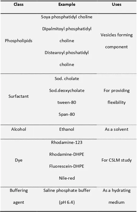
MATERIALS AND METHODS: Materials
commonly used for the preparation of transfersomes are summarized in Table 1. All the methods of preparation of transfersomes are comprised of two steps. First, a thin film is prepared hydrated and then brought to the desired size by sonication; and secondly, sonicated vesicles are homogenized by
extrusion through a polycarbonate
Available online on www.ijpsr.com 15
organic solvent (chloroform-methanol), organic solvent evaporated above the lipid transition temperature (room temp.
for pure PC vesicles, or 50 0C for
dipalmitoyl phosphatidyl choline) using rotary evaporator. Final traces of solvent were removed under vacuum for overnight. The deposited lipid films were hydrated with buffer (pH 6.5) by rotation
at 60 rpm min-1 fir 1 hr at the
corresponding temperature. The resulting vesicles were swollen for 2 hr at room temperature. To prepare small vesicles, resulting LMVs were sonicated at room temperature or 500C for 30 min. using a B-12 FTZ bath sonicator or probe sonicated at 40C for 30 min (titanium micro tip, Heat Systems W 380). The sonicated vesicles were homogenized by manual extrusion 10 times through a
sandwich of 200 and 100 nm
polycarbonate membrane15.
Characterization of Transfersomes: The characterization of transfersomes is generally similar to liposomes, niosomes and micelles.
Entrapment Efficiency: The entrapment efficiency is expressed as the percentage
entrapment of the drug added.
Entrapment efficiency was determined by first separation of the unentrapped drug by use of mini-column centrifugation method. After centrifugation, the vesicles were disrupted using 0.1% Triton X-100 or
50% n-propanol. The entrapment
efficiency is expressed as:
Entrapment efficiency= (amount
entrapped/ total amount added)*100
Vesicle Diameter: Vesicle diameter can be
determined using photon correlation
spectroscopy or dynamic light scattering (DLS) method. Samples were prepared in distilled water, filtered through a 0.2 mm
Table 1: Different additives used in formulation of transfersomes 9-14
membrane filter and diluted with filtered saline and than size measurement done by using photon correlation spectroscopy or
dynamic light scattering (DLS)
measurements (Gamal et al, 1999) 13.
Confocal Scanning Laser Microscopy
(CSLM) study: Conventional light
microscopy and electron microscopy both face problem of fixation, sectioning and staining of the skin samples. Often the
Class Example Uses
Phospholipids
Soya phosphatidyl choline
Dipalmitoyl phosphatidyl
choline
Distearoyl phoshatidyl
choline
Vesicles forming
component
Surfactant
Sod. cholate
Sod.deoxycholate
tween-80
Span-80
For providing
flexibility
Alcohol Ethanol As a solvent
Dye
Rhodamine-123
Rhodamine-DHPE
Fluorescein-DHPE
Nile-red
For CSLM study
Buffering
agent
Saline phosphate buffer
(pH 6.4)
As a hydrating
[image:4.612.310.537.191.538.2]Available online on www.ijpsr.com 16
structures to be examined are actually incompatible with the corresponding processing techniques; these give rise to misinterpretation, but can be minimized by Confocal Scanning Laser Microscopy (CSLM). In this technique lipophilic fluorescence markers are incorporated into the transfersomes and the light emitted by these markers used for following purpose:
For investigating the mechanism of
penetration of transfersomes across the skin,
For determining histological
organization of the skin (epidermal columns, interdigitation), shapes and architecture of the skin penetration pathways.
For comparison and differentiation of
the mechanism of penetration of
transfersomes with liposomes,
niosomes and micelles.
Different fluorescence markers used in CSLM study are as –
1. Fluorescein- DHPE (1, 2-
dihexadecanoyl- sn- glycero- 3-
phosphoethanolamine- N- (5 -
fluoresdenthiocarbamoyl), triethyl-
ammonium salt)
2. Rhodamine- DHPE (1, 2-
dihexadecanoyl- sn- glycero-
3ogisogietgabikanube- N-
LissamineTmrhodamine- B- sulfonyl), triethanol- amine salt)
3. NBD- PE (1, 2- dihexadecanoyl-
sn-glycero- 3- phosphoethanolamine- N-
(7-nitro- Benz- 2- oxa- 1, 3- diazol- 4- yl) triethanolamine salt)
4. Nile red.
Degree of Deformability or Permeability Measurement: In the case of transfersomes, the permeability study is one of the important and unique parameter for characterization. The deformability study is done against the pure water as standard.
Transfersomes preparation is passed
through a large number of pores of known size (through a sandwich of different microporous filters, with pore diameter between 50 nm and 400 nm, depending on the starting transfersomes suspension). Particle size and size distributions are noted after each pass by dynamic light scattering (DLS) measurements.
In vitro Drug Release: In vitro drug release study is performed for determining the permeation rate. Time needed to attain
steady state permeation and the
permeation flux at steady state and the information from in-vitro studies are used to optimize the formulation before more expensive in vivo studies are performed. For determining drug release, transfersomes suspension is incubated at 320C and samples are taken at different times and the free drug is separated by mini column
centrifugation (Fry et al., 1978)15. The
amount of drug released is then calculated indirectly from the amount of drug entrapped at zero times as the initial amount (100% entrapped and 0% released).
Available online on www.ijpsr.com 17
outermost skin layers, transfersomes reach the deeper skin layer, the dermis. From this latter skin region they are normally washed out, via the lymph, into the blood circulation and through the latter throughout the body, if applied under suitable conditions. Transfersomes can thus reach all such body tissues that are accessible to the subcutaneously injected liposomes. The kinetics of action of an epicutaneously applied agent depends on the velocity of carrier penetration as well as on the speed of drug (re) distribution and the action after this passage. The most important single factors in this process are:
1. Carrier in-flow
2. Carrier accumulation at the targets
site
3. Carrier elimination
The onset of penetration-driving force depends on the volume of the suspension medium that must evaporate from the skin surface before the
sufficiently strong trans-cutaneous
chemical potential chemical potential or water activity gradient is established. Using less solvent is favourable in this respect. The rate of carrier passage across the skin is chiefly determined by the
activation energy for the carrier
deformation. The magnitude of the penetration driving force also plays a big role. This explains, for example, why the occlusion of an application site or the use of too strongly diluted suspension hampers the penetration process.
Carrier elimination from the sub cutis is primarily affected by the lymphatic flow, general anaesthesia or any other factor that affects this flow, consequently, is prone to modify the rate of transcutaneous carrier transport. While it has been estimated that approximately 10% of the cardiac blood flow pass through each gram of living skin tissue, no comparable quotation is available for the lymph. Further, drug distribution is also sensitive to the number of carrier used, as this may affect the rate of vehicle degradation and / or filtration in the lymph nodes.
The lag between the time of application and the time of drug appearance in the body, therefore, is always quite long, complex and strongly sensitive to the type of drug and formulation administration. In the best case, the skin penetration lag amounts to
approximately 15 min. if rapidly
exchanging agents such as local analgesics are detected right under the skin
permeability barrier16. Less rapidly
exchanging molecules or molecules measured in the blood compartment are typically detected with a lag time between 2 and 6 hr. depending on the details of drug formulation. Molecules that do not diffuse readily from the carriers or agents delivered with the suboptimal carriers normally fall in this
category. The kinetics of vesicle
Available online on www.ijpsr.com 18
biological assays in which vesicle associated drugs exert their action directly under the skin surface. Local analgesics are useful for this purpose, for determining the kinetics of penetration, various lidocaine loaded vesicles were left to dry out on the intact skin. Corresponding subcutaneous injection is used as control. The animal's sensitivity to pain at the treated site after each application was then measured as a function of time. Dermally applied standard drug carrying liposomes or simple lidocaine solution have never caused any analgesic effect. It was
necessary to inject such agent
preparations to achieve significant pain suppression. In contrast to this, the
lidocaine-loaded transfersomes were
analgesic ally active even when applied dermally.
Maximum analgesic effect with the latter type of drug application was typically observed 15 minutes after the drug application. A marked analgesic effect was still noticeable after very long time. The precise reach as well as kinetics of transfersomes penetration through the skin are affected by: drug carrier interaction, application condition or form, skin characteristics, applied dose.
Salient features and limitations of Transfersomes: Transfersomes possess an infrastructure consisting of hydrophobic and hydrophilic moieties together and as a result can accommodate drug molecules
with wide range of solubility.
Transfersomes can deform and pass through narrow constriction (from 5 to 10 times less than their own diameter)
without measurable loss. This high deformability gives better penetration of intact vesicles. They can act as a carrier for low as well as high molecular weight
drugs e.g. analgesic, anaesthetic,
corticosteroids, sex hormone, anticancer, insulin, gap junction protein, and albumin.
They are biocompatible and
biodegradable as they are made from
natural phospholipids similar to
liposomes. They have high entrapment efficiency, in case of lipophilic drug near to 90%. They protect the encapsulated drug from metabolic degradation. They act as depot, releasing their contents slowly and gradually. They can be used for both systemic as well as topical delivery of drug. Easy to scale up, as procedure is simple, do not involve lengthy procedure and unnecessary use or pharmaceutically unacceptable additives.
Limitations of Transfersomes:
Transfersomes are chemically unstable because of their predisposition to oxidative degradation. Purity of natural phospholipids is another criteria militating against adoption of transfersomes as drug
delivery vehicles. Transfersomes
formulations are expensive.
Available online on www.ijpsr.com 19
This is due to high flexibility of the transfersomes membrane and is achieved by judiciously combining at least two
lipophilic/amphiphilic components
(phospholipids plus bio surfactant) with
sufficiently different packing
characteristics into a single bilayer. The high resulting aggregate deformability permits transfersomes to penetrate the skin spontaneously. This tendency is supported by the high transfersomes surface hydro-philicity that enforces the search for surrounding of high water activity. It is almost certain that the high
penetration potential of the
transfersomes is not primarily a
consequence of stratum corneum
fluidization by the surfactant because micellar suspension contains much more
surfactant than transfersomes
(PC/Sodium cholate 65/35 w/w %, respectively). Thus, if the penetration enhancement via the solubilization of the skin lipids was the reason for the superior penetration capability of transfersomes, one would expect an even better penetration performance of the micelles. In contrast to this postulate, the higher surfactant concentration in the mixed micelles does not improve the efficacy of material transport into the skin. On the contrary, mixed micelles stay confined to the topmost part of the stratum corneum
even they are applied non occlusively 7.
The reason for this is that mixed micelles are much less sensitive to the trans-epidermal water activity gradient than transfersomes. Transfersomes differ in at least two basic features from the mixed micelles, first a transfersomes is normally by one to two orders of
magnitude (in size) greater than standard lipid micelles. Secondly and more importantly, each vesicular transfersomes contains a water filled core whereas a micelle is just a simple fatty droplet. Transfersomes thus carry water as well as fat-soluble agent in comparison to micelles that can only incorporate lipoidal
substances 8, 9. To differentiate the
penetration ability of all these carrier
systems10 proposed the distribution
profiles of fluorescently labelled mixed
lipid micelles, liposomes and
transfersomes as measured by the Confocal Scanning Laser Microscopy (CSLM) in the intact murine skin. In all these vesicles the highly deformable transfersomes transverse the stratum corneum and enter into the viable epidermis in significant quantity.
Chapman & Walsh11 also showed
that the former two types of aggregates are confined to the outer half of the horny layer, where the cellular packing and intercellular seals are already compromised by the desquamation process. Pure lipid vesicles or micelles seem to have access to the low-resistance pathway only and thus very seldom reach the lower stratum corneum or even get into the viable part of the skin in significant quantities.
CONCLUSION: Transfersomes are
specially optimized particles or vesicles, which can respond to an external stress by rapid and energetically inexpensive, shape transformations. Such highly deformable particles can thus be used to
bring drugs across the biological
Available online on www.ijpsr.com 20
tested in artificial systems. Transfersomes can pass through even tiny pores (100 mm) nearly as efficiently as water, which is 1500 times smaller. Drug laden transfersomes can carry unprecedented amount of drug per unit time across the
skin (up to 100mg cm2h-1). The systemic
drug availability thus mediated is frequently higher than, or at least approaches 80-90%. The bio-distribution of radioactively labelled phospholipids applied in the form of transfersomes after 24 h is essentially the same after an epicutaneous application or subcutaneous injection of the preparations. When used under different application conditions, transfersomes can also positioned nearly exclusively and essentially quantitatively into the viable skin region.
REFERENCES:
1.Shaw JE and Chandrasekaran SK: In pharmacology of the skin, Greaves. Springer- Verlag, Berlin 1999; 115-122. 2.Cevc G: Isothermal lipid phase. Transitions Chemistry and
Physics of Lipids 1991; 57:293-299.
3.Schatzlein A and Cevc G: Skin penetration by phospholipids vesicles, Transfersomes as visualized by means of the Confocal Scanning Laser Microscopy, in characterization, metabolisim, and novel biological applications. Champaign. AOCS Press 1995; 191-209. 4.Panchagnula R: Transdermal delivery of drugs Indian
Journal Pharmacology 1997; 29:140-156.
5.Bain KR, Hadgkraft AJ, James WJ, and Water KA: Prediction of percutaneous penetration STS Publishing, Cardiff 1993; 3b: 226-234.
6. Cevc G, Blume G, Sehatzlein A, Gebauer D and Paul A: The skin- a pathway for systemic treatment with patches and lipid-based agent carriers Advance Drug Delivery Reviews 1996; 18:349- 378 7. Chapman SJ and Walsh A: Arch. Dermatol Res 1998;
282:304-320.
8. Gompper G and Kroll DM: Driven transport of fluid vesicles through narrow pores Physical Reviews 1995; E.52: 4198-4211.
9. Wearner RR, Myers MC and Taylor DA: Electron Probe Analysis of Human Skin: Determination of the Water Concentration Profile Journal of Investigative Dermatoogy 1988; 90:218-224.
10.Rand RP and Parsegian VA: Biochem. Biophys Acta.1989; 988: 351-377.
11.Cevc G, Blume G and Schatzlein A: Transfersomes-mediated transepidermal delivery improves the regio-specificity and biological activity of corticosteroids in vivo. Journal of Controlled Release 1997; 45:211-226. 12.Cevc G, Grbauer D, Schatzlein A and Blume G: Biochem.
Acta 1998, 1368:201-215
13.Gamal M, Maghraby M, Williams AC, Barry BW: Skin Delivery of Oestradiol from Deformable and Traditional Liposomes- Mechanistic Studies. Journal of Pharmacy & Pharmacology 1999; 51:1123-1134.
14.Schatzlein A and Cevc G: Non-uniform cellular packing of the stratum corneum and permeability barrier function of intact skin: a high-resolution confocal laser scanning microscopy study using highly deformable vesicles (Transfersomes). British Journal of Dermatology 1998; 138:583-598.
15.Fry DW, White JC and Goldman ID:Rapid separation of low molecular weight solutes from liposomes without dilution, Journal of Analytical Biochemistry 1978; 90:809-815.