IJPSR (2015), Vol. 6, Issue 11 (Research Article)
Received on 02 May, 2015; received in revised form, 24 July, 2015; accepted, 10 September, 2015; published 01 November, 2015
EVALUATION OF ANTIULCER ACTIVITY OF HYDROALCOHOLIC FRUIT PULP EXTRACT OF CUCUMIS SATIVUS
Satish Narra, K. S. Nisha and H. S. Nagesh
Department of Pharmacology, Gautham College of Pharmacy, Bangalore, Karnataka, India.
ABSTRACT: The study was aimed to investigate the activity of hydroalcoholic extract of Cucumis sativus in gastric ulcerated rats. Anti ulcer activity was evaluated via oral administration of HACS at the dose 250, 500 and 1000mg/kg thrice in a day for 5 days before pyloric ligation ulceration, indomethacin and ethanol induced ulceration. Various parameters such as ulcer index, gastric volume, pH of gastric juice, total acidity, estimation of total proteins, total carbohydrates, total hexoses, hexosamine, fucose, salic acid and histopathological parameters were studied in the control group and pretreated groups with hydroalcoholic extract of fruit pulp of Cucumis sativus and standard drug ranitidine. In indomethacin, ethanol and pyloric ligation model, the pretreatment with HACS and ranitidine significantly reduce the ulcer index, free acidity as compared with controlled group. The % protection of ulcers were evaluated. The extract showed significantly (P<0.05) increase in pH with significant decrease in volume of gastric juice, free and total acidity. Histopathological findings also confirm the antiulcer activity of Cucumis sativus fruit pulp extract in albino wistar rats. The present study showed that HACS have anti ulcer activity in 3 models.
INTRODUCTION: Gastric ulcers are very common disease human suffering today, it is a localised loss of gastric as well as duodenal mucosa. Peptic ulcer mainly seen in the path of GIT tract which is exposed to gastric acid and secretion, mainly 2 types of ulcers are peptic ulcer and duodenal ulcer. The major factors lead to ulcer is infection with gram negative, helicobacter pylori, increased HCl secretion inadequate mucosal defense against gastric acid. The peptic ulcers occurs due to imbalance between aggressive (acid, pepsin, bile, H. Pylori) and defensive factor (prostaglandin, bicarbonate secretion, etc.) 1.
QUICK RESPONSE CODE
DOI:
10.13040/IJPSR.0975-8232.6(11).4712-20
Article can be accessed online on:
www.ijpsr.com
DOI link: http://dx.doi.org/10.13040/IJPSR.0975-8232.6(11).4712-20
Now a days, number of medicinal plants have therapeutic role for the treatment of disease in human. Cucumis sativus belongs to the family Cucurbitaceae commonly known as cucumber, it is found throughout the Indian forest land and riversides. The various parts of the plant like leaves, tender shoots have also been used in the traditional system of medicine. The reported pharmacological are antibacterial, antioxidant, analgesic, antidiabetic, hepatoprotective. The fruit is used for skin whitening, skin irritation, bedsore, burns, sunburn. Our present study was carried out to find out anti ulcer activity of HACS in Pylorus ligation, indomethacin and ethanol induced ulcer model in rats.
MATERIALS AND METHODS: Plant materials:
The fruits were purchased from local market of Karnataka and was identified confirmed and Keywords:
HACS- Hydro Alcoholic Extract of
Cucumis sativus, PL- Pylorus ligation, CMC- Carboxy Methyl
Cellulose
Correspondence to Author: Satish Narra
Department of Pharmacology, Gautham College of Pharmacy, Bangalore, Karnataka, India.
authenticated by Dr. MV Rajanna, Dept of botany UAS, GKVK.
The whole fruit was cut into small pieces and shade dried at room temperature and the dried materials were pulverised separately into coarse powder by mechanical grinder. The resultant powder was then used for extraction. The powder was extracted directly with 70% v/v ethanol using soxhlet extractor. The extract was subjected to qualitative phytochemical analysis.
Phytochemical analysis:
Phytochemical analysis of HACS was carried out by using standard procedures of Treas and Evans and Kokate, C.K., Purohit A.P., Gokhale S.B 2, 3.
Animals:
Albino wistar rats weighing 150-250 gm were used for this study. The rats were procured from biogene, bangalore. Animals were maintained at 25±2oC and kept in well ventilated animal house under natural photoperiodic condition in polypropylene cages with paddy husk as bedding with free access to food and water ad libitum.
Acute toxicity studies:
Different doses of HACS were administered orally to seven groups of rats consisting of 6 rats in a group. The animals in group 1 served as control and received 1.0 ml of physiological saline. The animals in groups 2, 3, 4, 5, 6 and 7 received 250, 500, 1000, 1500 and 3000 and 6000 mg/kg body weight respectively through oral administration. They were all placed under observation for 24 h after which the number of dead animals was recorded 4.
Experimental Setup:
Pylorus Ligation (PL) induced ulcer model: Animals were divided into 5 groups each comprising of 6 rats. Group I animals received suspension of 1% CMC in distilled water for 5 days. Group II animals received ranitidine for 5 days. Groups III, IV, V received 250, 500, 1000 mg/kg .b.w. of HACS for 5 days. On the sixth day animals were kept for overnight fasting. Pyloric ligation was done according to the method Shay et.al 5.
Animals were anesthetized using phenobarbital and abdomen was cut open through a middle incision. Expose the stomach, a thread passed around the pylorus and ligated after 4 hours of pylorus ligation the rats were cervical decapitation and the stomachs were removed, the gastric content was collected and the volume was noted. The gastric contents were used for the estimation of physical and biochemical parameters.
Physical parameters: Ulcer index:
The no of ulcers were counted and scoring was undertaken according to the reported method Lakshmi et. Al 6. The ulcer index were determined by following formula:
Ulcer index = 10/xand where x= total ulcerated area/total mucosal area.
The % of ulcer protection were determined by using the formula
Ulcer protection (%) = (Uc-Ut/Uc) x100.
Uc= Ulcer index of treated group
Ut= ulcer index of disease control group
Gastric volume:
4 hours after ligation, animals were killed by cervical dislocation, stomachs were dissected out and the contents were collected into graduated tube and were centrifuged at 3000rpm for 10 minutes, the volume of centrifuged volume was expressed as ml/100gm b.w.
pH of gastric juice:
Ph of gastric juice was measured by pH meter.
Determination of total acidity:
corresponds to total acidity. Acidity can be expressed in terms of mEq/lti.e 7.
Estimation of Total Proteins:
The dissolved protein in gastric juice was estimated in the alcoholic precipitate obtained by adding 90% alcohol with gastric juice 9:1 ratio respectively. Then 0.1 ml of alcoholic precipitate of gastric juice was dissolved in 1ml of 0.1N NaOH and from this 0.05 ml was taken in another test tube. To this 4 ml of alkaline mixture was added and kept for 10 minutes. Then 0.4 ml of phenol reagent was added and again 10 minutes was allowed for colour development. Absorbance was measured against blank prepared with distilled water at 610 nm using spectrophotometer. The protein content was calculated in terms of g/ml of gastric juice 8.
Estimation of Total Carbohydrates:
The dissolved mucosubstances in gastric juice were estimated in the alcoholic precipitate obtained by adding 1ml of gastric juice to 9 ml of 90% alcohol, the mixture was kept for 10 minutes, and the supernatant was discarded. The precipitate separated was dissolved in 0.5 ml of 0.1N sodium hydroxide. To this 1.8ml of 6N, HCL was added. This mixture was hydrolyzed in the boiling water bath for 2 hrs. The hydrolysate was neutralized by 5N sodium hydroxide using phenolpthalin as indicator and the volume was made up to 4.5ml with distilled water and used for the estimation of total hexoses, hexosamine and fucose as described below 9.
Estimation of Total Hexoses:
To 0.4 ml of hydrolysate, 3.4 ml of orcinol reagent was added. The mixture was then heated in the boiling water bath for 15 min. this was then cooled under running tap water and intensity of the colour was read in spectrophotometer at 540 nm against the blank by using distilled water instead of hydrolysate. Total hexoses content was determined from the standard curve of D (+)-galactose-mannose and has been expressed in g/ml of gastric juice 10.
Estimation of Hexosamine: 0.5 ml of the hydrolysate fraction was taken. To this 0.5 ml of acetylacetone reagent was added. The mixture was heated in boiling water bath for 20 minutes and
then cooled under running tap water. 1.5 ml of 90% alcohol was then added followed by an addition of 0.5 ml of Ehrlich’s reagent. The reaction was allowed for 30 minutes. The colour intensity was measured in spectrophotometers at 530 nm against the blank prepared by using distilled water instead of hydrolysate. Hexosamine content of the sample was determined from the standard curve of D (+)-galactose-mannose and concentration has been expressed in µg/ml of gastric juice 11.
Estimation of Fucose: In this method, three test tubes were taken. In one tube 0.4ml of distilled water was taken to serve as control and in each of the other two tubes 0.4 ml of hydrolysate were taken. To all three tubes 1.8 ml of sulphuric acid and water in the ratio of 6:1 was added by keeping the tubes in ice-cold water bath to prevent breakage due to strong exothermic reaction. This mixture was then heated in boiling water bath for exactly 30minutes. The tubes were then taken out and cooled. In the blank and one of the hydrolysate containing tubes (unknown) 0.1ml of cysteine reagent was added, while cysteine reagent was added to the last test tube containing the hydrolysate (unknown), it was then allowed for 90mm to complete the reaction. The reading was taken in Hitachi 15-20 spectrophotometer at 396 and 430 nm setting the zero with the distilled water.
The optical density for the fucose in the hydrolysate was calculated from the differences in the reading obtained at 396 and 430 nm and subtracting the values without cysteine. This was read against standard curve prepared with D (+)-fucose. The fucose content was expressed in terms of µg/ml of gastric juice 12.
pipette out and intensity of colour was measured using spectrophotometer at 550 nm. The sialic content of the sample was determined from the standard curve of sialic acid and has been expressed in µg/ml of gastric juice 13.
Estimation of Free Radical Generation:
The fundic part of the stomach was homogenized (5%) in ice cold 0.9% saline with a glass homogeniser for 30 sec. The homogenate was then centrifuged at 800 g for 10 min followed by centrifugation of the supernatant at 12,000 g for 15 min and the obtained fraction was used for the following estimations.
1. Lipid peroxidase activity (LPO): LPO
product malondialdehyde (MDA) was
estimated using 1,1,3,3-tetraethoxypropane as the standard and is expressed as nmol/mg protein. The glandular portion of the gastric mucosa was homogenized with cold 0.15 M Tris-HCl (pH 7.4) to give a 10% (w/v) homogenate. After 10min, 0.2 ml of tissue homogenate, 0.2 ml of 8.1% sodium dodecyl sulphate (SDS), 1.5 ml of 20% acetic acid and 1.5 ml of 8% TBA were added. The volume of the mixture was made up to 4 ml with distilled water and then heated at 95 on a water bath for 60 min using glass balls as condenser. After incubation the tubes were cooled to room temperature and final volume was made to 5 ml in each tube. 5.0 ml of butanol: pyridine (15:1) mixture was added and the contents were vortexed thoroughly for 2 min. After centrifugation at 3000 rpm for 10 min, the upper organic layer was taken and its OD read at 532 nm against an appropriate blank without the sample 13.
2. Superoxide dismutase (SOD) activity: SOD estimation involve the inhibition of reduction of nitro blue tetrazolium (NBT) to blue coloured Formosan in presence of phenazine
methasulphate (PMS) and NADH was
measured at 560 nm using n-butanol as blank. One unit of enzyme activity was defined as the amount of enzyme that inhibits rate of reaction by 50% in one min under the defined assay conditions and the results have been expressed as units (U) of SOD activity/mg protein 13.
3. Catalase (CAT) activity: 100µl of supernatant was added to cuvette containing 1.9 ml of 50 mM phosphate buffer (pH 7.0). Reaction was started by the addition of 1.0 ml of freshly prepared 30 mM H2O2. Decrease in absorbance
was read at 240 nm for 3min at interval of 30 sec. The activity was calculated using
extinction coefficient of H2O2
0.041micromole/cm2. Results were expressed as micromole of H2O2 utilized/min/gm tissue.
The rate of Decomposition of H2O2 was
measured spectrophotometrically from changes in absorbance at 240 nm 13.
Indomethacin induced ulcer model:
Animals were divided into 6 groups each comprising of 6 rats and animal were fasted for 24 h. prior to the experiment. The group Iand II animals were treated with 1% CMC in distilled water thrice a day for 5 days. Group III treated with ranitidine 5mg/kg thrice a day for 5 days. Group IV, V, VI treated with HACS at the dose of 250, 500, 1000 mg/kg thrice a day for 5 days respectively. On the 6th day the animal grouped II, III, IV, V, VI were treated with indomethacin (20mg/kg p.o.) once. On the 6th day, animals were sacrificed by cervical dislocation after 12 hrs of last dose. The stomach was removed out and cut open along the greater curvature of stomach. The stomach washed under the running tap water and the no of ulcers per stomach were noted and examined the no. of ulcers under the dissecting microscope 14.
Ethanol Induced Ulcer Model:
minutes. The stomach was dissected out, gastric juice was collected and its volume was measured. The stomach was opened along the greater curvature, rinsed under a stream of water and pinned flat on a corkboard. Erosions formed on the glandular portion of stomach were counted and each was given a severity rating on 1-3 scale, based on diameter of the ulcer. The overall total diameter of ulcers in one stomach divided by factor 10 was designated as ulcer index (UI) 15, 16.
Histopathological Evaluation:
After the standard processing, the ulcerated gastric tissues were examined under the microscope for histopathological changes such as inflammation, infiltration, and erosion.
RESULTS:
The HACS revealed the presence of flavonoids, polyphenols, alkaloids, glycosides, steroids and tannins. The acute toxicity studies, after oral administration of various doses of HACS (100, 200, 500, 1000 and 2000 mg/kg) indicated no behavioral changes or mortality up to 72 h after treatment. HACS 250, 500, 1000 mg/kg, given orally, thrice daily for 5 days showed dose-dependent cytoprotective effect against gastric ulcers induced by indomethacin sodium, pyloric ligation and ethanol.
Pylorus ligation induced ulcer: The extract showed a reduction in the gastric volume at 250, 500 and
1000 mg/kg dosage levels. Other aggressive factors like free acidity, total acidity levels and protein content in comparison to positive control group decreased in all the three dose levels. Ulcer score and carbohydrate/protein ratio also supported the result. But only the highest dose 1000 mg/kg caused significant reduction in above parameter which was comparable to standard drug ranitidine (Table 1).
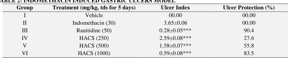
Indomethacin induced ulcer: It was observed that the treatment with HACS (250, 500 and 1000 mg/kg) and ranitidine (50 mg/kg) significantly reduced the lesion index, and provide the cytoprotection in comparison with indomethacin treated group (P< 0.05) (Table 2). Ethanol induced ulcer: Pretreatment with HACS showed significant reduction (P<0.05) in ulcer index. While Ranitidine (50 mg/kg) standard also showed significant decrease in the above parameter (Table 3).
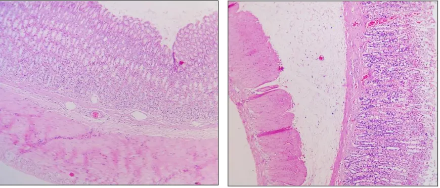
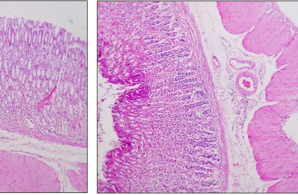
Histopathological evaluation: Pylorus ligation induced ulceration caused lesions including hemorrhagic erosion, oedema and lymphocytes infiltration of gastric mucosal layer. Pretreatment with HACS at doses 250, 500, 1000 mg/kg, showed dose dependent significant protection against all observed damages in gastric mucosa. Ranitidinde 50 mg/kg showed significant protection against the observed damages in gastric mucosa (Fig. 1).
TABLE 1: EFFECT OF HACS ON PYLORIC LIGATION INDUCED GASTRIC ULCERS IN RATS
Group Treatment (mg/kg, tds for 5 days) Ulcer Index Ulcer Protection (%)
I Vehicle 6.05±0.14 00.00
II Ranitidine (50) 0.39±0.7*** 93.5
III HACS (250) 4.07±0.11*** 32.8
IV HACS (500) 2.62±0.28*** 56.6
V HACS (1000) 1.012±013*** 83.2
[image:5.612.55.556.627.728.2]Data are mean±S.E.M, n = 6 in each group ***Significant variation as compared to pyloric ligated group (P<0.05).
TABLE 2: INDOMETHACIN INDUCED GASTRIC ULCERS MODEL
Group Treatment (mg/kg, tds for 5 days) Ulcer Index Ulcer Protection (%)
I Vehicle 00.00 00.00
II Indomethacin (30) 3.65±0.06 00.00
III Ranitidine (50) 0.28±0.05*** 90.4
IV HACS (250) 2.59±0.08*** 27.6
V HACS (500) 1.58±0.07*** 55.8
VI HACS (1000) 0.59±0.08*** 83.5
TABLE 3: ETHANOL INDUCED GASTRIC ULCERS MODEL
Group Treatment (mg/kg, tds for 5 days) Ulcer Index Ulcer Protection (%)
I Vehicle 5.733±0.1626 00.00
II Ranitidine (50) 0.683±0.1167*** 88.09
III HACS (250) 4.138±0.2257*** 40.71
IV HACS (500) 3.221±0.1116*** 57.39
V HACS (1000) 1.917±0.1456*** 79.13
Data are mean±S.E.M, n = 6 in each group ***Significant variation as compared to pyloric ligated group (P<0.05).
TABLE 4: EFFECT OF PRETREATMENT OF DRIED FRUIT PULP OF CUCUMISSATIVUS ON pH, GASTRIC VOLUME, FREE ACIDITY AND TOTAL ACIDITY IN PYLORIC LIGATED ANIMALS
Treatment (mg/kg, tds for 5 days) Gastric volume pH Free acidity Total acidity
Vehicle 5.91±0.29 1.66±0.66 94.33±1.38 119.7±1.74
Ranitidine (50) 1.61±0.11*** 4.35±0.06*** 32.5±1.17*** 57.67±1.56***
HACS (250) 3.85±0.12*** 2.31±0.07*** 51.83±1.66*** 80.5±1.25***
HACS (500) 3.35±0.28*** 2.36±0.07*** 47.17±0.94*** 73.17±1.04***
HACS (1000) 2.66±0.36*** 3.31±0.08*** 40.17±0.70*** 67.67±1.28***
Data are mean±S.E.M, n = 6 in each group ***Significant variation as compared to pyloric ligated group (P<0.05)
TABLE: 5. EFFECT OF EXTRACT ON LPO, SOD AND CAT LEVEL IN PYLORIC LIGATED RAT
Treatment (mg/kg, tds for 5 days) SOD CAT LPO
Vehicle 9.63±0.02 6.43±0.24 45.10±0.79
Ranitidine (50) 1.41±0.01*** 42.57±0.79*** 112.87±0.87***
HACS (250) 7.51±0.01*** 8.94±0.92*** 73.64±0.66***
HACS (500) 5.54±0.01*** 22.32±0.69*** 63.15±0.66***
HACS (1000) 2.47±0.01*** 34.1±0.63*** 53.66±0.94***
Data are mean±S.E.M, n = 6 in each group ***Significant variation as compared to pyloric ligated group (P<0.05)
TABLE: 6. EFFECT OF EXTRACT ON GASTRIC JUICE MUCOPROTEIN AND MUCOSAL GLYCOPROTEIN IN 4HRS PYLORIC LIGATED RAT
Treatment (mg/kg, tds for 5 days)
Proteins Hexosamine Hexoses Fucose Salic acid
Vehicle 333.64±2.13 119.5±5.87 88.57±5.87 36.98±2.34 12.43±2.86
Ranitidine (50) 166.16±6.24*** 195.39±1.27*** 363.32±6.34*** 178.51±3.56*** 55.64±2.23*** HACS (250) 323.56±4.13*** 134.76±1.28*** 99.66±4.32*** 45.67±3.12*** 14.26±2.78*** HACS (500) 309.33±3.63*** 145.43±1.33*** 114.57±4.98*** 59.89±3.89*** 18.72±2.54*** HACS (1000) 247.85±5.42*** 163.66±5.12*** 133.46±5.12*** 67.32±2.97*** 25.77±2.89*** Data are mean±S.E.M, n = 6 in each group ***Significant variation as compared to pyloric ligated group (P<0.05).
Histopathology Studies:
[image:6.612.96.531.546.732.2]
FIG.3: GROUP IV (HACS 500 MG/KG B.W.) FIG.4: GROUP V (HACS 1000 MG/KG B.W.)
DISCUSSION: The present study investigated antiulcer activity of hydroalcoholic extract of Cucumis sativus in different experimental gastric ulcer models.
The results of acute toxicity test shows that Cucumis sativus is a safe drug. The hydroalcoholic extract of Cucumis sativus showed significant antiulcer effect against pyloric ligation induced gastric ulcer, indomethacin induced gastric ulcers and ethanol induced gastric ulcers in rats. The pathogenesis of ulcer remains controversial but its causes is known to be aggravated due to an imbalance between the aggressive factors (i.e. acid and pepsin) and factors that maintain the mucosal integrity (i.e. mucus, bicarbonate and prostaglandins) 17.
Pylorus ligation induced ulcer was used to study the effect of fruit extracts on gastric acid secretion and mucus secretion. The ligation of the pyloric end of the stomach causes accumulation of gastric acid in the stomach. This increase in the gastric acid secretion causes ulcers in the stomach. HACS in PL-induced ulcers have showed marked protective effect via reduction in acid secretion, free & total acidity with simultaneous increase in acidic pH (Table 4). HACS also significantly increased the total carbohydrate content such as hexosamine, sialic acid, hexoses, fucoseand decreased the proteins of the gastric juice, found to be defensive factor in ulcers (Table 6).
Ulcers are caused due to imbalances between offensive (gastric acid secretion) and defensive
(gastric mucosal integrity) factors 18 and hence the effects of HACS can be explained based on these factors. In pyloric ligation model, the role of free radicals in gastric ulcerations is well documented
11
. HACS remarkably reduced lipid peroxidation in rat gastric mucosa (Table-5). HACS has been reported to possess significant anti-oxidant activity in vitro 19. SOD scavenges the super oxide radical one of the reactive oxygen species (ROS) responsible for lipid peroxidation. This reaction leads to increase in generation of peroxyl radical (RO2), which is also capable of producing more
oxidative damage 20. Pyloric ligation-induced ulceration involves damage by ROS apart from acid and pepsin related factors 21. The anti-oxidant activity in gastric mucosal was observed from decrease in LPO may be due to increase in SOD and CAT levels (Table 5).
HACS significantly increased SOD & CAT levels in all the three groups with simultaneous decrease in LPO level, suggesting decrease in oxidative damage. This may be due to restoration of balance between free radical scavenging enzymes SOD and CAT in the gastric mucosa, effectively counteracting the free radicals generated by cascade of reactions. Thus, the anti-ulcerogenic activity of HACS may also be due its anti-oxidant effects. The anti-ulcerogenetic activity of HACS can also be confirmed from the histopathological studies.
along the long axis of the glandular stomach as described in other studies. Treatment with HACS at three times in a day for five days (250, 500 & 1000 mg/kg) to different groups showed significant decrease in the intensity of gastric mucosal damages induced by the necrotizing agent indomethacin sodium compared with vehicle treated group (Table 2). Cytoprotective action by drugs has been considered to be due to the generation of prostaglandins or blockade of back diffusion of H+ ions 22 may be the major mechanism which is responsible for anti- ulcer activity.
Ethanol has been shown to increase the risk of ulcer in humans but produces potent ulceration in rats 23. Oral administration of ethanol interrupts the mucosal defense system, thereby aggravating mucosal damage that might bring about necrosis as well as apoptosis of gastric mucosal cells 24 and significant production of oxygen free radicals leading to increased lipid peroxidation, which causes damage to cell and cell membrane 25. Ethanol induces ulcers by the reduction of gastric mucosal blood flow and mucus production in the gastric lumen, a decrease in endogenous glutathione and prostaglandins levels and increase of ischemia, gastric vascular permeability, generation of free radicals, and production of leukotrienes. It has been found that oxygen-derived free radicals are implicated in the mechanism of acute and chronic ulceration and scavenging these free radicals can play an appreciable role in healing these ulcers. Ethanol induced generation of free radicals elevate the lipid peroxide level and reduces the cysteine, which is required for glutathione synthesis, thereby depleting glutathione levels. Reduced glutathione is found in high concentration in gastric mucosa of rats and humans. Glutathione is important for the maintenance of mucosal integrity and depletion of glutathione from the gastric mucosa induces macroscopic mucosal ulceration.
Different therapeutic agents including plant extracts are used to inhibit the gastric acid secretion, or to stimulate the mucosal defense mechanism by increasing the mucus production protecting the surface epithelial cells, or interfering with the PG synthesis 26. HACS significantly
healed the penetrating ulcers induced by ethanol after 5 days of treatment (Table 3). Hence, the cytoprotective and ulcer healing effect of HACS may be associated with its phytoconstituents that has predominant effects on both mucosal offensive and defensive factors.
The phytoconstituents like flavonoids, tannins, terpenoids and saponins have been reported in the extract. In several antiulcer literatures as possible gastro protective agent flavonoids, tannins and triterpenes are among the cytoprototecive active materials for which antiulcerogenic efficacy has been extensively confirmed 27.
CONCLUSION: In conclusion, the hydroalcoholic fruit extract of Cucuims sativus has a gastroprotective property against experimentally induced ulcers in rats and the research has to be done to know the exact gastroprotective mechanism.
ACKNOWLEDGMENT: The authors are grateful to the management of Gautham College of Pharmacy, Bangalore for providing necessary facilities to carry out the experiments and Green Chem, Bangalore for providing the herbal extract.
REFERENCES:
1. Dashputre L, Naikwade NS: Evaluation of anti-ulcer
activity of methanolic extract of Abutilon indicum Linn leaves in experimental rats. International journal of pharmaceutical sciences and drug research 2011; 3(2):97-100.
2. Trease GE and Evans WC: Trease and Evans
Pharmacognosy. Bailliere Tindal, London, Edition 16, 2009:27-39.
3. Kokate CK, Purohit AP, Gokhale SB: Phytochemical tests,
in: Pharmacognosy, Nirali Prakashan, Pune, Edition 35, 2006: 510-512.
4. http://www.oecd-library.org/2010.
5. Bharti S, Wahane VD and Kumar VL: Protective effect of
Calotropisprocera latex extracts on experimentally
induced gastric ulcers in rat. Journal of
Ethnopharmacology2010 Feb3; 127(2):440-444.
6. Lakshmi V, Singh N, Shrivastva S, Mishra SK, Dharmani
P, Mishra V and Palit G: Gedunin and photogedunin of
Xylocarpusgranatum show significant anti-secretory effects and protect the gastric mucosa of peptic ulcer in rats. Phytomedicine 2010 jul; 17 (8-9):569-574.
7. Vinod N, Albina A, Gopalakrishna HN, Dorababu P,
Mirshad PV, Divya B and Dipsanker C: Evaluation of the
anti-ulcer activity of NR-ANX-C (a polyherbal
8. Roy SP, Niranjan CM, Jyothi TM, Shankrayya MM, Vishawanath KM, Prabhu K, Gouda VA and Setty RS: Antiulcer and anti-inflammatory activity of aerial parts
Enicostemmalittorale Blume. Journal of Young Pharmacists 2010 Oct-Dec; 2(4):369-373.
9. Jince MJ, Kandhasamy S and Sellamuthu M: Protective effects of methanolic extract of Hedyotispuberula (G. Don) R. Br. ex Arn. against experimentally induced gastric ulcers in rat. Journal of Ethnopharmacology2010 Aug 19; 131(1):216-219.
10. Bhagawati S, Sairam K and Sanjay S: Gastroprotective potential of risperidone, an atypical antipsychotic, against stress and pyloric ligation induced gastric lesions. Chemico-Biological Interactions 2011 Apr25; 190(2-3):155-164.
11. Rakesh K, Sindhu, Pradeep K, Jagdeep K, Ashok K and Sandeep A: Investigation into the antiulcer activity of
Rheum ribes Linn. leaves extracts. International Journal of Pharmacy and Pharmaceutical Sciences 2010; 2(4):90-93.
12. Kar DM, Maharana L, Si SC, Ka MK and Sasmal D:Anti
ulcer activity of ethanolic extract of fruit of
Trapabispinosa Roxb in animals. Der Pharmacia Lettren 2010; 2(2):190-197.
13. Sathaye S, Bagul Y, Gupta S, Kaur H, Rdkar R:
Hepatoprotective effects of aqueous leaf extract and crude isolates of Murrayakoenigii against in vitro
ethanol-induced hepatotoxicity model. Experimental and
Toxicologic Pathology 2011 Sep; 63(6):587-591.
14. Bavani EM, Surendran S, Vijayakumar M, Ojha S, Rawat
AKS, Rao ChV: Gastroprotective activity of
Cinnamomumtamala leaves on experimental gastric ulcers in rats. Journal of Ethnopharmacology 2010 Mar24; 128(2):537-540.
15. Ghosh, MN: Fundamentals of Experimental
Pharmacology. Hilton and Company, Kolkata, Edition 3, 2005:153-158.
16. Hollander D, Taranawski A, Krause WJ, and Gergely H: Protective effect of sucralfate against alcohol-induces gastric mucosal injury in the rat. Gastroenterology 1985; 88:366–374.
17. Berardi RR and Welage SL: Peptic Ulcer Disease. In: Dipiro TJ, Talbert RL, Yees GC, Matzke GR, Wells GB and Posey ML: Pharmacotherapy- a pathophysiologic approach. McGraw-Hill, Edition 6, 2005:632-648.
18. Laine L, Takeuchi K, Tarnawski A: Gastric mucosal
defense and cytoprotection: bench to bedside.
Gastroenterology 2008; 135:41–60.
19. Kumar D, Kumar S, Singh J, Nagender, Rashmi, vashistha
BD and Singh N: Free Radical Scavenging and Analgesic Activities of Cucumis sativus L. Fruit extract. Journal of Young Pharmacist 2010 Oct-Dec; 2(4):365-368.
20. Parameswari RP, Vasanthkumar M, Gayathri V,
Manikandamathavan VM, Ramakrishnan G, Sangeetha
MK, Vijayakumar V, Balaji H, Raghavendran,
Chamundeeswari D and Vasanthi HR: Prophylactic Role of a Herbomineral Drug “Thamira Parpam” Against Cysteamine-Induced Oxidative Stress in Liver and Duodenum of Rats. Biological Trace Element Research 2010; 138(1-3):212-225.
21. Muthusamy P, Suresh AJ and Balamurugan G: Antiulcer Activity of Azimatetracantha: A Biochemical Study Research Journal of Pharmacy and Technology 2009; 2(2): 344-48.
22. Rakesh AK, Gulecha VS, Mahajan MS, Mundada AS and
Gangurde HH: Evaluation of antiulcer activity of
polyherbal formulation. International Journal of
Pharmaceutical Research and Development 2009; 1(10):1-6.
23. Robert A, Nezamis JE, Lancaster C and Hanchar AJ: Cytoprotection by prostaglandins in rats: Prevention of gastric necrosis produced by alcohol, HCl, NaOH, Hypertonic NaCl and thermal injury. Gastroenterology 1979; 17:433-443.
24. Kandhasamy SB, Chinb NK: Antioxidant and antiulcer effects of ethylacetate fraction of Merremia tridentate (L.) Hallier F. Root. Agriculture and agricultural science procedia 2014; 2:406-414
25. Vinothapooshan G, Sundar K: Antiulcer activity of
Mimosa pudica leaves against gastric ulcer in rats. Research journal of pharmaceutical, biological and chemical sciences. 2010 Oct-Dec; 1(4):606-614.
26. Ghangale GR, Mahale T and Jadhav ND: Evaluation of Antiulcer Activity of Ocimum sanctum in Rats, Veterinary World 2009; 2(12):465-466.
27. Kumar K, Mruthunjaya K, Kumar S, Rajendran M:
Antiulcer activity of ethanol extract of the stem bark of
Careyaarborea Roxb. International current pharmaceutical journal 2013 Feb; 2(3):78-82.
All © 2013 are reserved by International Journal of Pharmaceutical Sciences and Research. This Journal licensed under a Creative Commons Attribution-NonCommercial-ShareAlike 3.0 Unported License. This article can be downloaded to ANDROID OS based mobile. Scan QR Code using Code/Bar Scanner from your mobile. (Scanners are available on Google Playstore)
How to cite this article: